DI Exam 1
1/123
There's no tags or description
Looks like no tags are added yet.
Name | Mastery | Learn | Test | Matching | Spaced | Call with Kai | Chat |
|---|
No analytics yet
Send a link to your students to track their progress
124 Terms
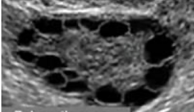
Polycystic ovary
What is this and what type of scan
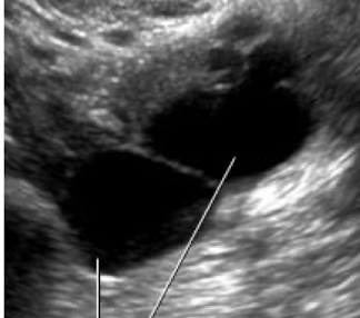
Ovarian Cyst, Pelvic US
What is this
Ovarian Cancer
What is this
Bladder Cancer
What is this and what type of scan
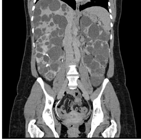
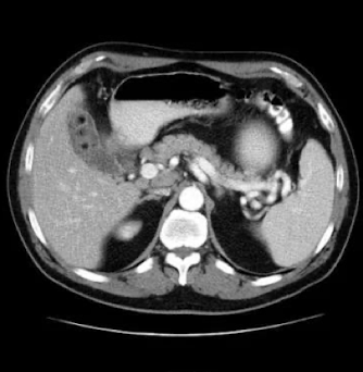
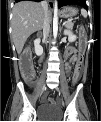
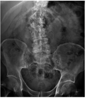
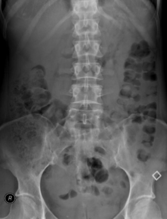
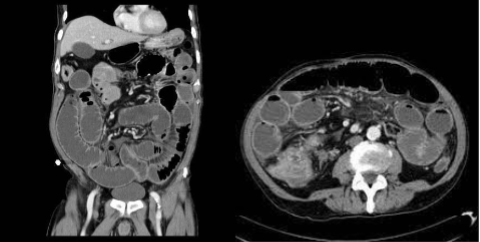
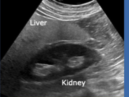
Polycystic kidney disease, CT scan

What is this and what type of scan
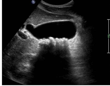
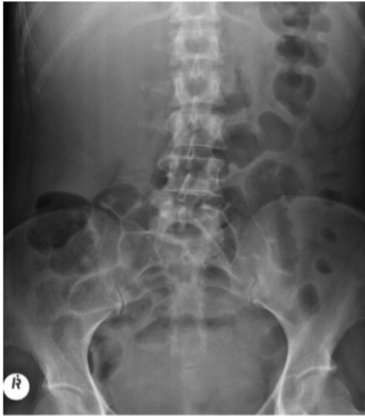
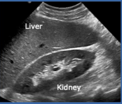
Polycystic kidney disease, US
What is this
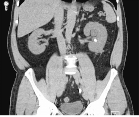
Renal Calculi
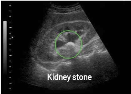
What is this and what type of scan
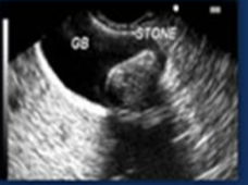
Renal Calculi, US
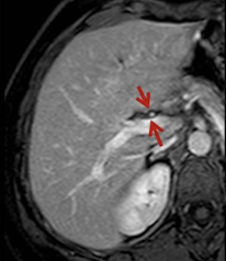
What is this and what type of scan
Cirrhotic liver, CT
What is this and what type of scan
Normal liver, CT
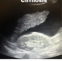
What is this and what type of scan
Liver Cirrhosis, US
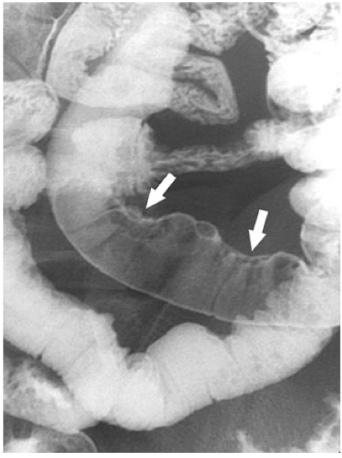
What is this and what type of scan
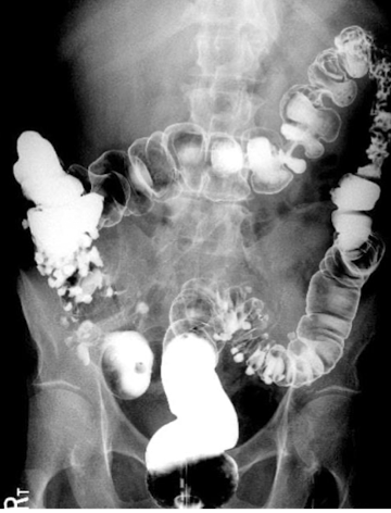
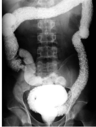
colon polyp, barium enema
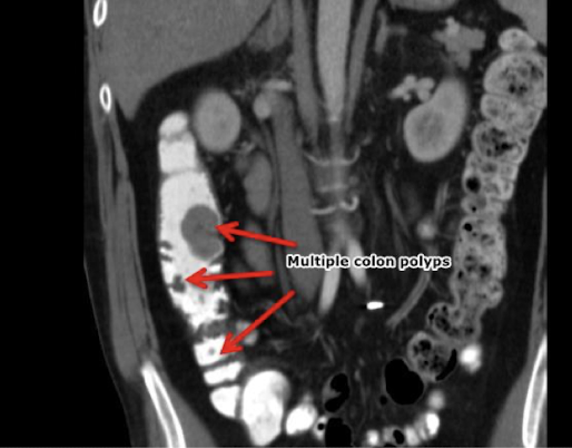
What is this and what type of scan
Colon polyp, CT
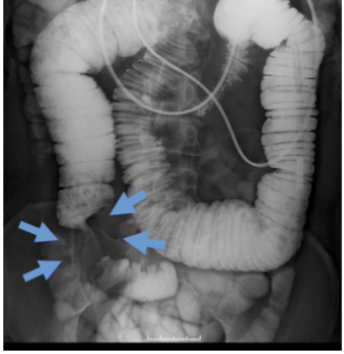
What is this and what type of scan
Diverticulosis, Barium Enema
What is this and what type of scan
Diverticulosis, CT
What is this and what type of scan
Fatty liver, US
What is this and what type of scan
Perforation, Barium Enema
What is this and what type of scan
Appendicits, US
What is this and what type of scan
Appendicits, CT
What is this and what type of scan
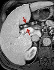
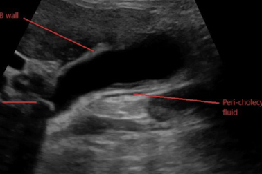
Cholecystitis, US
What is this and what type of scan
Cholecystitis, CT
What is this
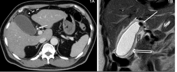
Cholelithiasis
What is this and what type of scan
Cholelithiasis, CT
What is this
Ulcerative colitis
What is this
Ulcerative colitis
What is this and what type of scan
crohns, CT
What is this and what type of scan
Crohns, Upper GI w sm bowel follow through
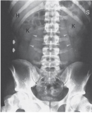
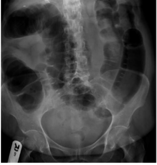
What are H, S, K what type of scan is this
H: Hepatic angle, S: spleen, K: kidney, Normal Abdominal X-ray
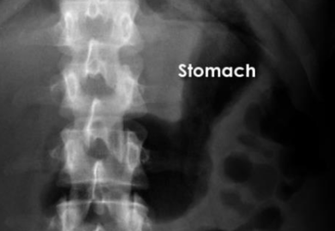
What is this and what type of scan
Stomach, AXR
What is this
small bowel
What is this and what type of scan
Large bowel, AXR
What is this and what type of scan
Liver, AXR
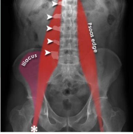
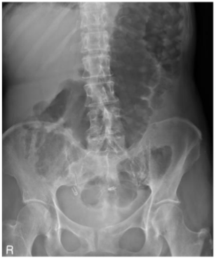
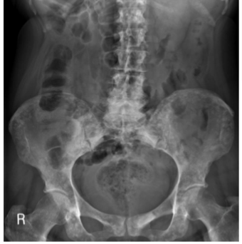
Identify Psoas muscles, lumbar vertebrae, femur, illiacus
yes
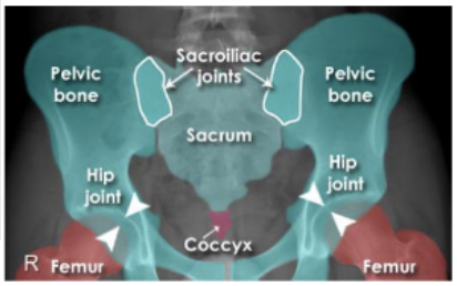
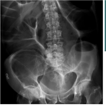
Identify parts of pelvis (6)
yes
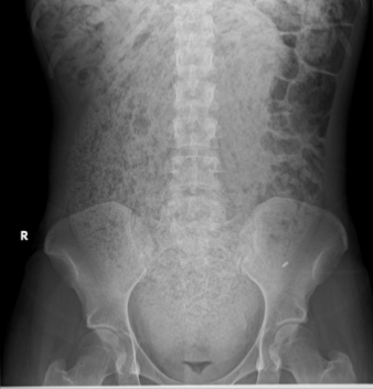
What is this and what type of scan
Calcifications, AXR
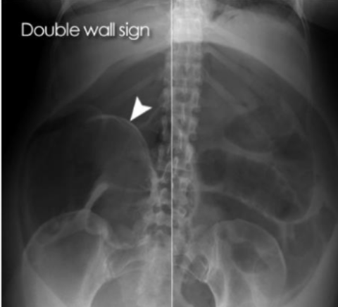
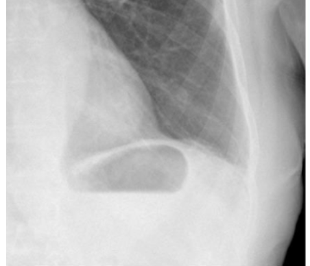
What is this and what type of scan
free gas under the diaphragm 2/2 perforated duodenal ulcer (abnormal bowel gas pattern), ABX
What is this and what type of scan
free air in bowel 2/2 perforated bowel, abx
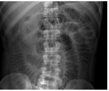
What is this and what type of scan
normal stomach bubble, axr
What is this and what type of scan
small bowel obstruction/ileus, abx
What is this
Large bowel obstruction
What is this and what type of scan
sigmoid volvulus (twisted) “bird beak sign or coffee bean sign”, abx
What is this and what type of scan
Bowel wall inflammation/toxic mega colon, abx
What is this and what type of scan
chronic constipation, abx
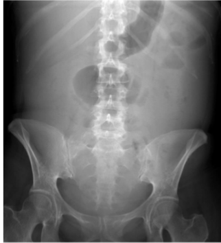
What is this and what type of scan
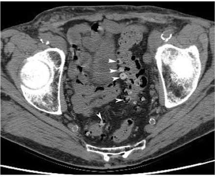
AAA, Abx
What is this and what type of scan
Hepatomeagly, abx
What is this and what type of scan
Splenomegaly
What is this and what type of scan
ascites, abx
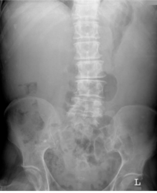
What is this and what type of scan
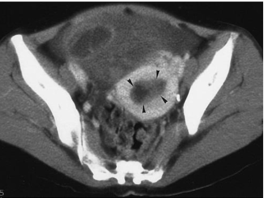
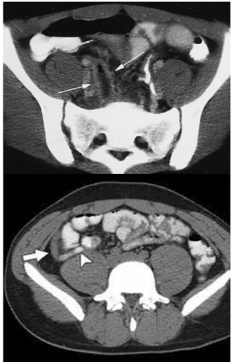
small pelvic mass, abx
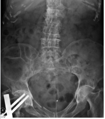
What is this and what type of scan
fracture and osteoarthritis, abx
What is this and what type of scan
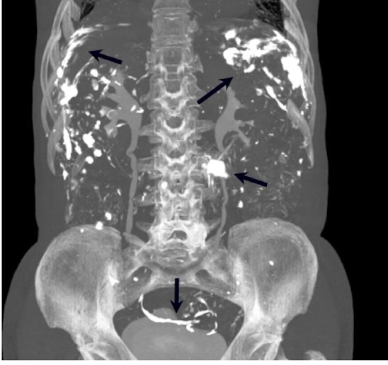
bone mets, abx
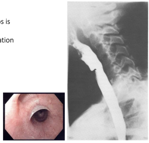
What is this
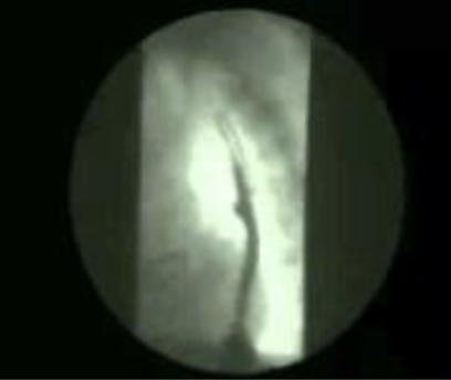
Achalasia
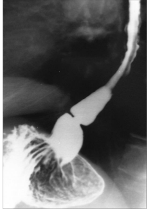
What is this
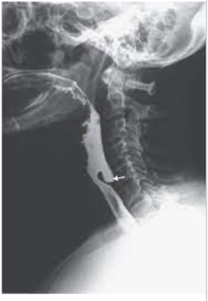
Zenkers Diverticulum
What is this
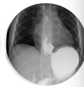
Hiatal hernia
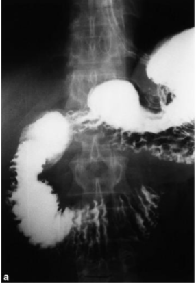
What is this and what type of scan
GERD, barium swallow
What is this
Esophageal ca
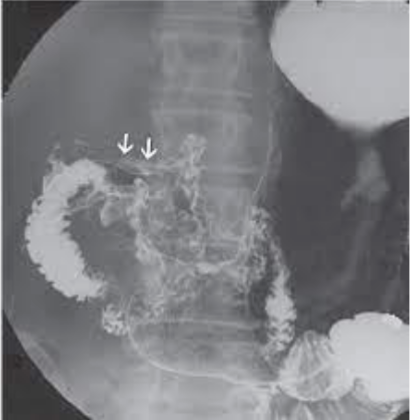
What is this
Esophageal web
What is this
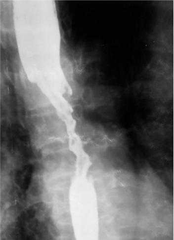
Schatzki ring
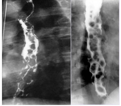
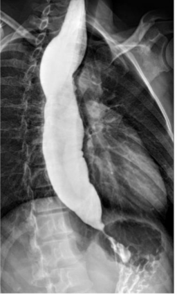
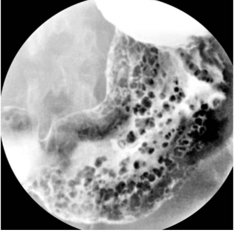
What is this and what type of scan
Varices, Barium swallow
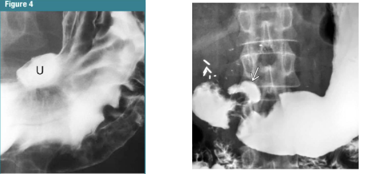
What is this and what type of scan
Peptic ulcer disease, barium swallow
What is this
Pancreatic ca
What is this
duodenal cancer
What is this and what type of scan
gastric polyps, barium swallow
What is this
small bowel obstruction, barium swallow
What is this
small bowel obstruction
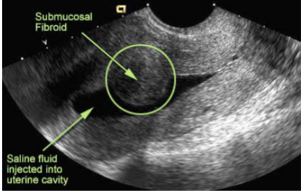
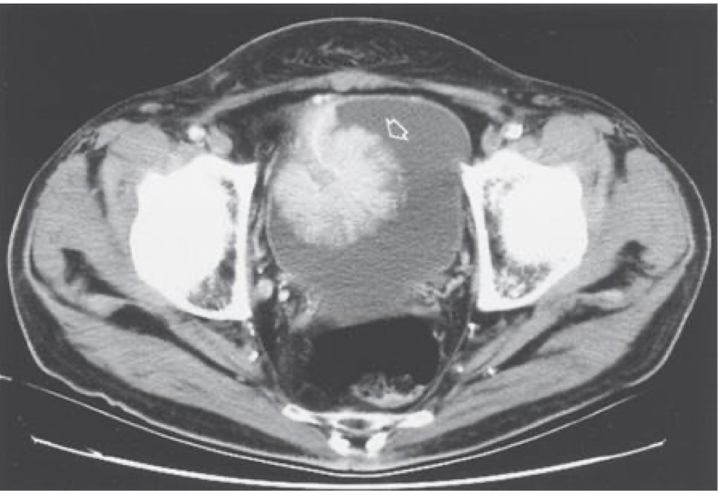
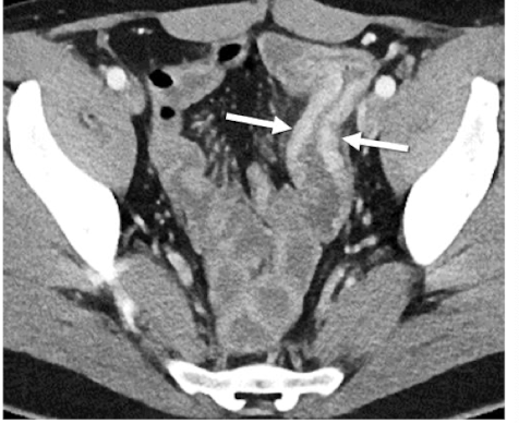
What is this and what type of scan
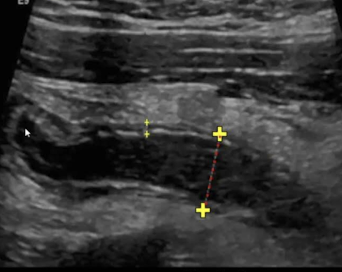
fibroids, transvaginal US
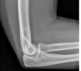
What is this
normal elbow

What is this
post + ant fat pad
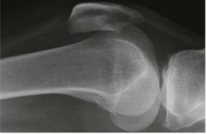
What is this and what type of scan
normal knee, x-ray
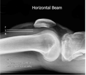
What is this and what type of scan
fat fluid level, x-ray
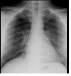
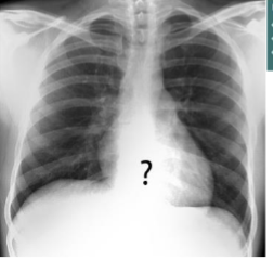
What is this and what type of scan
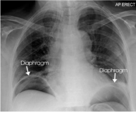
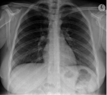
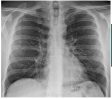
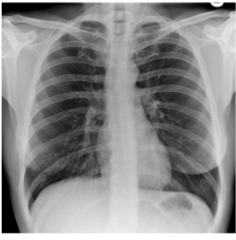
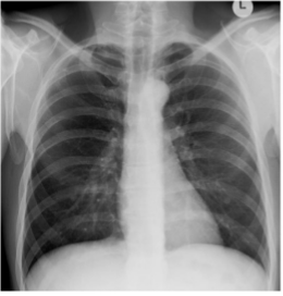
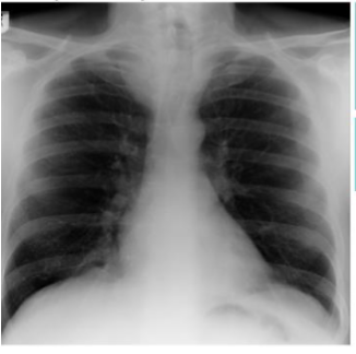
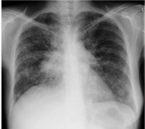
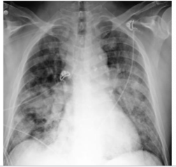
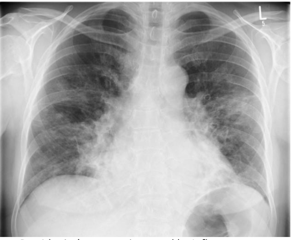
PA CXR
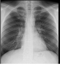
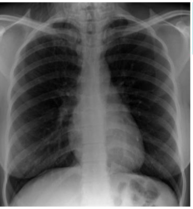
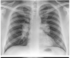
What is this
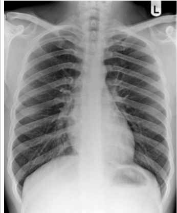
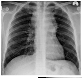
AP
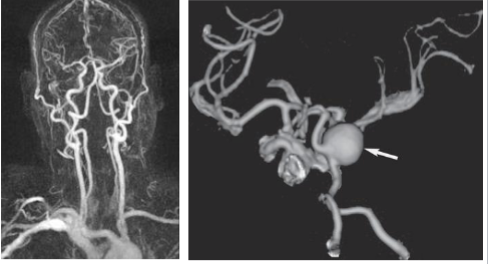
What is this and what type of scan
CT angio
Echogenicity of this
isoechoic
Echogenicity of this
Kidney = hypoechoic to liver
Liver = hyperechoic to kidney
Echogenicity of this
Gallbladder = anechoic
stone = hyperechoic
Echogenicity of this
tumor = hypoechoic to surrounding tissue
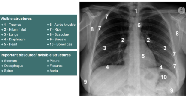
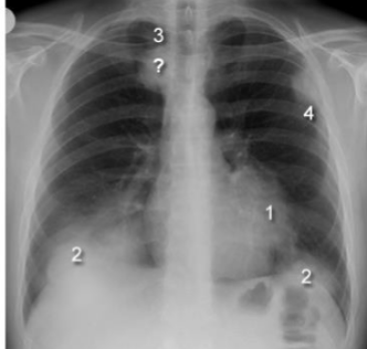
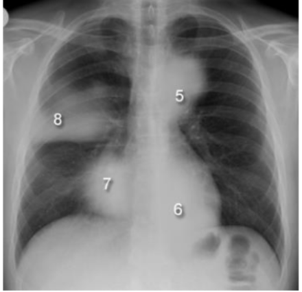
Label this CXR (10 things)
1) trachea 2) hilum- should have R and L 3) lungs 4) diaphragm 5) heart 6) aortic knuckle 7) ribs 8) scapulae 9) breasts 10) bowel gas
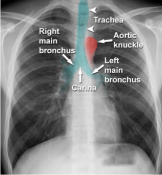
Identify trachei, bronchi, aortic nuckle
yes
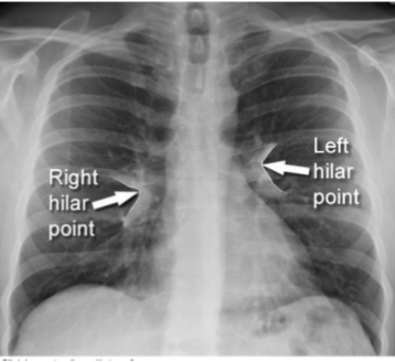
Idenfity R + L hila
yes
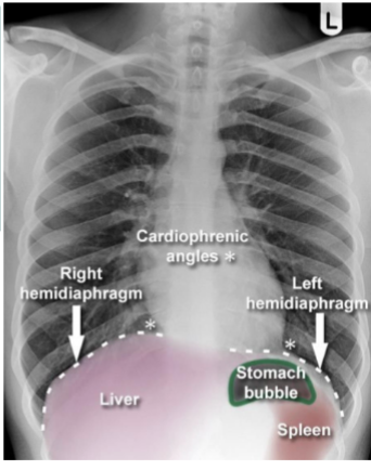
Identify cardiophrenic angle, R + L hemidiaphragms, stomach bubble, liver, spleen
yes
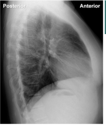
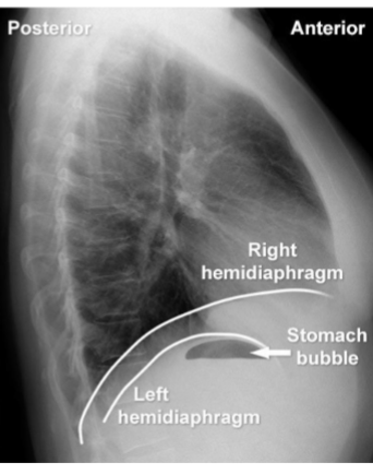
identify hemi diagrams (2) and stomach bubble
yes
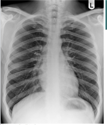
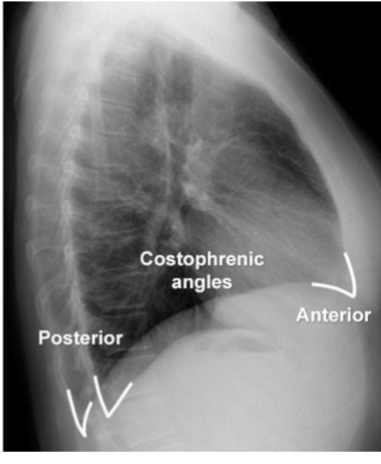
identify costophrenic angles
yes
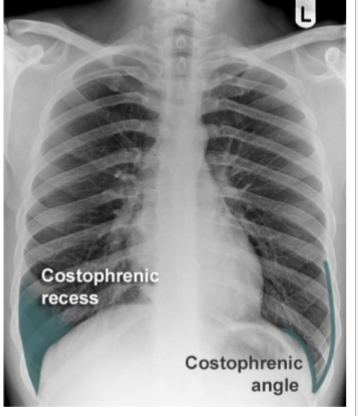
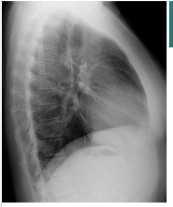
Identify costophrenic angles (post + ant)
yes
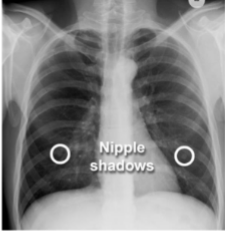
Identify soft tissues/what are you looking for?
nipples
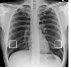
identify nipple shadows
yes
what do u see?
under penetration
what do you see?
hair
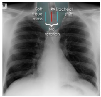
what is this?
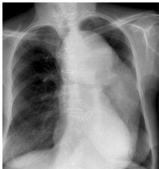
tracheal displacement
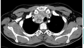
what is this and what kind of scan
tracheal displacement, CT
what is this? and what causes it
B/L hilar enlargement, Sarcoidosis
what is this? and what causes it
asymmetric hilar enlargement, breast ca, metastatic disease
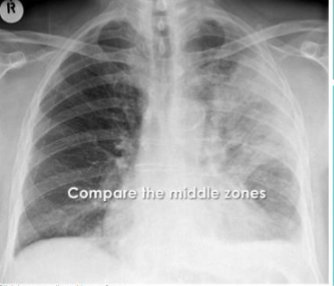
what is this? and what causes it
Mediastinal mass (aortic knuckle deviated), hodgkin lymphoma
what is this? and what causes it
Mediastinal mass, hodgkin lymphoma
what is this? and what causes it
Thoracic aortic aneurysm

what is this?
lobar pna
what is this?
multifactorial pna
What are these silhouette signs? what causes them?
1: L Heart boarder (lingula disease), 2: Hemidiaphragm (lower lobe lung disease), 3: paratracheal stripe (paratracheal disease), 4: chest wall (lung, pleural or rib disease)
What are these silhouette signs? what causes them?
5: aortic knuckle (anterior mediastinal or L upper lobe disease), 6: paraspinal line (post thorax disease), 7: R heart boarder (middle lobe disease) 8: density above horizontal fissure
What is this? and what causes it?
air space filling w air bronchograms, PNA
What is this? and what causes it?
viral pna caused by influenza