The thorax wall and pleura
 Call Kai
 Learn
Learn Practice Test
 Spaced Repetition
 Match
Match Flashcards
 Knowt Play
1/62

There's no tags or description
Looks like no tags are added yet.
Name | Mastery | Learn | Test | Matching | Spaced | Call with Kai | Chat |
|---|
No analytics yet
Send a link to your students to track their progress
63 Terms
What makes up the boundaries of the thoracic cage?
dorsally- thoracic vertebrae
laterally- ribs and intercostal mm.
ventrally- sternum
caudally- thoracic outlet
cranially- thoracic inlet
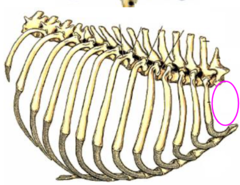
what makes up the boundaries of the thoracic inlet?
T1 vertebrae and longus colli m., first pair of ribs, and manubrium of sternum
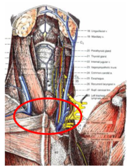
what structures pass through the thoracic inlet?
-esophagus
-trachea
-external jugular vv.
-common carotid artery
-vagosympathetic trunks
-recurrent laryngeal nn.
-roots of the phrenic nn.
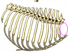
what are the boundaries of the thoracic outlet?
-last thoracic vertebrae
-last pair of ribs and costal arch
-xiphoid
-it is closed by the diaphragm

What are the external intercostal mm?
-Fill the ICS up to the CCJ
-Inspiratory
-FIBERS RUN CAUDOVENTRALLY
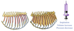
What are the internal intercostal muscles?
-Fill up the ICS up to the sternum
-Expiratory
-FIBERS RUN CRANIOVENTRALLY
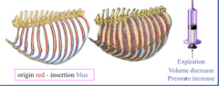
What are the inspiratory muscles?
1. Scalenus m
2. serratus dorsalis cranialis m.
3. diaphragm
4. rectus thoracis
5. external intercostal mm (not shown in image)
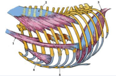
What are the expiratory muscles?
1. internal intercostal mm.
2. serratus dorsalis caudalis m.
3. abdominal muscles (not shown in image)

where is the attachment of the inspiratory muscles?
-muscle fiber direction = caudoventral
-insertion on ribs is caudoventral to origin

What is the action of the inspiratory muscles?
-pull the ribs craniodorsally
-widen the thoracic cavity
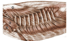
What innervates the inspiratory and expiratory muscles?
ventral branches of the thoracic spinal (intercostal) nerves

What is the attachment of the expiratory muscles?
-fibers directed cranioventral from the cranial border of the preceding ribs
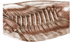
What is the action of the expiratory muscles?
-act to decrease the volume within the thoracic cavity by pulling ribs caudally

Where is the intercostal V.A.N located?
along the CAUDAL border of each rib
if you are doing surgery, do NOT make an incision caudal to the rib. Make it in the middle of the ICS. If you are doing a nerve block, put it just caudal to the rib

the intercostal nerves are the ________________ branches of the thoracic spinal nerves
VENTRAL

Where do the dorsal intercostal veins drain into?
the Azygous veins

Where do the ventral intercostal veins drain in to?
internal thoracic veins

When blocking nerves after thoracotomy how many intercostal nerves should you block?
you should block a minimum of two to three adjacent intercostal nerves due to overlapping nerve supply

what is the endothoracic fascia?
connective tissue (CT) lining the thoracic wall outside of the parietal pleura
it also forms the C.T. of the mediastinum where it surrounds nerves, vessels, and other structures within the mediastinum
in the middle of the mediastinum, it becomes dense (condenses) and forms the fibrous pericardium
what is the clinical significance of the endothoracic fascia being continuous with the deep fascia of the neck?
if you have an infection in the fascia of the neck, it can spread to the thoracic cavity
the diaphragm is innervated by the __________________
phrenic nerve
what does the diaphragm do?
contraction of the diaphragm increases the volume of the thoracic cavity --> main inspiratory muscle

true/false- it is normal to hear gut sounds under the costal regions (Intrathoracic part of the abdominal cavity)
TRUE- the abdomen pushes the diaphragm cranially and invades
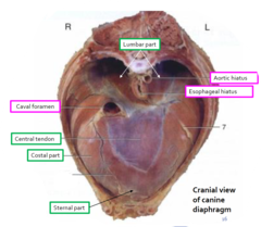
What are the parts of the diaphragm?
1. tendinous center (central tendon)
2. muscle (periphery) = lumbar, costal, and sternal part
What are the openings in the diaphragm?
1. aortic hiatus
2. esophageal hiatus
3. caval foramen
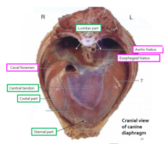
What is included in the aortic hiatus?
aorta, azygous, thoracic duct (lymphatic duct)
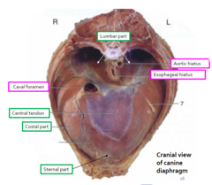
What is included in the esophageal hiatus?
esophagus, ventral and dorsal vagal trunks
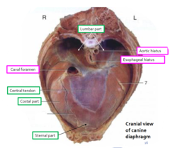
What is included in the caval foramen?
caudal vena cava

What is the cupula of the diaphragm?
the most cranial portion of the diaphragm
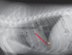
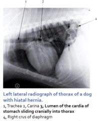
what happens when there is a hiatal hernia?
the stomach prolapses into the thoracic cavity
traumatic diaphragmatic ruptures usually involve the ___________ part
costal
the liver is the commonly herniated organ due to its position and attachment to the diaphragm
true/false- animals that lose one or both phrenic nerves causes a large effect on the animal's ability to breathe
FALSE- the diaphragm is a skeletal muscle supplied by the phrenic nerve. When an animal gets a phrenectomy it has minimal effects on the animal's ability to breathe. Pressure from the limbs, the intercostal muscles, and other inspiratory muscles hypertrophy in order to compensate for the paralyzed diaphragm
what is the pleura?
the serosa lining the thoracic cavity and the thoracic organs
what are the different names for the serosa in the various body cavities?
1. pleura = thorax
2. pericardium = surrounding the heart
3. peritoneum = in abdominal and pelvic cavities
4. vaginal tunic = testicles
the potential spaces between the parietal and visceral serous membranes contain ________________
serous fluid to prevent friction
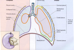
What are the four different types of pleura?
1. visceral pleura
2. parietal pleura
3. mediastinal pleura (covers mediastinum = place between lungs)
4. pericardial pleura (part of mediastinal pleura on either side of pericardium)

what are the two regions of the parietal pleura?
1. costal pleura: lines ribs and intercostal mm.
2. diaphragmatic pleura: covers diaphragm

why do dogs and cats rarely present with a unilateral penumothorax?
the pleura in the ventral part of the caudal mediastinum is fused. It is thin and devoid of intervening connective tissue. it is sometimes described as fenestrated.
due to the flimsy caudal mediastinal pleura, dogs and cats rarely present with a unilateral pneumothorax

What are the pleural cupulae?
"bubbles" of pleura at the cranial extent of each lung that protrude a short distance through the thoracic inlet into the neck
If you do surgery at base of neck BE CAREFUL... you could cause pneumothorax

the most cranial part of the pleural sac is the ___________________
cupula pleurae
The most caudal part of the pleural sacs is the __________________________
costodiaphragmatic recess
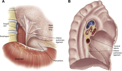
What is the pulmonary ligament?
the continuation/connection between the pulmonary and mediastinal pleura at the hilus of the lung. It is formed where the mediastinal pleura reflects to become the pulmonary pleura
AKA The pulmonary ligament is a double serosal sheath that extends from the lower lobe of the lung to the mediastinum and from the hilus to the diaphragm.
What is the plica venae cavae?
separate fold of mediastinal pleura on the right side that surrounds the caudal vena cava and right phrenic nerve extending into the diaphragm

what is the mediastinal recess?
a small pleural space medial to the plica venae cavae.
it is occupied by the accessory lobe of the lung
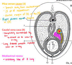
what are the three regions of the mediastinum?
1. cranial mediastinum (from thoracic inlet to the heart)
2. middle (cardiac) mediastinum
3. caudal mediastinum (between heart and diaphragm)

What is included in the dorsal portion of the cranial mediastinum?
esophagus, trachea, great vessels of the heart (brachiocephalic trunk and cranial vena cava) and their branches
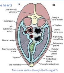
What is included in the middle/ventral portions of the cranial mediastinum?
thymus, internal thoracic aa and vv, cranial mediastinal lymph nodes

what is included in the dorsal portion of the middle mediastinum?
esophagus, tracheal bifurcation and root of lung, tracheobronchial lymph nodes, aortic arch

What is included in the contents of the middle/ventral portions of the middle mediastinum?
heart and pericardium, folds of pleural tissue
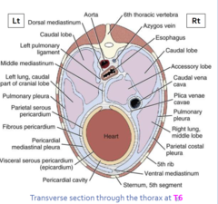
what is included in the contents of the dorsal portion of the caudal mediastinum?
esophagus, aorta, and right azygos vein
What is included in the middle portion of the caudal mediastinum?
caudal vena cava
What is included in the ventral portion of the caudal mediastinum?
the ventral portion is reduced to the potential space between the layers of pleura
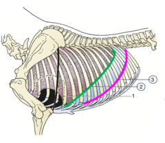
what is the line of pleural reflection?
where the costal pleura reflects onto the diaphragm forming the costodiaphragmatic recess (narrow space in the pleural cavity that's located where the costal and diaphragmatic pleura meet)

what is the basal (caudal) border of the lung
it is the caudal most extent the lung expands during inspiration
true/false- the region between the basal border of the lung and the line of pleural reflection is always devoid of lung
TRUE
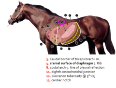
line of pleural reflection is shown in pink and the basal border of the lung is shown in green
what is the superficial thoracic "spur" vein?
it courses along the thoracic body wall at about the level of the olecranon, it should be avoided.
thoracocentesis (tapping the pleural cavity) is usually done just dorsal to the superficial thoracic vein. the needle is inserted between the 7th on the R side and 8th/9th ICS on the L side
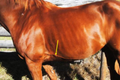
what is a pleural effusion?
fluid in the pleural space
excess fluid --> limits ling expansion --> lung collapse and difficult breathing
What is hydrothorax?
excess serous fluid
What is haemothorax?
blood in the pleural space
What is pyothorax?
pus in the pleural space
What is chylothorax?
Presence of lymphatic fluid in the pleural space.
why doesn't the elephant get pleural effusion?
it has no pleural space
What is pneumothorax?
air in the pleural cavity
air can enter in two ways: externally from penetrating wound or internally due to lung trauma