Edexcel IAL Biology Unit 2 - Topic 3: Cell Structure
1/43
There's no tags or description
Looks like no tags are added yet.
Name | Mastery | Learn | Test | Matching | Spaced | Call with Kai |
|---|
No study sessions yet.
44 Terms
What are all living things composed of?
eukaryotic and prokaryotic cells.
Ultrastructure of eukaryotic cells
Human cells are eukaryotic.
Contain a nucleus and membrane-bound organelle.
Most eukaryotic cells have the same internal organelles, but cell specialisation means cells often differ in number and sometimes types of organelle present.
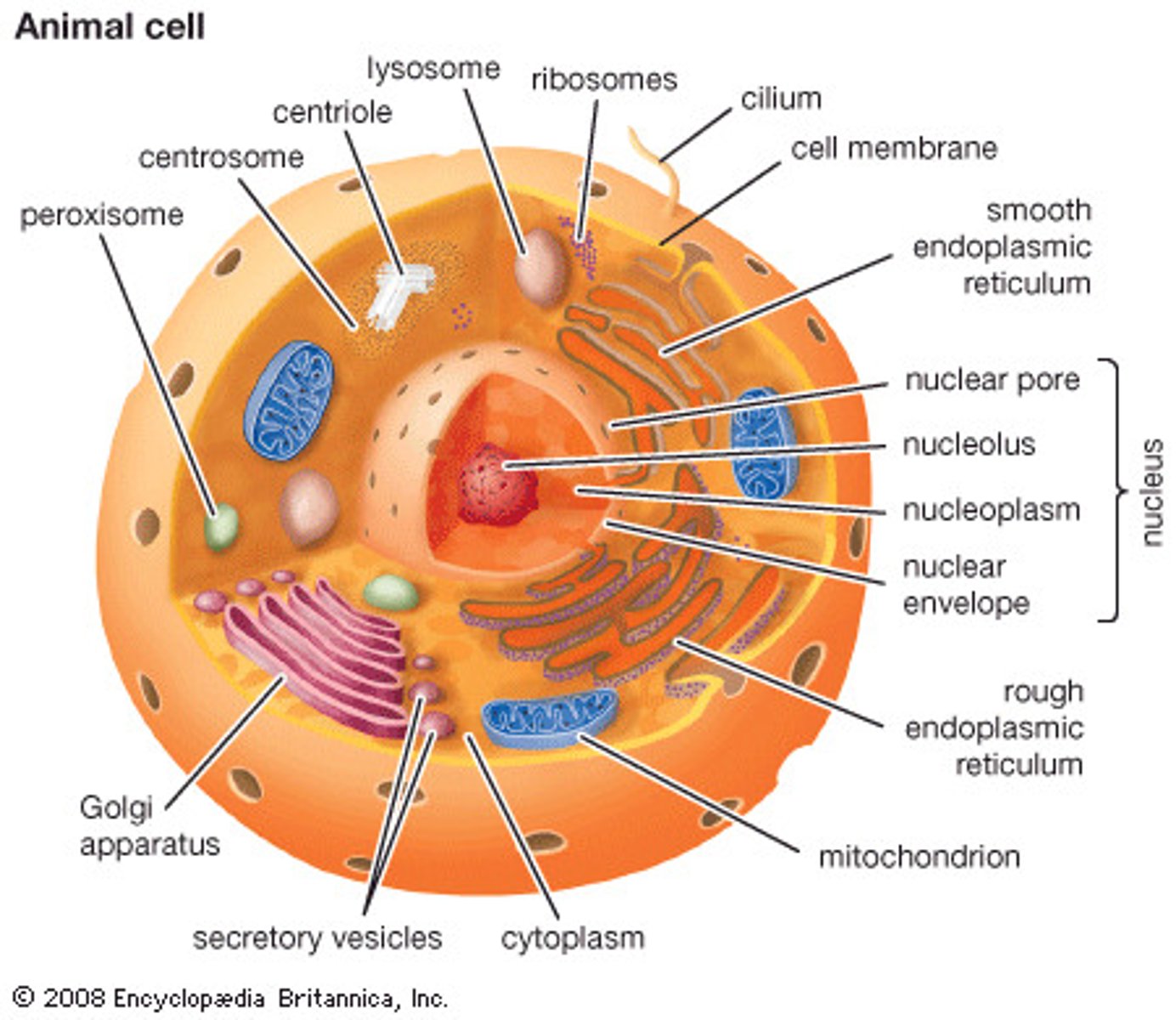
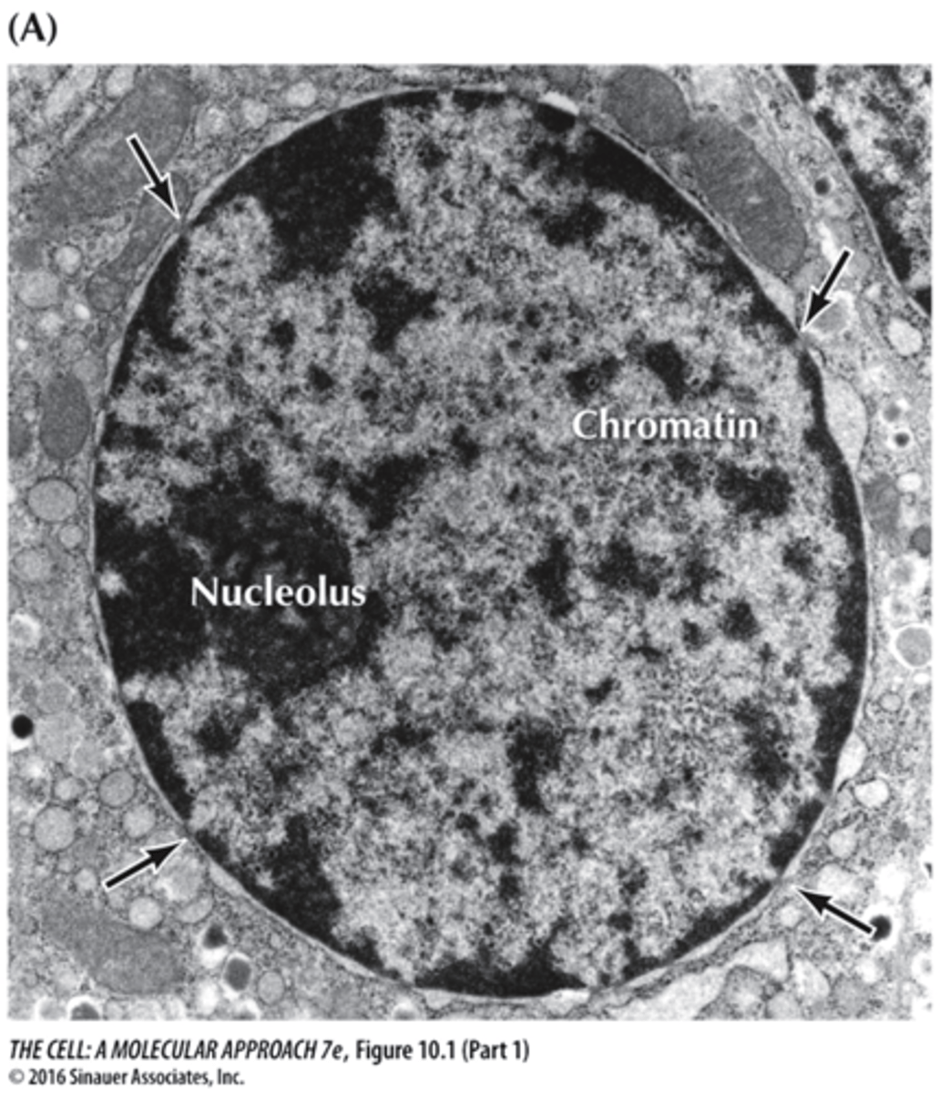
Structure and function of nucleus
double membrane bound called the envelope containing pores which enable molecules to enter and leave the nucleus.
has linear DNA which dictates protein synthesis
site of transcription of DNA
nuclear Membrane enables compartmentalisation and keeps reactions occuring in the nucleus and its contents separate from the cyptoplasm
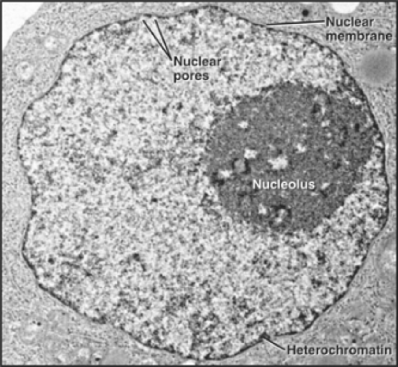
Structure and function of nucleolus
Site of ribosome production and rRNA
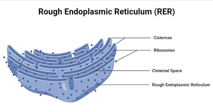
Structure and function of rough endoplasmic reticulum
ribosomes attached to its outer surface and has single membrane bound cisternae
increases S.A for attachment of ribososmes.
polypeptide chain moves into the cisternae of the RER and folds to assume a tertiary structure.
protein is packaged into vesicles which bud off from the RER and go fuse with the golgi
Structure and function of smooth endoplasmic reticulum
A system of membrane bound sacs.
Produces and processes lipids.

Structure and function of ribosomes
Composed of two subunits made of RNA + protein
Site of translation of mRNA
not membrane bound
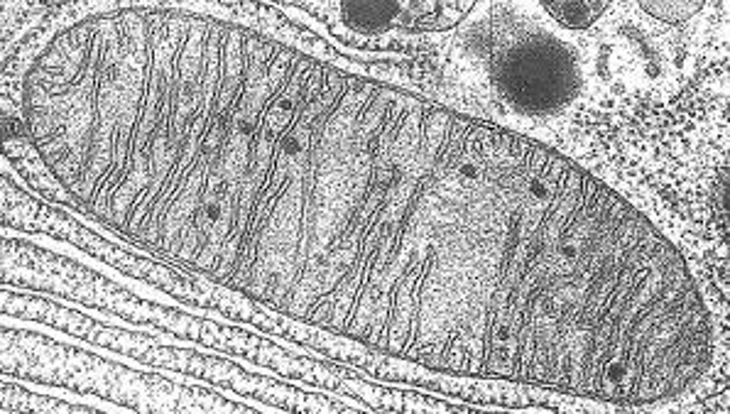
Structure and function of mitochondria
bound by a double membrane called the envelope.
Inner membrane is highly folded to increase surface area for attachment
with a matrix on the inside, containing all the enzymes needed for respiration.
70s ribosomes present
site of aerobic respiration which forms ATP providing energy for active transport
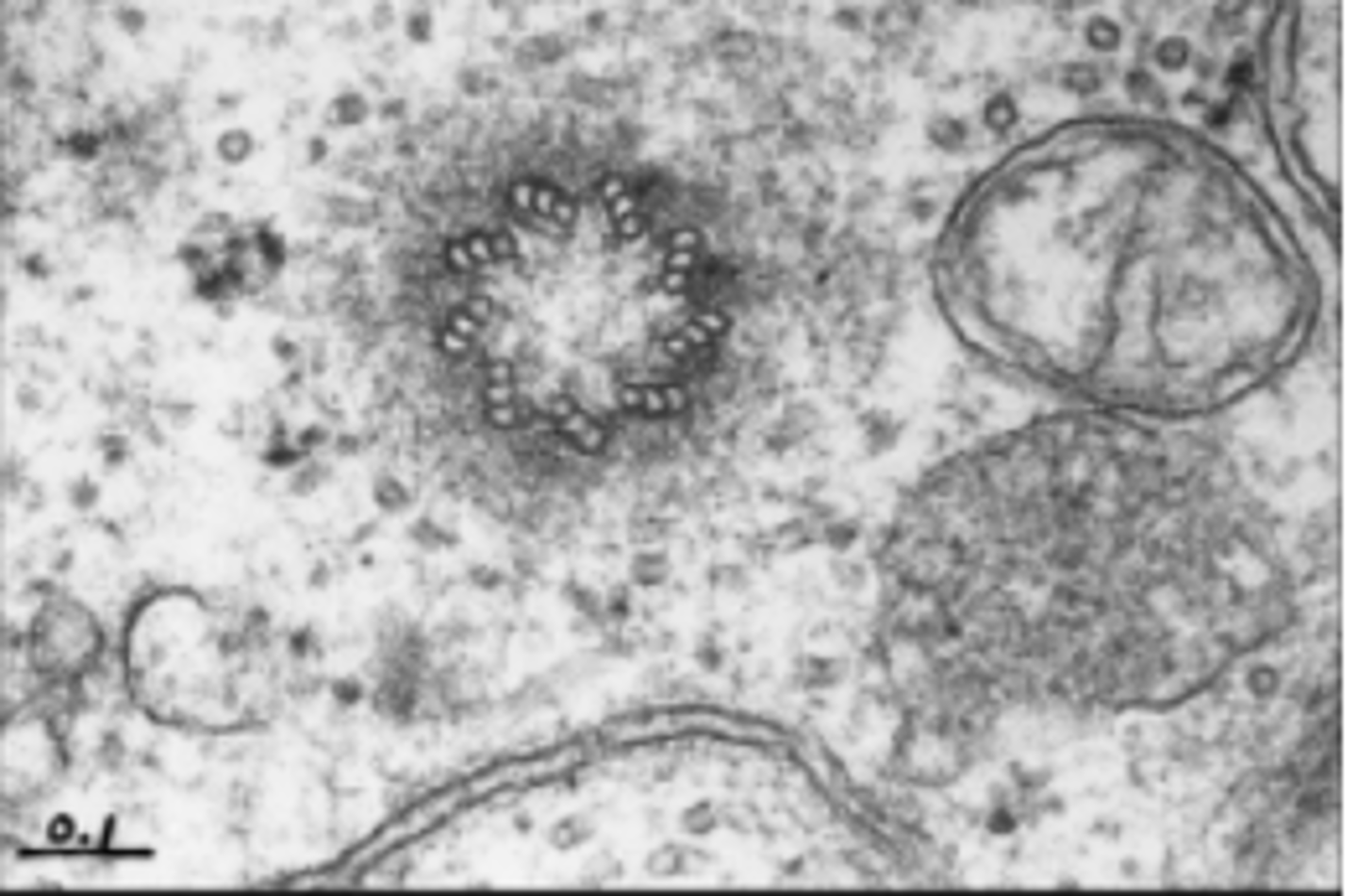
Structure and function of centrioles
Hollow cylinders containing a ring of microtubules arranged at right angles to each other.
Involved in cell division lay down the spindle fibres which shorten to separate chromatids
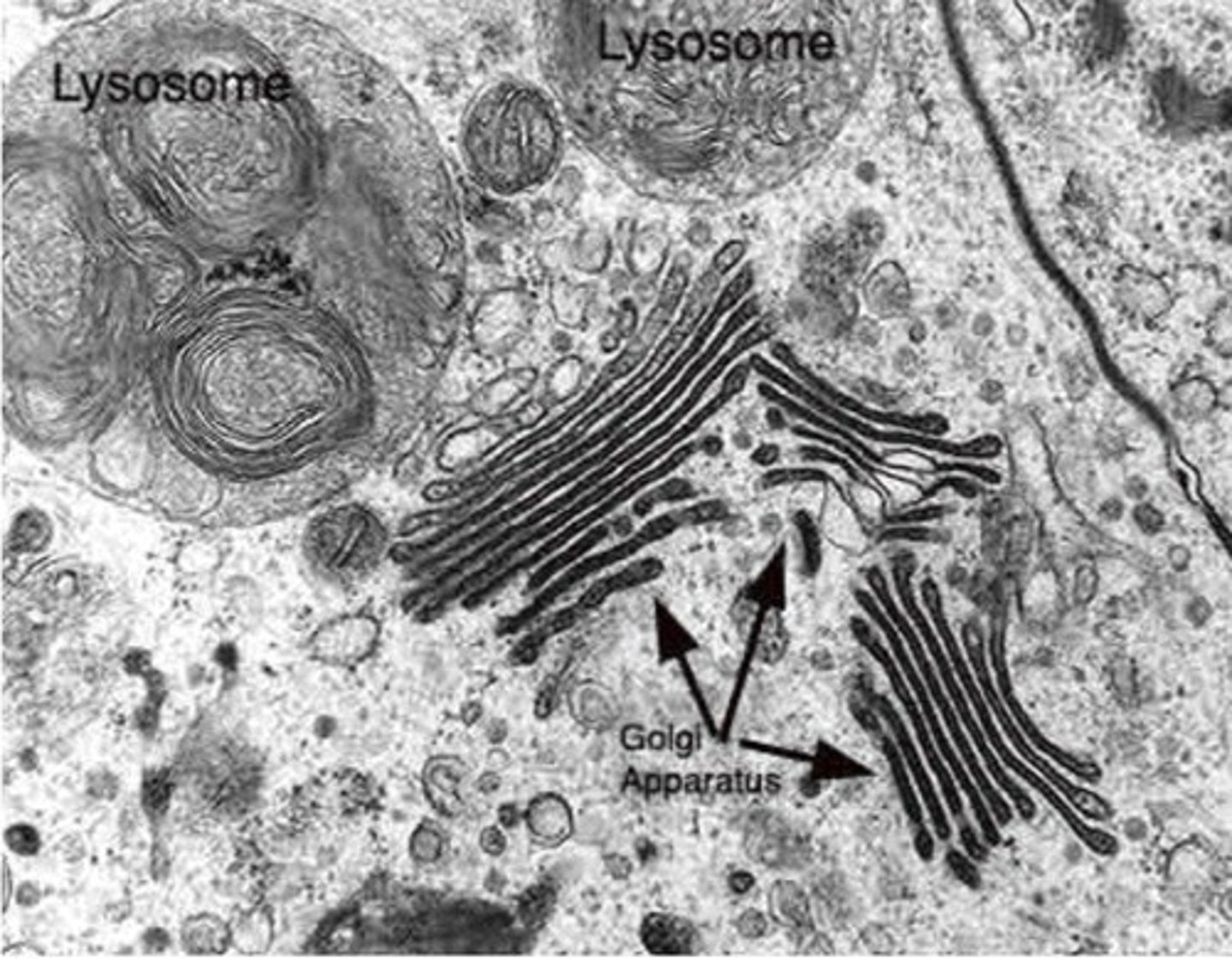
Structure and function of lysosomes
A vesicles containing digestive enzymes bound by a single membrane.
digest bacteria or worn out organelles
carries out autolysis

Structure and function of Golgi apparatus
3 to 4 curved cisternae single membrane bound. many vesicles associated with it
Golgi apparatus modifies proteins by adding a lipid or a carb
Produces lysosomes
packages protein into secretory vesicles
Role of the rER and the Golgi apparatus in the formation of extracellular enzymes
polypeptide chain moves into the cisternae of the RER and folds to assume a tertiary structure where it is packaged into a vesicle and buds off from the RER to fuse with the golgi
in golgi the proteins are modified, it is then packaged in a secretory vesicle which can then fuse with the cell surface membrane to release the enzymes outside of the cell by exocytosis
Ultrastructure of prokaryotic cells
Bacteria cells are prokaryotic cells.
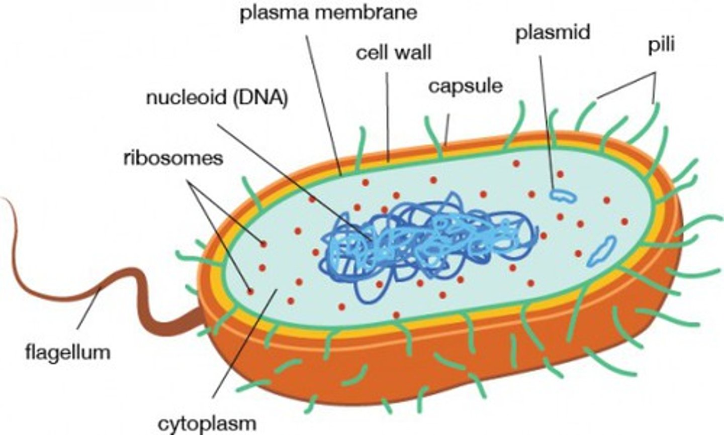
structures present in bacteria
slime capsule
plasmids
pilli
flagellum
mesosme
Function of capsule
Protective slimy layer which prevents dehydration of cell
Function of plasmid
small circular DNA molecules only present in some bacteria
Function of flagellum
A tail like structure which rotates to move the cell
Function of pili
help attach to other bacterial cells.
mesosme
infolding of the cell surface membrane which increases surface area for atachment of enzymes for aerobic respiration
Function of ribosomes
Site of protein production
How can magnification and resolution be achieved using light and electron microscopy?
The electron microscope uses a beam of electrons and their wave-like characteristics to magnify an object's image, unlike the optical microscope that uses visible light to magnify images.
To achieve the maximum resolution in a microscope system, each of the optical components should be of the highest NA available. In addition, using a shorter wavelength of light to view the specimen will increase the resolution
Magnification
The degree to which an image of an object is larger than the object itself.
Image size = Actual size x Magnification
Resolution
The degree to which it is possible to distinguish between 2 points that are close together
Optical microscopes
Uses light to form an image, light has a longer wavelength than electrons, so this microscope has a lower resolution than the electron microscopes.
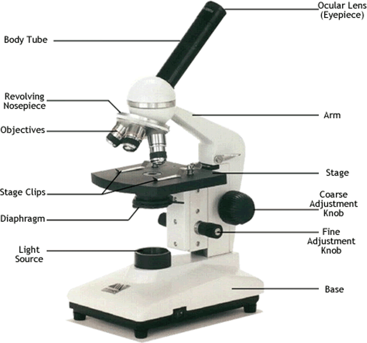
Advantages of using optical microscopes
Easy to use
Slides are easy to prepare
Can view live specimens
Disadvantages of using optical microscopes
Lowest resolution
Lowest maximum magnification
Only large organelles are visible
Scanning electron microscope
Scans a beam of electrons across the sample which knocks off electrons which then form an image
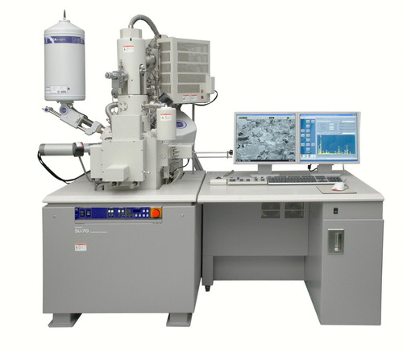
Advantages of using scanning electron microscopes
Higher resolution than optical microscope
Higher magnification than optical microscope
Forms 3D images
Can be used on thick specimens
Disadvantages of using scanning electron microscopes
Lower resolution than TEMs
Transmission electron microscope
Passes electrons through a thin specimen; denser regions absorb more electrons so less pass through creating a darker area on the image.
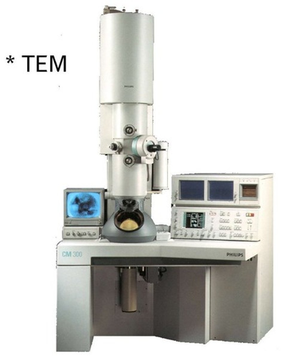
Advantages of using transmission electron microscopes
Highest resolution
Highest magnification
Can see internal structures of organelles
Disadvantages of using transmission electron microscopes
Needs very thin specimens
Slides are hard to prepare
Importance of staining specimens in microscopy
Stains and dyes are applied to tissue samples and bind to organelles making them easier to view.
Staining increases the contrast in the image formed, this can make it easier to see apart 2 objects that are close together, so it increases resolution.
different parts of the prokaryotic cells
cell wall
cell membrane
circular DNA
ribososmes (70s)
cell wall
structural su[ppport for cell contents
prevents osmotic bursting
cell membrane
controls what enters and exits the cyptoplasm which is the site of chemical reactions
circular DNA
carries genetic code that dictates protein synthesis
ribososmes 70s
site of translation of mRNA
differences between prokaryotic and eukarotic
prokaryotic cells have circular DNA while eukaryotic has Linear DNA
prokaryotic have 70s while eukaryoric has 80s and 70s
prokaryotes such as bacteria have no membrane bound organelles while eukaryotes have double and single bound organelles
cell
basic unit of structure and function in living organisms
Tissue
group of similar cells of the same origin performing a given function
organ
this is a group if different tissues and the organ can perform 1 or more functions
system
group of different organs working together in a coordinated manner
compare and contrast tissue and organ
same- both are made of cells
different- tissue is specialised similar cells whereas organ is amde of different tissues
tissues are specialized and so they perform 1 function but organ can perform more than 1 function