Module 1: Skeletal System, Articulations, and Muscles
 Call Kai
 Learn
Learn Practice Test
 Spaced Repetition
 Match
Match Flashcards
 Knowt Play
1/263

There's no tags or description
Looks like no tags are added yet.
Name | Mastery | Learn | Test | Matching | Spaced | Call with Kai | Chat |
|---|
No analytics yet
Send a link to your students to track their progress
264 Terms
Cartilage:
- tissue types?
- matrix?
- cell types?
- blood supply?
- function?
- Tissue types = hyaline, elastic, and fibrocartilage
- Matrix = gel-like composition
- Cell types = chondrogeneic, chondroblasts, and chondrycytes
- Blood supply = avascular/ gets nutrients via diffusion from surrounding tissues
- Function = support, resists compressive forces/ stressors
Bones:
- tissue types?
- matrix?
- cell types?
- blood supply?
- function?
- Tissue types = compact, and cancellous (spongy)
- Matrix = hardened matrix/ calcified
- Cell types = osteoprogenitors, osteoblasts, osteocytes
- Blood supply = highly vascularized/ own blood supply/ direct nutrient delivery to tissue
- Function = support, resists compressive forces/pulling forces/ stressors
What do hyaline cartilage and fibrous membranes serve as?
templates for bone formation
Four main functions of the skeletal system
1) Support the body/ structural support
2) Protect vital structures/ organs
3) Produce red blood cells
4) Act as levers for muscles/ allows for movement
Red marrow vs. Yellow marrow vs. Bony Tissue
-Red: found in spongy bone at the ends, produce RBC/ WBC, mostly in infants, site of blood cell formation
-Yellow: found in the middle of long bones and stores fat/ triglycerides, more abundant as we age
-Bony tissue: storage of minerals such as Ca2+ and PO4-3 that allow for mineralization/ the hardening of bones

What is bone made of?
Hardened tissue composed of multiple cell types / an extracellular matrix w/ mineral binding proteins
What is cartilage made of?
chondrocytes in lacunae/ have a higher percentage of water
hyaline cartilage
common?
where is it found?
function?
surrounded by?
made of?
- Most common type of cartilage; it is found on the ends of long bones, ribs, and nose
- helps reduce friction
- is surrounded by denser irregular connective tissue called the perichondrium
- made of primarily fine collagen fibers

fibrocartilage
is it surrounded by something?
strength?
function?
found where?
made of?
- lacks a perichondrium
- very strong/ resists intensive compressive forces
- it is weight bearing and shock absorbing
- found in the vertebra/ spine, pubic bone, and knee caps
- made of mostly thick collagen fibers

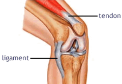
Ligaments vs Tendons, both?
- Ligaments = fibrous connective tissue connecting bone to bone
- Tendons = connective tissue joining bones and muscles
BOTH are dense / help anchor stuff together
Axial vs Appendicular Skeleton
axial = axis of gravity (vertebra, skull, thoracic cavity)
appendicular = appendages (upper and lower limbs)

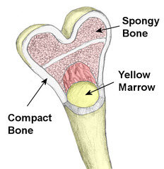
Compact bone:
- composition?
- extracellular matrix: secreted by/ makeup/ what can it do?
- what does compact bone surround?
- solid/ has a smooth homogenous composition
- ECM secreted by osteoblasts and consists of proteoglycans, collagen, etc. —> fibers in ECM bind inorganic minerals like calcium and phosphate
- compact bones always surround spongy bone

Spongy bone:
- composition/ structure?
- how is it arranged?
- spaces filled with?
- lattice of rods called trabeculae are arranged to resist stress and compression
- spaces between rods of bone are filled with marrow (red or yellow/ changes to mostly yellow as you age)
Perichondrium
Dense, irregula,r connective tissue membrane covering cartilage (hyaline)
Bone shapes
1. Sutural = extra bones located between cranial bones
2. Long = longer than they are wide/ serve as levers for movement.
3. Irregular = no distinct shape/ ie: vertebra
4. Flat = thin and usually covered
5. Short = cube shaped/ carpals + tarpals
6. Sesamoid = a type of short bone that forms in a tendon (the patella)
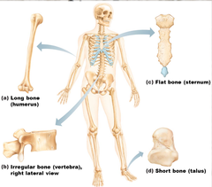
Composition of long bones vs flat, irregular, and short
Long:
- compact bone surrounding marrow filled cavity
- Spongy bone in both ends
- Hyaline cartilage lining areas of articulations
Flat, Irregular, and Short:
- compact bone layers surrounding spongy bone
- hyaline cartilage lining areas of articulations
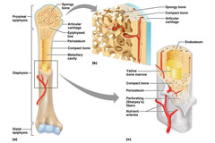
General anatomy of the long bone
compact bone; spongy bone; diaphysis; epiphysis; epiphyseal line; articular cartilage; periosteum; endosteum

Diaphysis/ shaft
- forms the axis
- is an outer layer of compact bone
- surrounds a medullary cavity
- aka diaphysis

metaphysis
- growth zone between the epiphysis and the diaphysis
- point of widening at end of diaphysis
- region of bone growth
- contains the epiphyseal plate

Epiphyseal plate
- disc of hyaline cartilage that allows bone to grow longitudinally during childhood
- once growth is complete, the cartilage converts to bone and is called the epiphyseal line
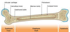
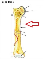
medullary cavity
cavity within the shaft of the long bones filled with bone marrow
nutrient foramen
small opening in the middle of the external surface of the diaphysis, through which an blood supply and nerves enters the bone to provide nourishment

proximal epiphysis
the end of the bone located nearest to the midline of the body

Distal epiphysis
end of the bone located farthest away from the midline/ point of attachment
need to be strong for sites of articulation

Epiphyses are covered by...
a layer of hyaline cartilage called articular cartilage (cushioning/ stress absorbing)
epiphyseal line
- remnant of epiphyseal plate
- think plate of compact bone in adults located at both epiphyses

The external and internal surfaces of bone are covered by...
membranes
Periosteum
outer fibrous/ Collagen layer
- outer membrane of compact bone
- has an outer layer of dense irregular connective tissue
- has an inner osteogenic layer of bone forming cells (osteogeneic cells)

osteogeneic cells
- stem cells that differentiate into osteoblasts
- found on inner layer of periosteum
Endosteum
Cellular layer
- inner membrane for bone
-membrane lining the medullary cavity of a bone
- contains connective layer w/ osteogenic cells
- lines the inner surface of compact bone and the trabeculae of spongy bone

Collagen fibers extend from...
the outer layer of periosteum into the bony matrix
Sharpey's fibers
-collagen fibers that connect periosteum to compact bone/ anchor membrane
- very thick at points of tendon/ ligament attachment
- can pull away from the matrix (shin splints)
Multiple cells are important in the formation and maintenance of...
bone tissue
Osteoprogenitor cells
what do they become?
where are they found?
- stem cells that differentiate into osteoblasts
- line both the inner layer of periosteum and endosteum

Osteoblasts
what do they make?
shape?
division?
lifespan?
- matrix synthesizing cell responsible for bone growth
- cuboid shaped cells that produce the extracellular matrix
- non mitotic (don't divide, instead encase themselves in the matrix of bony tissue)
- most undergo apoptosis/ die once matrix formed OR become a mature bone cell (osteocyte)
Osteocytes
Derived from what?
What do they do?
Shape importance?
Location?
- derived from osteoblasts and maintain the tissue environment/ maintains the bone matrix
- have many projections that allow communications with other bone cells
- located between plates of bone and are connected to one another
Osteoclast
Derived from?
What do they do?
Shape?
- derived from bone marrow cells
- bone reabsorbing cells/ degrade the bony matrix
- have a ruffled membrane to increase surface area for reabsorption of nutrients

What is compact bone composed of on a geometrical level?
rings of hardened matrix connected by small passageways
Osteon
what is it?
what does it consist of?
- structural unit of compact bone
- consist of long pillars of hardened matrix that run lengthwise to bone

concentric lamellae
- layers of calcified matrix that make up plates of bone

circumferential lamellae
location?
- located deep to periosteum and superficial to endosteum and extend around entire circumference of the diaphysis and resist twisting of long bone

Collagen fibers of the matrix:
- direction + importance?
- they run in opposite directions, allowing bones to withstand stress in multiple directions
"Twister Resister" —> resists breaking of bones w/ twisting motion
Central (Haversian) Canal
what is it/ what does it do?
what wraps around it?
- opening in the center of an osteon, carries blood vessels and nerves
- plates of hardened matrix wrap around this
- runs lengthwise to bone

Perforating (Volkmann's) canals
location?
connect what?
- At right angles to the central canal
- Connects medullary cavity to central canal
- connects nerves and blood supply from periosteum to central canal

Canaliculi:
What do they connect?
What are they filled with?
Extensions of?
How are they formed?
- little channels that connect lacunae
- small passageways filled with tissue fluid
- extensions of osteocytes
- formed by bone hardening that forms between places where osteocytes project/ connect to eachother

Osteocytes are connected by
gap junctions
Compact bone is composed of...
rings of hardened matrix
The matrix is secreted by...
osteoblasts
Lacunae formation
mineralization occurs and osteoblasts are walled off into lacunae —> becomes osteocytes
Organic components vs Inorganic components of compact bone
1. Organic:
- provides flexibility in bony tissue
- collagen, glycosaminoglycans, proteoglycans
2. Inorganic: hydroxyapatites
- provides structure/ hardening
- calcium phosphate, calcium carbonate
Collagen fibers allow for....
How?
- allow for ben but not break of bones
- get flexibility from sacrificial bonds
Sacrificial bonds
in or between collagen molecules that stretch and break to dissipate energy and prevent fractures

When do bones break?
when sacrificial bonds encounter a force too big to dissapate
Osteogeneis imperfecta
Defect in what?
Leads to what?
- brittle bone disease
- defect in collagen protein
- leads to lack of tensile strength/ bones break under stress
embryonic skeleton begins as... and form....
membranes and cartilage/ form the scaffolds for bone development
2 kinds of ossification (bone formation)
intramembranous, endochondral
endochondral ossification
what is it?
template?
what bones?
- Process of transforming cartilage into bone
- bone formation using hyaline cartilage template
- all bones below base of skull (other than clavicles)
intramembranous ossification
what is it?
template?
what bones?
- process of bone formation/ fibrous membrane replaced by bone tissue
- using mesenchymal template
- bones of skull and clavicle
mesenchymal stem cells
produce both chondrocytes and osteoblasts/ stem cell tissue
Steps of intramembranous ossification
1. ______ condense and differentiate into _____. ______ centers form.
2. ____ differentiate into ____ which secrete _____
3. ______ become _____ by _____ and the ____ undergoes ____
4. The bone is ______ and not _____. This provides a ____ for ____ to form around blood vessels in the area. The ____ also forms.
5. _____ continue to secrete new bone onto _____ and convert _____ into ____
1. Mesenchymal cells condense + differentiate into osteoprogenitor cells (ossification centers form)
2. Osteoprogenitors differentiate into osteoblasts which secrete bony matrix
3. osteocytes become surrounded by hard bone aka osteoid undergoes calcification
4. bone is immature and not highly organized ( woven bone) —> provides template for trabeculae to form around blood vessels in the area/ periosteum forms
5. Osteoblasts contine to secrete new bone onto trabeculae + convert woven bine into spongy bone

Embryonic ossification
intramembranous and endochondral ossification

Bone collar formation:
1. ____ secrete ___ and results in a collar made of ____
kind of growth?
- osteoblasts secrete bone matrix and results in a collar made of compact bone
- appositional growth (chondroblasts under perichondrium)

zones of cartilage in epiphyseal plate
1. Resting cartilage: typical hyaline cartilage, small chondrocytes in lacunae, light colored matrix
2. Proliferating cartilage: chondrocytes in rows, undergoing mitosis, dark colored matrix
3. Hypertrophic cartilage: chondrocytes increase in size, stop cell division, chondrocytes mature
4. Calcified cartilage: chondrocytes begin to calcify matrix and die
5. Ossification: osteoprogenitor cells + blood vessels enter the spaces left behind, new bone deposited

Fetal cartilage:
based off what?
surrounded by what?
- hyaline based
- surrounded by perichondrium
Primary ossification center:
a region deep in the ___
where ___ starts during ___ ossification
region, deep in the periosteal collar, where bone development starts during endochondral ossification
Endochondrial ossification:
1. ______ invade the the proximal and distal ______
2. _____ centers form
3. same thing occurs and ______ replaces _____
4. ______ enter the _____ to differentiate into _____
5. ______ lay down bone on ______ —-> converted to _____ over time
1. epiphyseal blood vessels invade the proximal and distal epiphyses
2. secondary ossification centers form
3. same thing happens —> bone replaces cartilage
4. Osteoprogenitor cells enter the ossification center to differentiate into osteoblasts
5. osteoblasts lay down bone on cartilage template —> converted to bone over time
What starts to form after a child is born
Epiphases (makes lots of spongy bone)
When does longitudinal growth stop?
- when epiphyseal plates harden and become epiphyseal lines
- only remaining hyaline cartilage in bone is the articular cartilage
How do bones grow in width?
____ increases due to activity of ____ cells in the _____
through a process called appositional growth (girth increases due to activity of osteoprogenitor cells in periosteum)
Appositional growth:
bone is deposited by __ on ___ side of the bone?
bone is reabsorbed by ___ on the ___ side of the bone?
- growth in width
- bone deposited by osteoblasts on periosteal side of the bone
- bone resorbed by osteoclasts on the endosteal side of bone
What helps regulate bone growth, maintenance, and remodeling
hormones
Somatomedins
- secreted in response to?
- derived from?
- importance?
- a liver derived hormone secreted in response to growth hormone
- aid in body growth and maintinence/ the most potent stimulator of cartilage, bone and skeletal muscle growth
Growth hormone
hormone secreted by anterior pituitary gland that stimulates growth of bones
Sex hormones:
induce what?
what does it drive the division of?
when do they slow down?
- induce rapid growth of bones
- drives the division of cartilage cells in the epiphyseal plate
- after puberty, sex hormones slow down at the epiphyseal plates —> epiphyseal plates harden and become epiphyseal lines —> longitudinal growth stops
Major regulator of bone remodeling
estrogen
Estrogen:
- where does act?
regulates bone modeling continually at the surface of the periosteum + endosteum
When is spongy vs compact bone replaced?
spongy: every 3-4 years
compact: every 10 years
What is similar vs different regarding Endochondral vs. Intramembranous ossification?
Endochondral:
- has osteocytes, osteoblasts, and osteoclasts
- creates spongy + compact bone
- has a hyaline cartilage template
- is the bones below the skull
Intramembranous:
- has osteocytes, osteoblasts, and osteoclasts
- creates spongy + compact bone
- has a mesenchymal cell template
- is the skull + clavicle
Osteoporosis:
what is it?
what does it cause?
what is it correlated to?
men vs women?
- occurs when resorption of bone is greater than deposition of bone
- causes mass to decline despite normal matrix composition
- correlated to a decline in estrogen levels, so post-menopausal women are at high risk
- men can get it but men have larger bones, so it takes longer for them to become symptomatic AND testosterone can be converted into estrogen
Other risk factors of osteoporosis:
poor diet, vitamin d deficiency, smoking, sedentary lifestyle
What kind of bone is most affected by osteoporosis?
spongy bone
What do light regions of osteons represent:
older osteons/ mineralized
What do dark regions of osteons represent:
newer osteons/ undergoing mineralization
Fracture classifications:
-Closed = bone stays inside skin
-Open = bone comes out of skin
-Incomplete = bone breaks partially
-Complete and displaced = broken/ doesn't stay in anatomical alignment
-Complete and non-displaced = broken but bone stays in anatomical alignment
*also determined by the direction of the fracture

Avulsion
- complete severing of body part (typically toe or finger)

Colles
- fracture of the distal end of the lateral forearm bone (radius) / produces a dinner fork deformity

Comminuted
- bone is splintered into several small pieces between main parts
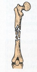
Complete
- bone broken into two or more pieces
Compound (open)
- broken ends of bone protrude through the skin
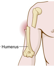
Compression fracture
- bone is squashed/ crushed (may occur in a vertebra during a fall)

Displaced fracture
- fractured bone parts are out of anatomic alignment
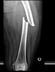
Depressed fracture
- broken part of bone forms concavity

Epiphyseal fracture
- epiphysis is separated from the diaphysis at the epiphyseal plate

Greenstick
- partial fracture/ one side of bone breaks, the other side is bent

Hairline fracture
- fine crack in which sections of bone remain aligned
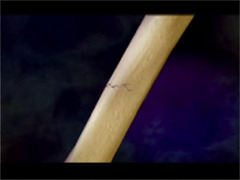
Impacted fracture
- one fragment of bone is firmly driven into the other

Incomplete fracture
- partial fracture extends only partway across the bone

Linear fracture
- fracture is parallel to the long axis of the bone

Oblique fracture
- diagonal fracture is at an angle

Pathologic
- weakening of a bone caused by disease process