Anatomy & Physiology 1 Exam 1
 Call Kai
 Learn
Learn Practice Test
 Spaced Repetition
 Match
Match Flashcards
 Knowt Play
1/115

There's no tags or description
Looks like no tags are added yet.
Name | Mastery | Learn | Test | Matching | Spaced | Call with Kai | Chat |
|---|
No analytics yet
Send a link to your students to track their progress
116 Terms
Anatomy
The study of the structures of the body and their relationships to each other, the oldest medical science (looking at a feature of the body and describing/understanding it in terms of its position, form, shape, and whether its connected to other features/organs)
Physiology
The study of the functions of the body parts and how they work to carry out their life-sustaining activities (what it does)
Compare and contrast anatomy and physiology
Anatomy dictates physiology and Physiology dictates anatomy (form and function go together)
Ex. Adaptations: Talons of an eagle are sharp (anatomy) so they can be used to kill prey (physiology)
Gross Anatomy
Studying anatomy without a microscope. Using dissections, we can study where organs are, relative to size, shape, and position
Microscopic Anatomy
Studying anatomy with a microscope. Using microscopic, we can study structures that are too small to see with the naked eye, like cells or tissues.
Molecular Physiology
Studying the function of individual molecules. Ex. Protein function is related to protein shape
Cellular Physiology
Studying the function of individual cellsS
Systemic Physiology
Studying the function of individual organ systems. Ex. A gastroenterologist studies the digestive system
Pathophysiology
Studying how diseases and aging alter physiology of organs and organ systems
Structural Organization
Atoms → Molecules → Organelle → Cells → Tissue → Organ → Organ System → Organism
Nine Characteristics of Life
Separation of external and internal environment is necessary (Ex. the plasma membrane separates cells from each other and from their environment, the skin seperates the body’s internal environment from the external environment)
Movement is necessary (Motility is when substances move through the body [cardiac and smooth muscle do this]; Locomotion is when whole body parts move [skeletal muscles do this])
Living things respond to stimuli (stimuli are changes in the internal or external environment; once they are detected, reactions occur to address them)
Digestion and absorption are necessary (Digestion is breaking down ingested substances, Once large molecules are broken down, small molecules can be absorbed)
Metabolism is necessary (Metabolism is the sum of all the chemical reactions that keep an organism: Catabolism - the reactions that break down large molecules, Anabolism - the reactions to build large molecules)
Excretion is necessary (Waste products like feces, urea, and CO2 must leave the body)
Growth and repair are necessary (Growth is an increase in the size or number of cells)
Reproduction is necessary (Reproduction is the production of offspring, *not vital to individual organisms to reproduce, *is vital for species to reproduce)
Homeostasis (Maintenance of stable internal conditions, the central principle of physiology)
Homeostasis
A relatively constant set of internal conditions that stay the same even when external conditions change
Homeostasis is dynamic, meaning it changes to keep things in balance
All the body’s organ systems work together to maintain homeostasis
Homeostasis is maintained using negative and positive feedback loops
Negative Feedback Loops
Goal: to get the body back to the original set point as quickly as possible
The response reduces (or completely cancels) the impact
Examples: thermoregulation (body temperature), chemical balance (pH or blood salinity), hormone levels (insulin and glucagon)
Positive Feedback Loops
Goal: to create a “new temporary set point”
The response increases the effect of the stimulus
Positive feedback loops control things that are NOT constantly being adjusted
Examples: blood clotting, labor and delivery, neuron signaling
Organ System Functions
Integumentary - The external body covering that makes Vitamin D and has receptors and glands
Muscular - The system that enables movement, maintains posture, and generates heat
Skeletal - The system that protects and supports organs and provides a framework for muscle attachment
Nervous - The control system that responds to internal and external stimuli
Cardiovascular - The system that pumps blood (with oxygen, carbon dioxide, nutrients, and waste)
Lymphatic - The system that collects fluid from tissues and houses white blood cells
Respiratory - The system that supplies oxygen to the blood and removes carbon dioxide
Endocrine - The glands that secrete hormones to regulate growth, reproduction, and metabolism
Digestive - The system that breaks down food into absorbable units (that enter the bloodstream) and eliminates waste
Urinary - The system that regulates water, electrolyte, and acid-base balance in the blood
Reproductive - The system that is used to generate offspring
Body Cavity
All of the body’s organs are found inside cavities
Cavities provide protection to the organs inside them
Cavities can be open or closed
Open Cavities
The oral cavity
The digestive cavity
The nasal cavity
The orbital cavity
The external ear
Closed Cavities
Dorsal Body Cavity (Cranial Cavity + Vertebral Cavity)
Thoracic Cavity (Superior mediastinum, Pleural cavity, Pericardial)
Abdominopelvic Cavity (Abdominal Cavity + Pelvic Cavity)
Ventral Body Cavity (Both Thoracic and Abdominopelvic Cavities)
Major Right Upper Quadrant (RUQ) Organs
Liver (right portion)
Gallbladder
Duodenum (first part of the small intestine)
Pancreas (head)
Right kidney and right adrenal gland
Major Left Upper Quadrant (LUQ) Organs
Stomach
Spleen
Left kidney and left adrenal gland
Pancreas (body and tail)
Liver (left portion)
Major Right Lower Quadrant (RLQ) Organs
Appendix
Cecum (first part of the large intestine)
Right ureter
Right reproductive organs (right ovary and fallopian tube)
Major Left Lower Quadrant (LLQ) Organs
Descending and sigmoid colon
Left ureter
Left reproductive organs (left ovary and fallopian tube)
Regional Terms
Cephalic (Head)
Fronta
Orbital
Nasal
Oral
Mental
Cervical (throat)
Thoracic (chest)
Sternal
Axillary
Mammary
Abdominal
Umbilical
Pelvic
Inguinal (groin)
Pubic (genital
Upper Limb
Manus (Hand)
Lower Limb
Pedal (foot)
Cephalic
Otic
Occipital (back of the head)
Back (dorsal)
Scapular
Vertebral
Lumbar
Sacral
Gluteal
Perineal
Anatomical Position
Body is erect
Palms facing forward
Feet slightly separated
Superior
above
Inferior
below
Lateral
away from the midline
Medial
closer to the midline
Proximal
closer to the attachment point (arms and legs)
Distal
farther from the attachment point (arms and legs)
Anterior
closer to the front
Posterior
closer to the back
Ventral
closer to the front
Dorsal
closer to the back
Prone
laying facing down
Supine
laying face up
Right Lateral Recumbent
lying on right side
Left Lateral Recumbent
lying on left side
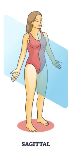
Midsagittal (medial) Plane
Divides the body into right and left sections
Frontal (coronal) Plane
divides the body into anterior (front) and posterior (back) sections

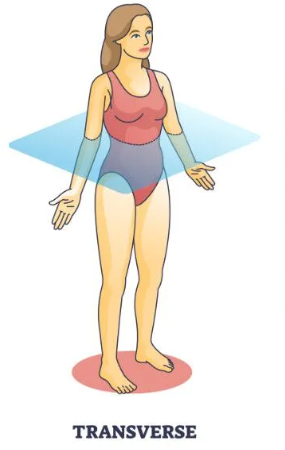
Transverse Plane
divides the body into superior (top) and inferior (bottom) sections
Tissue
a group of cells (and extracellular materials) that have similar structures (anatomy) and similar functions (physiology)
Epithelial Tissue
found on external surface and internal cavities, prevents leakage from one cavity to another, protects, secretes, absorbs, and filters. Contains Lining of digestive tract organs and other hollow organs, glands (e.g., pancreas), and skin surface (epidermis)
Connective Tissue
supports, protects, and binds other tissues together. Contains bones, tendons, fat, and other soft padding tissue
Muscle Tissue
cells that contract and cause movement. Contains muscles attached to bones (skeletal), heart (cardiac), and walls of hollow organs (smooth)
Nervous Tissue
receives, processes, and transmits messages (internal communication). Contains brain, spinal cord, and nerves
Functions of epithelial tissue
Protection - against microorganisms, UV light, mechanical damage
Absorption - transmission of molecules into cells
Filtration - separation of materials
Excretion - elimination of waste materials
Secretion - movement of substances
Sensory Reception - detection of changes in external environment
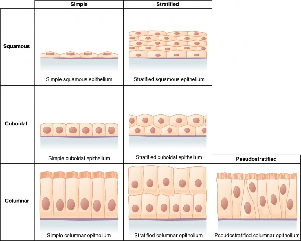
Epithelial cell shapes
Squamous = flat or “squashed”
Cuboidal = cube-shaped
Columnar = column-shaped
Epithelial cell layers
Simple = 1 layer
Stratified = more than 1 layer
Pseudostratified = 1 layer, but appears to be many
Transitional = several layers

Characteristics of epithelial tissue
Epithelial tissue has apical-basal polarity
the apical surface (“top”) is exposed to the outside
the basal surface (“bottom”) is attached to lower tissue layers (e.g., basement membrane)
T or F: Epithelial tissue is always on top of connective tissue
True
T or F: Epithelial tissue is avascular (no blood vessels).
True
Tight junctions
Form a leak-proof barrier between neighboring epithelial cells (found in areas where extracellular fluids need to be kept separate, like the blood-brain barrier)
Adherens junctions
Indirectly link cell cytoskeletons to provide strong connections
Gap junctions
Provide a direct channel between neighboring epithelial cells (so they can have a shared chemical environment)
Desmosomes
Provide structural reinforcement between epithelial cells in areas that stretch
Hemidesmosomes
Anchor epithelial cells to underlying connective tissue
Simple Squamous Epithelium
Locations:
Lining capillaries (small blood vessels)
Alveoli (air sacs)
Glomeruli (kidney blood vessels)
The mesothelium (around abdominopelvic organs)
Functions:
Allowing diffusion and absorption
Simple Cuboidal Epithelium
Locations:
Kidney tubules
Ducts of glands
Ovaries
Seminiferous tubules
Functions:
Secretion
Absorption
Simple Columnar Epithelium
Locations:
Non-ciliated: the digestive tract, ducts of glands
Ciliated: the uterus, the oviducts
Functions:
Non-ciliated: secretion and absorption
Ciliated: movement
Pseudostratified Columnar Epithelium
Locations:
Upper respiratory tract
Reproductive system (e.g., vas deferens, epididymis)
Functions:
Secretion
Movement
Stratified Squamous Epithelium
Locations:
Keratinized (hard, structural protein): skin
Non-keratinized: mouth, vagina, rectum
Functions:
Protection from abrasion (friction)
Stratified Cuboidal Epithelium
Locations:
Salivary glands
Sweat glands
Mammary glands
Functions:
Secretion
Strengthens glands
Stratified Columnar Epithelium
Locations:
Pharynx (throat)
Urethra (urinary tract)
The conjunctiva (white of eye)
Functions:
Protection
Secretion
Transitional Epithelium
Locations:
Urinary bladder
Functions:
Stretching
Protection from urine
Endocrine Glands
Secrete into the bloodstream (hormones)
Exocrine Glands
Excrete out of the body or an organ (sweat, saliva, mucus, digestive enzymes) via ducts
Extracellular Matrix (ECM)
Surrounding material that supoorts the cell
Connective Tissue Components - Fibers
Collagen Fibers → thick fibers that are very strong
Elastic Fibers → thin, stretchable fibers
Reticular Fibers → delicate, net-like fibers
Connective Tissue Cells
-blast cells are immature (still building ground substance and proteins)
-cyte cells are mature (can only maintain the existing matrix)
Fibroblasts → fiber-building cells in many connective tissues
Chondrocytes → mature cartilage cells
Osteocytes → mature bone cells
Erythrocytes and Leukocytes → mature blood cells
Types of Connective Tissue

General Functions of Connective Tissue
Binding
Areolar loose connective tissue
Dense regular connective tissue
Dense irregular connective tissue
Protection
Bone tissue
Blood
Adipose tissue
Energy storage
Adipose tissue
Bone
Support
Bone
Hyaline cartilage
Elastic cartilage
Fibrocartilage
Insulation
Adipose tissue
Transport
Blood
Dense elastic connective tissue
Areloar Connective Tissue
Important Features
All three fiber types are seen in this tissue
Fibroblasts (fiber-building cells) and leukocytes (white blood cells) are visible
Location
Surrounding organs
Below the epithelium
Function
Wraps and cushions organs
Retains extracellular fluid
Adipose Connective Tissue
Adipose tissue (fat) is made of adipocytes
Their nucleus is pushed to the side of the cell
Locations
Abdominal cavity
Under skin
Breasts
Functions
Lipid storage
Protection
Insulation
Reticular Connective Tissue
Reticular connective tissue only has reticular (net-like) fibers
Location
Lymph nodes
The spleen
Liver
Bone marrow
Functions
Supports red and white blood cells
Assists in filtration
Dense Regular Connective Tissue
The many collagen fibers in this tissue are arranged regularly (fibroblasts are squished flat in between the fibers)
Location
Tendons (attach muscle to bones, “thick rubber band”)
Ligaments (attach bone to bone)
Aponeuroses (attach muscle to bone, “thin sheet”)
Function
Strong attachment for bones and muscle
Withstands pulling in one direction
Dense Irregular Connective Tissue
The many collagen fibers in this tissue are arranged irregularly (the collagen fibers are also thicker than in dense regular)
Location
The reticular layer of the dermis
Capsules around joints and organs
Function
Withstands pulling in multiple directions
Dense Elastic Connective Tissue
Elastic fibers that appear dark purple when stained
Location
Walls of large arteries
Walls of the bronchiole tubes in the lungs
Function
Stretching and recoiling (like a rubber band)
Cartilage
Three types
Hyaline
Elastic
Fibrocartilage
Functions
Strong and flexible to protect joints
Helps keep joint shape when moving
Absorbs shock
Anatomical features
Tough, dense flexible matrix
Chondrocytes - sparsely distributed cells in lacunae
Surrounded by perichondrium - dense irregular connective tissue
Hyaline Cartilage
This is the most abundant cartilage in the body
the cells are called chondrocytes, found in lacuna (“little house” or bubble), appears “glass-like”
Location
The fetal skeleton
Some joints
Nose
Function
Template for bone
Support and reinforcement
Elastic Cartilage
Location
External ear
Epiglottis (prevents choking)
Function
Maintains shape
Allows flexibility
Fibrocartilage
Has thick collagen fibers that are visible, stain blue on slides
Location
Intervertebral discs of the spine
Knee
Temporomandibular joint
Function
Strong support
Absorbing shock
Compact Bone
The ECM of bone contains calcium phosphate + collagen
Location
The outside of bones
Function
Support and protection
Muscle attachment site
Calcium storage
Spongy Bone
Spongy bone has trabeculae (little columns that distribute force)
Location
Beneath compact bone
Function
Hematopoiesis (making red blood cells)
Fat storage
Cutaneous membrane
The skin
Mucous membrane
Cavities open to the environment
Serous membrane
On organs in the ventral body cavities
Integument
The skin or cutaneous membrane is the barrier between the internal and external environments
Major functions of the integument
Barrier to environmental microbes
Prevents water loss
Thermoregulation
Detecting sensation
Vitamin D synthesis
Excretion
Layers of the skin
Epidermis
Keratinized stratified squamous epithelium
Dermis
Papillary layer of the dermis
Areolar loose connective tissue
Reticular layer of the dermis
Dense irregular connective tissue
Subcutaneous (hypodermis)
Adipose loose connective tissue
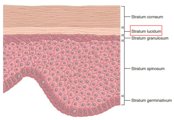
Layers of the Epidermis (in order from top to bottom)
Stratum corneum
Stratum lucidum (only present on the palms of hands and soles of feet)
Stratum granulosum
Stratum spinosum
Stratum basale
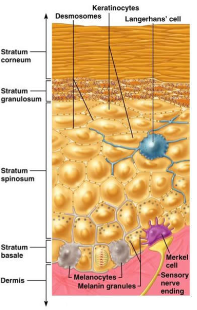
Stratum basale
The deepest layer of the epidermis, closest to the dermis
Includes melanocytes (produces melanin) and Merkel cells (tactile cells) stimulates the Merkel disc (sensory nerve cell)
Stratum spinosum
Keratinocytes in this layer have spiny cell junctions
Desmosomes strongly attach these cells to one another
Langerhans cells and melanin granules are also in this layer
Most abundant (thickest) layer of living cells

Stratum granulosum
The dark spots in this layer are lamellar granules
Function: prevents water loss from the skin
Cells start to keratinize here
Cells begin to die in the upper region of this layer

Stratum corneum
This is the outermost layer of dead/non-living cells
Increased keratinization provides protection against abrasion and microbes
These cells are constantly being shed, generating replacement

Stratum lucidum (clear layer)
Only seen in thick skin
It is superficial to (closer to body surface) stratum granulosum
It is deep to (farther away from body surface) stratum corneum
Thick skin is present on the palms of the hands and the soles of the feet
It does not have sebaceous (oil) glands or hair
Cells of the Epidermis
Keratinocytes are the most abundant cell type
Function: replenish epithelium and make keratin
Keratinocytes on the outer skin surface (corneum) are dead
Langerhans cells are the “immune system” of the epidermis
Function: phagocytose foreign substances
These cells migrate to the epidermis from the red bone marrow
Melanocytes are the melanin-producing cells
Darkens skin
Function: protecting the skin from UV radiation
Melanin is stored in melanosomes which are transferred to keratinocytes
Merkel cells are rare
Function: detecting touch and proprioception (spatial position)
Interacts with the Merkel disc in the underlying dermis (a sensory nerve cell)
Skin Pigments
Melanin is a reddish-yellow/brownish-black pigment
Made by melanocytes in the epidermis
Everyone has the same number of melanocytes, but not the same amount of melanin
Exposure to UV radiation stimulates the activity of melanocytes
Carotene is a yellow/orange pigment obtained from plants
Carotene is used to make Vitamin A (retinol)
It can accumulate in stratum corneum
Hemoglobin leads to red/pink color
It is found in red blood cells of the dermis
When melanin levels are low, hemoglobin’s color is more visible
Blushing is due to vasodilation
Alterations to skin color
Erythema: redness due to increased blood
Pallor: paleness due to decreased blood
Bruising: changing color to pooling blood under skin
Albinism: melanocytes unable to produce melanin
Cyanosis: blueness due to low oxygenated blood
Jaundice: yellowing due to liver malfunction
Vitiligo: certain areas lose ability to produce melanin
Addison’s Disease: bronze patches due to adrenal gland malfunction
Papillary Layer of the dermis
Attached to the epidermis
Dermal papillae are ridges with collagen fibers and nervous structures
The pattern of these ridges determine a person’s fingerprint
Fingerprints = “friction ridges”
Fingerprints assist with grip and the sense of touch