Patient Care in Radiography Chapter 1 Key Terms
 Call Kai
 Learn
Learn Practice Test
 Spaced Repetition
 Match
Match Flashcards
 Knowt Play
1/28

There's no tags or description
Looks like no tags are added yet.
Name | Mastery | Learn | Test | Matching | Spaced | Call with Kai | Chat |
|---|
No analytics yet
Send a link to your students to track their progress
29 Terms
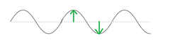
amplitude
with respect to a sine wave, the distance between peak and trough.
anode
the electrode at which oxidation occurs; also, the electrically positive, target end of an x-ray tube.
attenuation
the process of reduction, such as the reduction of x-ray beam intensity when it penetrates matter.
bucky
a moving grid that limits the amount of scatter radiation reaching an image receptor, thereby increasing the image contrast.

cathode
negative electrode; the negative side of the x-ray tube, which consists of the focusing cup(s) and the filament(s).
collimator
a device for limiting the size and shape of an x-ray beam

detent
a mechanism that tends to stop a moving part in a specific location; are built into x-ray tube supports to facilitate tube placement at standard locations.

electromagnetic energy
the range of energies of electromagnetic radiation, which varies from wavelengths 0.000001 nm to more than 5 miles
electron stream
the flow of negatively charged particles across an x-ray tube, formed when the high positive electrical potential at the target attracts electrons of the space charge, which move rapidly across the tube.
filament
the source of electrons found at the electrically negative cathode end of the x-ray tube. It is made of the element tungsten.
fluoresce
to produce light; characteristic of certain crystals to emit light of one wavelength when exposed to energy of a different, usually shorter wavelength; such light emission.
fluoroscope
a device used to produce a radiographic image on a fluorescent screen or cathode ray tube monitor for visual examination of internal structures in motion in real time.
focal spot
a small area on an x-ray tube target where the electron stream strikes the target and x-rays are produced.
frequency
with respect to the sine wave, the number of times per second that a wave crest passes a given point.
grid
a device placed between the patient and the image receptor during a radiographic examination to limit the amount of scatter radiation reaching a radiographic image receptor, thereby increasing the image contrast.
grid cap
a grid mounted in a frame that can be attached to the front of an image receptor for mobile radiography and other special applications.
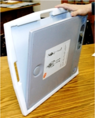
image intensifier
an electronic device used to produce a fluoroscopic image with a low radiation exposure. A beam of x-rays passing through the patient is converted by a special vacuum tube into a pattern of electrons. The electrons are accelerated and concentrated onto a small fluorescent screen, where they present a bright image that is usually displayed on a television monitor.
image receptor (IR)
any device that captures a radiographic image. Examples include radiographic film, phosphorescent screens, computed radiography imaging plates, and digital detectors.

latent image
the unobservable image stored in the silver halide emulsion of film or the photostimulable phosphors in computed radiography image receptors; it is made visible by processing.
photon
the smallest quantity of electromagnetic energy. It has no mass and no charge and travels at the speed of light. Can occur in the form of light rays, x-rays, gamma rays, and other electromagnetic energy
photostimulable phosphor
crystals that store the energy of the remnant x-ray beam and release it as light when stimulated by a laser; components of computed radiography IRs.
quantum (pl. quanta)
a group or bundle of photons
remnant radiation
the radiation that remains after the x-ray beam passes through an object to produce an image; same as exit radiation.
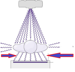
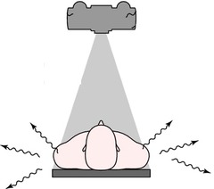
scatter radiation
ionizing radiation that occurs as a result of the interaction between the primary x-ray beam and matter. It is emitted from the matter in all directions during the exposure, tending to fog the image and produce a radiation hazard in the room. The patient is the principal source of scatter radiation that occurs during radiography.
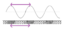
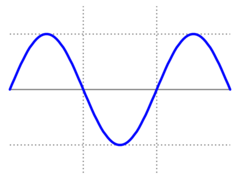
sine wave
a visual graphic of changes in the radiation field that occur in the form of a repeating wave.
space charge
Electron cloud around the filament. Free electrons provide the needed electrons for x-ray production.
target
tungsten disk located at the electrically positive anode end of the x-ray tube, the end opposite the filament. The smooth, hard surface of the target is the site to which the electrons travel and is where the x-rays are generated.
trendelenburg position
a supine position in which the patient's head is lower than the feet

wavelength
with respect to sine wave, the distance between a given point on one wave cycle and the corresponding point on the next successive wave cycle