205: The Heart
 Call Kai
 Learn
Learn Practice Test
 Spaced Repetition
 Match
Match Flashcards
 Knowt Play
1/150

There's no tags or description
Looks like no tags are added yet.
Name | Mastery | Learn | Test | Matching | Spaced | Call with Kai | Chat |
|---|
No analytics yet
Send a link to your students to track their progress
151 Terms
0.5 heart width / thoracic width
(widest length of heart is about 1/2 the length of widest part of thorax)
What is the cardiothoracic (CT) ratio?
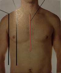
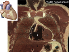
Mid-sternal line
Identify the line
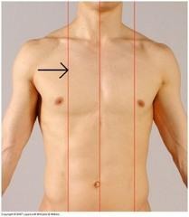
Mid-clavicular line
Identify the line
Right atrium
What is shown at #1?
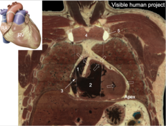
Right ventricle
What is shown at #2?
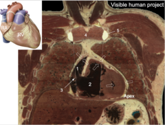
Border of sternum
What is shown at #3?
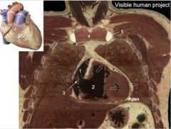
Manubrium
What is shown at #4?
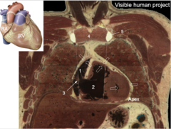
Costal cartilage
What is shown at #5?
Right atrium
What is shown at #1?
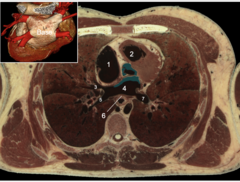
Right ventricle
What is shown at #2?
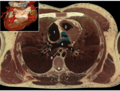
Pulmonary vein
What is shown at #3, 5, and 7?
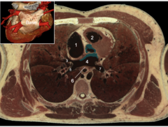
Left atrium
What is shown at #4?
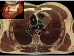
Base of heart
What is shown at #6?
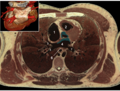
Superior mediastinum
What mediastina is the following found in:
roots of great vessels, aortic arch, ligamentum arteriosum, thoracic duct
Through the sternal angle
Where is the transverse thoracic plane defined?
Anterior mediastinum
What mediastina is the following found in:
Thymus, retrosternal fat, nerves
Posterior mediastinum
What mediastina is the following found in:
descending aorta, esophagus, vagus n.
T4-T5 interspace
The superior mediastinum is found above what vertebrae space?
Phrenic nerve
Identify the nerve
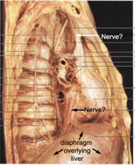
C3, 4, 5
What vertebrae does the phrenic nerve come from?
right ventricle
what heart chamber lie directly behind the sternum?
middle mediastinum
which mediastina does the heart reside?
pericardium
what structure encases the heart?
phrenic n.
what nerve runs between the percardium and the parietal pleura of the lungs and anterior to the roots of the heart?
vagus n.
what nerve runs posterior to the roots of the heart?
left
2/3 of the heart sits on the _____ side of the sternum
pericardiacophrenic vessels
what vessels supply the pericardium?
Parietal pericardium
What is shown at #1 and 4?
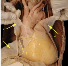
Visceral pericardium
What is shown at #2?
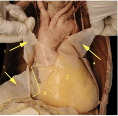
Pericardial cavity
What is shown at #3?
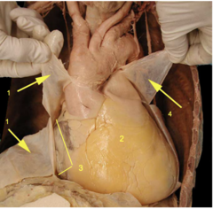
To reduce friction
Why do mesothelial layers produce serous fluid?
pericardial effusion
fluid in pericardial cavity
pericardial effusion
patient presents with muffled heart sounds , weakened beat, jugular distension, low arterial pressure. what is the diagnosis?
pericardial effusion
identify the pathology
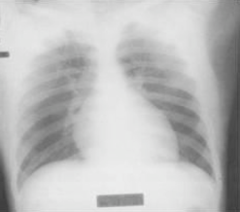
-muffled heart sounds due to blood insulation, weakened beat
-jugular distension due to reduced venous return
-low arterial pressure due to reduced stroke volume/cardiac output
Becks triad:
cardiac temponade
acute build up of excess fluid in pericardial cavity:
jugular distention
pressure on the heart's right atria from fluid in the pericardial sac will cause:
low atrial pressure
reduced stroke vol/cardiac output will cause =
transverse pericardial sinus
permits expansion of great vessels during systole, should, during bypass surgery, these vessels should be clamped off
aorta
pulmonary trunk
what two vessels are isolated by the transverse pericardial sinus?
Transverse sinus
What permits expansion of great vessels during systole?
thebesian veins
these structures are capillary beds within the myocardium that connect adjacent arteries and veins:
myocardium
thebesian veins are located in which heart layer?
oblique sinus
permits expansion of left atrium during exhalation
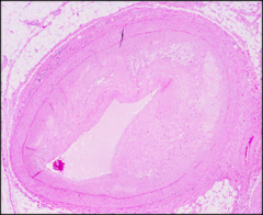
coronary artery disease
identify the pathology within the lumen of the artery:
left atrium
what heart chamber lies as the base of the heart?
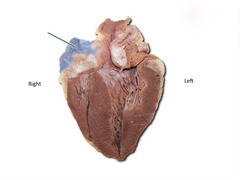
right atrium
identify the structure
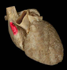
right auricle
identify the structure
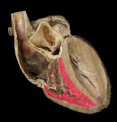
right ventricle
identify the structure
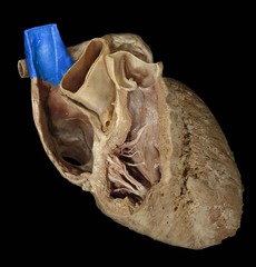
superior vena cava
identify the structure
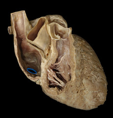
inferior vena cava
identify the structure
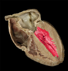
left ventricle
identify the structure
left auricle
identify the structure
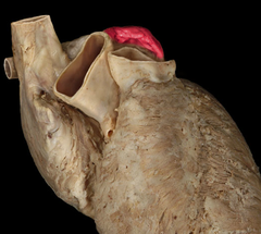
left atrium
identify the structure
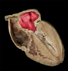
pulmonary veins
identify the structure
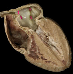
pulmonary trunk
identify the structure
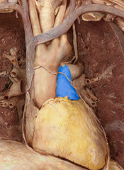
Apex of heart
identify the structure
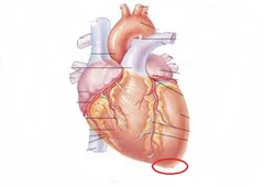
pulmonary arteries
identify the structure
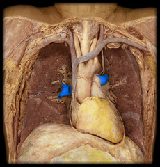
right coronary artery
identify the structure
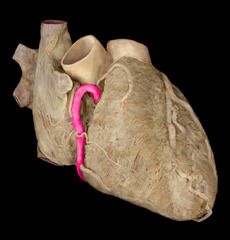
Ligamentum arteriosum
identify the structure
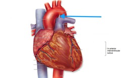
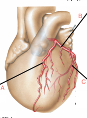
left coronary artery
Identify structure B
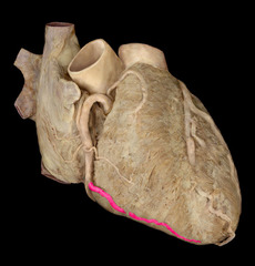
right marginal artery
identify the structure
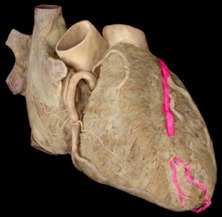
anterior interventricular artery
identify the structure
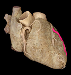
left marginal artery
identify the structure
left circumflex artery
identify the structure
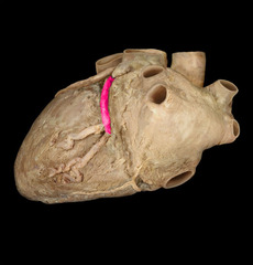
posterior interventricular artery
identify the structure
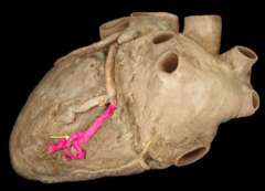
atrial branch of RCA
identify the structure
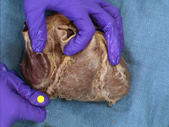
SA nodal branch of RCA
identify the structure
Oblique vein of LA
identify the structure
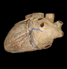
small cardiac vein
identify the structure
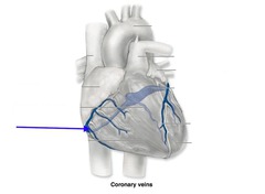
small cardiac vein
what vein runs with the right marginal artery?
right marginal artery
what artery runs with the small cardiac vein?
great cardiac vein
identify the structure
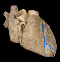
great cardiac vein
what vein runs alongside the anterior interventricular artery
anterior interventricular artery
what artery runs alongside the great cardiac vein?
middle cardiac vein
identify the structure
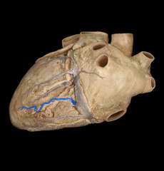
middle cardiac vein
what vein runs alongside the posterior interventricular artery?
posterior interventricular artery
what artery runs alongside the middle cardiac vein?
coronary sinus
identify the structure
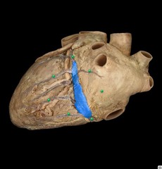
anterior cardiac veins
identify the structure
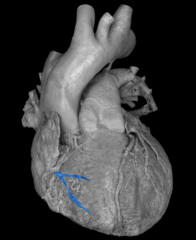
Terminology: cardiac is used for veins (with the exception of coronary sinus) and coronary is used for arteries
Terminology: cardiac is used for (arteries/veins) and coronary is used for (arteries/veins)
right atrium
all veins that drain the heart will drain into the:
coronary artery disease
damage to coronary arteries, most
commonly due to buildup of cholesterol
deposits (atherosclerotic plaque)
- Leading cause of death in US for men and women
1. Coronary artery collateralization (new capillary growth)
2. Reverse blood flow occurs in thebesian veins under conditions of low perfusion pressure
3. "Endogenous" bypass via vasa vasorum ("vessel of a vessel")
What are the three reperfusion strategies for coronary artery disease?
anterior interventricular artery
what is the most common artery to suffer coronary occlusion?
right coronary artery
what is the 2nd most common artery to suffer coronary occlusion?
great saphenous vein
internal thoracic artery
radial artery
name 3 blood vessels that can be used in coronary bypass grafts:
Left anterior descending artery ~40-50%
Right coronary artery ~30-40%
Circumflex branch ~15-20%
What are the coronary artery occlusion percentages?
Reverse the direction bc of valves
When using the great saphenous vein, what must be done for CABG?
Radial artery
What vessel has the greatest longevity for CABG?
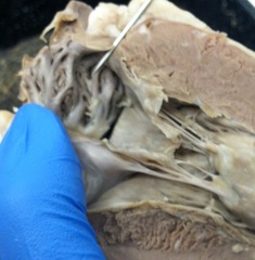
Pectinate muscles
identify the structure
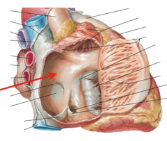
Sinus venarum
identify the structure
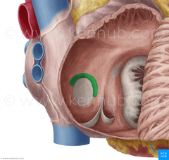
Limbus of fossa ovalis
identify the structure
valve of inferior vena cava
identify the structure
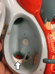
coronary sinus valve
identify the structure
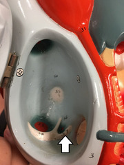
Right atrioventricular valve (tricuspid)
identify the structure
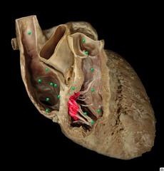
crista terminalis
identify the structure
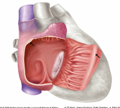
fossa ovalis
identify the structure
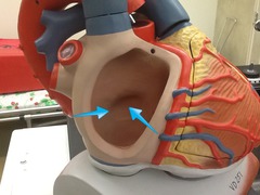
foramen ovale
a fetal shunt from right atrium to left atrium
ductus arteriosus
a fetal shunt from pulmonary truck to aorta