amino acids
 Call Kai
 Learn
Learn Practice Test
 Spaced Repetition
 Match
Match Flashcards
 Knowt Play
1/37

There's no tags or description
Looks like no tags are added yet.
Name | Mastery | Learn | Test | Matching | Spaced | Call with Kai | Chat |
|---|
No analytics yet
Send a link to your students to track their progress
38 Terms
amino acids
monomers that build the protein polymers, through polymerization.
Oligopeptides and polypeptides
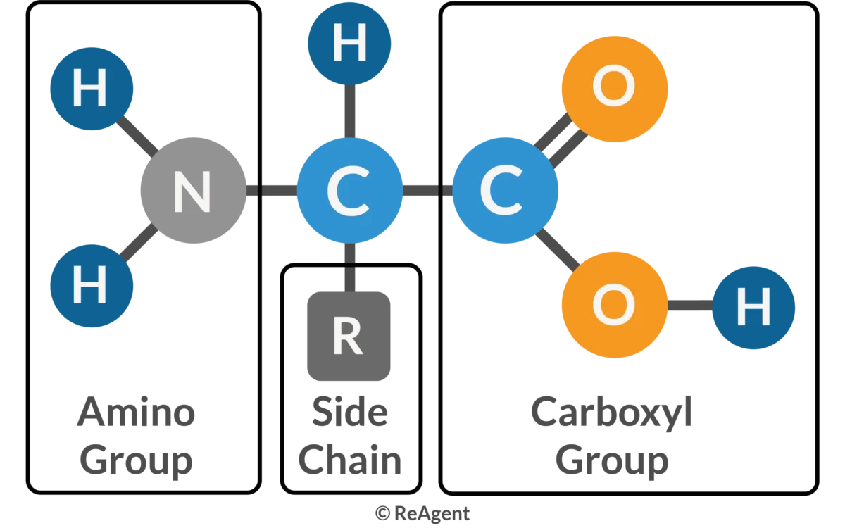
amino acid structure
amino group attached to an a-carbon, the a-carbon is also attached to a carboxylic acid group.
every amino acid has an R group attached to a-carbon
The pKa of the carboxylic acid and amino groups of the α-amino acids are about 2 and 10
true
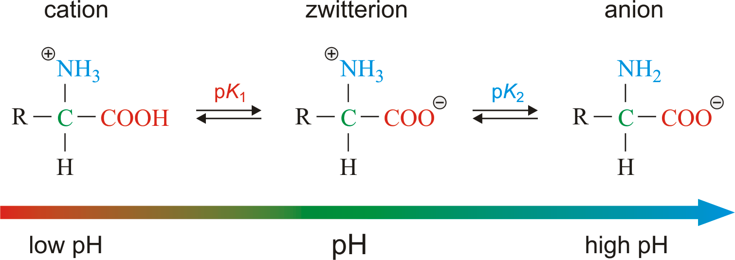
zwitterion
is a single molecule that carries both a positive and a negative charge at the same time, but whose overall net charge is zero.
This zwitterionic form is the dominant form of amino acids around their isoelectric point (pI)
At physiological pH (~7.4) inside cells, standard amino acids exist almost entirely as zwitterions
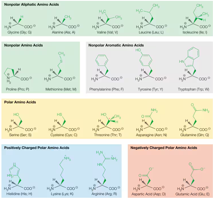
how many amino acids are there
There are 20 common α-amino acids that are the major building blocks of proteins
selenocysteine and pyrrolysine, are found in small numbers of proteins
chiral
a carbon atom has four different substituents attached to it ____ or a stereocenter, or (preferably) an asymmetric carbon
enantiomers
Stereoisomers that are nonsuperimposable mirror images of each other.
if a molecule is chiral (asymmetric ) then it contains an enantiomers
for example alanine, it has an L and D.
Are L- and D-amino acids chemically different in a test tube, and why do biological systems exclusively use L?
Chemical Identity: In isolation (at pH 7), L- and D-amino acids are identical mirror images with the exact same chemical properties and reactivity; neither is inherently "better."
Biological Standard: Evolution standardized on L-amino acids, meaning our cellular machinery, enzymes, and protein folding patterns are custom-built exclusively for L-enantiomers.
Structure & Drugs: Individual L- and D-monomers have flipped 3D "handedness." Because our proteins are L-based, drugs or molecules must match that L-shape to fit into our asymmetric biological binding sites
All amino acids except glycine can exist in D and L form
true, two of its four groups bonded to the a-carbon are the same.
The variety of side chains on amino acids allows proteins enormous versatility in structure and function.
true
Amino Acids with Nonpolar Aliphatic Side Chains
Glycine, alanine, valine, leucine, and isoleucine have aliphatic side chains.
hydrophobic and typically hide in the interior of water-soluble proteins
Glycine
R-H
t is frequently found on the surfaces of proteins due to its ability to form tight turns in protein structures. Such turns are sites in the protein structure where the protein polymer folds back on itself to maintain a compact structure

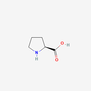
Proline
non polar, has an amino group, found on the surfaces of protein
Amino Acids with Nonpolar Aromatic Side Chains
Three amino acids phenylalanine, tyrosine, and tryptophan have nonpolar aromatic side chains.
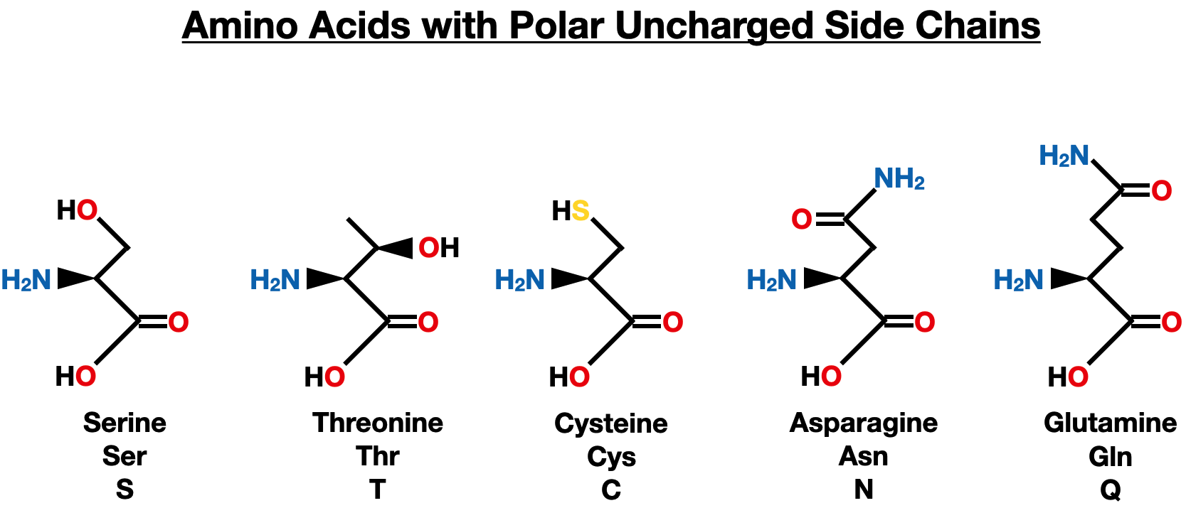
Amino Acids with Polar Side Chains
Serine, threonine, asparagine, and glutamine have polar side chains, and they can form multiple H bonds with water molecules and/or other good H bond donors and acceptors.
these five amino acids are most often found on the surfaces of proteins, where they can contact the aqueous environment in cells or in circulation.

cysteine
—SH group, good nucleophiles and often play key roles in enzyme activity.
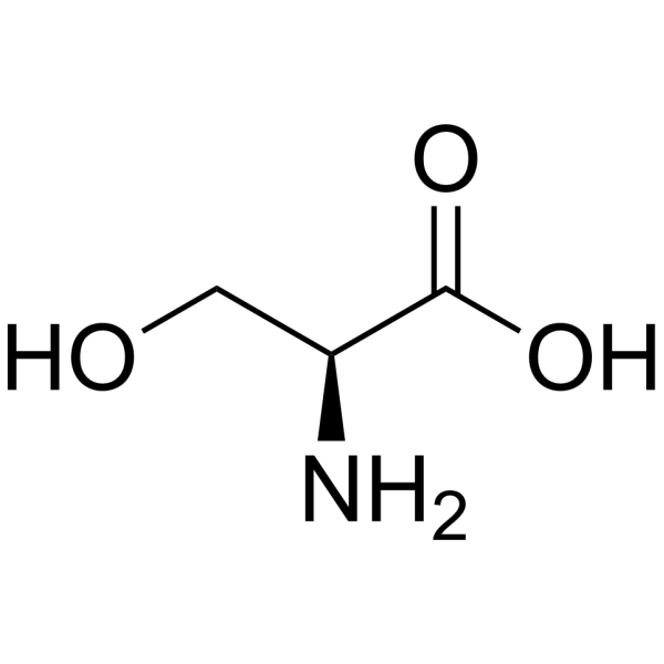
serine
-OH group, good nucleophiles and often play key roles in enzyme activity.

the oxidation of two cysteine
yields a disulfide bonds that stabilize the active structure of a protein that contains them.

Amino Acids with Positively Charged (Basic) Side Chains
Histidine, lysine, and arginine have basic groups in their side chains.

Amino Acids with Negatively Charged (Acidic) Side Chains
Aspartic acid and glutamic acid

Rare Genetically Encoded Amino Acids
Selenocysteine (“Sec”) and pyrrolysine (“Pyl”) are sometimes referred to as the 21st and 22nd amino acids.
a repurposed stop codon
posttranslational modification
Sometimes amino acid side chains are modified after being incorporated into a protein
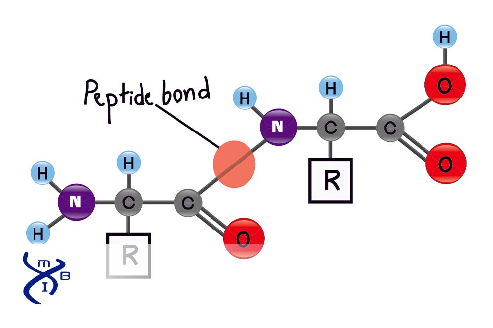
Peptide Bond
it consists of an amide bond between the α-carboxyl group of one amino acid and the α-amino group of the next.
The Structure of the Peptide Bond
invariably the amide carbonyl (C═O) and amide N—H bonds are nearly parallel
There is little twisting possible around the peptide bond because the C—N bond has a substantial fraction of double-bond character
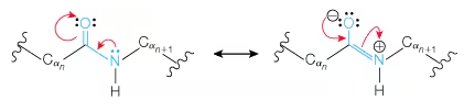
Peptide Bond resonance
Why the trans form is favored in peptide bonds
The trans form is highly favored because the R groups on adjacent α-carbons can sterically interfere in the cis configuration.
why do polypeptide hydrolysis?
because they are metastable, thermodynamically unstable but kinetically persistent.
hydrolysis of Peptide bond
if done uncatalyzed it is very slow at physiological pH and Temp, so a catalysts needs to be present
Harsh chemical conditions (e.g., boiling in strong mineral acid such as 6 M HCl, which can also damage some side chains).
Catalysis by proteolytic enzymes (proteases),
proteases
Enzymes that cleave peptide bonds in a polypeptide. Many show specificity for a particular amino acid sequence.
done under mild conditions, including in digestion and in biochemical analysis.
oligopeptides
A molecule composed of amino acid residues linked by peptide bonds that is smaller than a protein, which is called a polypeptide.
N-terminus
The end of a polypeptide chain that carries an unreacted amino group.
many proteins have N-termini blocked by N-formyl or N-acetyl groups
C-terminus
(also called carboxyl terminus) The end of a polypeptide chain that carries an unreacted carboxyl group
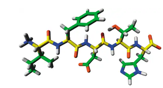
main chain
composed of the atoms that make up the peptide bonds—namely, the α-NH, the and the α-C═O groups of each amino acid residue in the peptide.
polyampholytes
A molecule (like a polypeptide) containing both positively and negatively charged groups. charge–charge interactions within the polypeptide chain
Near Negative Charges (Glu or Asp):
Effect: Unfavorable charge–charge repulsion makes the group less likely to lose a proton.
Result: Apparent pKa increases (becomes less acidic).
Near Positive Charges (Lys, His, or Arg):
Effect: Favorable charge–charge attraction stabilizes the deprotonated form, making the group more likely to lose a proton.
Result: Apparent pKa decreases (becomes more acidic).
why do neighboring charges influence an amino acid's pKa
Near Negative Neighbors (Crowded): The group keeps its proton because losing it adds more negative charge to an already crowded, repulsive area. (Unstable higher pKa).
Near Positive Neighbors (Hungry): The group loses its proton because the nearby positive charge stabilizes that newly formed negative charge, making the whole situation energetically happy. (Stable lower pKa).
Amino acids, peptides, and proteins are ampholytes; each has an isoelectric point.
true
In the formation of a peptide bond, which functional groups link together, and in what direction does protein synthesis occur?
Functional Groups: The carboxylic acid group of the first (or growing chain's) amino acid links to the amino group of the incoming second amino acid.
Direction: Amino acids are exclusively added to the free C-terminal end of the growing peptide chain.
amino acid residue
The portion of each amino acid remaining in the chain