Lecture 6 - Pediatric Infectious Disease
 Call Kai
 Learn
Learn Practice Test
 Spaced Repetition
 Match
Match Flashcards
 Knowt Play
1/113

There's no tags or description
Looks like no tags are added yet.
Name | Mastery | Learn | Test | Matching | Spaced | Call with Kai | Chat |
|---|
No analytics yet
Send a link to your students to track their progress
114 Terms
What temperature defines a fever in pediatric patients?
A rectal temperature of 38.0°C (100.4°F) or higher.
How may neonates present temperature-wise despite a significant infection?
They may present as hypothermic.
Why is aspirin contraindicated in pediatric patients with fever?
Due to the risk of developing Reye syndrome.
Does the height of a fever or the response to antipyretics distinguish bacterial from viral infection?
No, neither distinguishes the cause of fever.
What are the three categories of fevers in children?
Short duration with localizing signs, fever without a focus, and fever of unknown origin.
What defines a fever without a focus?
Common in children
What defines a fever of unknown origin (FUO)?
Daily fever >14 days without etiology on routine workup, or >1 week on intense workup.
What drives the algorithms of fever without a focus?
Age-based risk stratification (
What are typical bacterial causes of sepsis in neonates?
Group B Streptococcus, Listeria monocytogenes, and Escherichia coli.
What are typical bacterial causes of sepsis in children 2 to 36 months old?
Streptococcus pneumoniae, Hib, Neisseria meningitidis, and non-typhoidal Salmonella.
Why is fever in infants under 2 months of age particularly dangerous?
Immature immune system and permeable blood-brain barrier increase serious bacterial infection risk.
What does an immature immune system increase risk for?
Higher risk of serious bacterial infection (SBI) leading to sepsis.
What are common SBI's in
Bacteremia, meningitis, pneumonia, UTI, HSV, bacterial diarrhea, osteomyelitis, or septic arthritis. Often present with only fever and poor feeding, without localizing signs.
What percentage of febrile infants ≤28 days harbor invasive bacterial infections?
Approximately 4% of febrile infants.
What are the key differences between Early-Onset and Late-Onset Neonatal Sepsis?
Early-onset (0-7 days) is vertically transmitted; Late-onset (8-28 days) is horizontally acquired (community/hospital) or vertically acquired.
What organisms typically cause early-onset neonatal sepsis?
GBS, E. coli, Klebsiella, and Listeria monocytogenes.
What organisms typically cause late-onset neonatal sepsis?
Same as early PLUS H. influenzae, S. pneumoniae, N. meningitidis.
How does neonatal sepsis manifest in early onset vs late onset?
Early: respiratory failure, shock, meningitis (30%), DIC
Late: lethargy, poor feeding, hypotonia, seizures, bulging fontanelles.
What are the risk factors of early-onset sepsis?
Maternal GBS colonization, prolonged ROM (>18 hours), chorioamnionitis, prematurity.
What defines a toxic-appearing child aged 0 to 36 months?
Lethargic, limp, or inconsolable crying.
What is the management for any toxic-appearing child aged 0-36 months with fever >100.4°F?
Full sepsis work-up, including hospital admission, CBC, cath UA, blood/urine/CSF cultures, and broad-spectrum parenteral antibiotics.
What key change was introduced in the 2021 AAP guidelines for infants 22-60 days old?
When initial labs are reassuring, some infants may NOT need lumbar puncture or empiric abx, and may be monitored at home with shared decision making. Based on age and lab results! ONLY IF 22 days+.
What is the mandatory management for ALL febrile infants 0-21 days old?
Full sepsis work-up, empiric IV antibiotics (Ampicillin + Ceftazidime/Gentamicin), and hospital admission.
When should empirical acyclovir be added to a febrile infant's regimen?
In infants
What is the 3-step management process for febrile non-toxic infants 22-28 days old?
1. Determine risk
2. If all-low risk criteria met: obtain urine cx, blood cx and inflammatory markers. Maybe LP, maybe IV/IM abx. Observe in hospital
3. If NOT low risk: with abnormal UA but NORMAL inflammatory: maybe perform LP, administer abx, observe in hospital
with abnormal inflammatory markers: perform LP. If CSF and UA normal give IM abx and observe at home OR observe in hospital +/- IV/IM abx.
If CSF or UA abnormal: give IV/IM abx and observe in hospital.
What are the low-risk criteria?
Uncomplicated history, well-appearing, normal labs, negative UA, inflammatory markers normal, normal stool studies (if diarrhea)
What is the 3-step management process for febrile non-toxic infants 29-60 days old?
1. Determine risk
2. If all low-risk criteria met: obtain CBC, blood/urine cx and inflammatory markers. NO need for LP or abx. Observe at home with close f/u within 24-36 hours.
3. If NOT low risk: abnormal UA but normal inflammatory, gie PO abx and observe at home with 12-24 hrs f/u. With abnormal inflammatory, may perform LP. if CSF normal give IV or PO abx, and observe in hospital or home. If CSF abnormal give IV abx and admit. If CSF not available, give IV abx and admit or go home.
What single biomarker is best for risk-stratifying invasive bacterial infections in infants?
Procalcitonin.
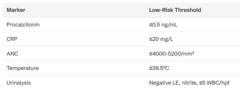
If procalcitonin is unavailable, which alternative parameters are used for risk stratification?
CRP, ANC, and temperature.
Which organisms pose an increased bacteremia risk in unimmunized febrile children 2-36 months?
Encapsulated organisms: S. pneumoniae, Hib, and N. meningitidis.
Why are febrile children with sickle cell disease at higher risk for severe infection?
Functional asplenia increases risk for encapsulated bacteremia and Salmonella/S. aureus osteomyelitis.
What initial management is required for any child with sickle cell disease presenting with fever?
Obtain blood culture, CBC, reticulocyte count, and administer IV Ceftriaxone.
What is the initial management for a well appearing child with a high grade (>102.2) fever?
If unimmunized: blood cx, CBC, urine
If immunize: urine IF male < 6 months, all females.
What is the primary difference in duration between Fever Without Source (FWS) and Fever of Unknown Origin (FUO)?
FWS lasts
What is the most common cause of Fever Without Source in children?
Self-limited viral infections (about 75% of cases).
What is the definition of meningitis?
Inflammation of the leptomeninges.
What defines aseptic meningitis and what is its most common cause?
Negative CSF bacterial cultures; primary cause is viral (enteroviruses and parechoviruses).
What are the most common bacterial causes of meningitis in neonates (
Group B Streptococcus, E. coli, and other enteric gram-negatives.
What are the most common bacterial causes of meningitis in children 1-3 months?
S. pneumoniae, gram negative bacilli, GBS.
What are the most common bacterial causes of meningitis in children >3 months old?
Streptococcus pneumoniae and Neisseria meningitidis.
What clinical signs of meningitis are seen in older children (>12 months)?
Headache, irritability, nausea, nuchal rigidity, photophobia, lethargy, vomiting, fever usually present. Kernig and Brudzinski signs often positive.
What clinical signs of meningitis are seen in young infants (
Signs may be minimal - irritability, depressed mental status, temperature instability, poor feeding, bulging fontanelle (sign of increased ICP)
What are red flag symptoms indicating rapid disease progression in meningitis?
Focal neurologic signs, seizures, petechial/purpuric lesions, sepsis, shock, and coma.
When should a lumbar puncture (LP) be delayed or avoided in suspected bacterial meningitis?
Cardiovascular instability or evidence of increased ICP (risk of brain herniation).
What can PCR panels do?
Diagnose bacterial vs viral meningitis (identifies pathogen) especially after prior abx, but can't tell susceptibility.
Describe the expected CSF findings in acute bacterial meningitis.
High opening pressure, cloudy appearance, WBC >1,000 (neutrophils), high protein, low glucose (
Describe the expected CSF findings in viral meningitis.
Normal opening pressure, clear appearance, WBC 10-1,000 (lymphocytes), normal protein, normal glucose.
What CSF phenomenon can occur early in viral meningitis (
PMN predominance initially, which later shifts to lymphocyte predominance.
What is the empiric antibiotic treatment for bacterial meningitis in neonates (0-28 days)?
Ampicillin plus Cefotaxime (or Ampicillin plus Gentamicin).
What is the empiric antibiotic treatment for bacterial meningitis in >28 days?
Ceftriaxone plus vancomycin.
Why is ampicillin specifically included in the neonatal empiric meningitis regimen?
To cover Listeria monocytogenes.
Why is vancomycin added to meningitis empiric therapy for patients >28 days old?
Due to increasing resistant Streptococcus pneumoniae strains.
What is the role of adjunct dexamethasone in bacterial meningitis treatment?
Reduces hearing loss in H. influenzae meningitis when given before or with first antibiotic dose.
What major post-recovery evaluation must ALL children with meningitis receive before hospital discharge?
A formal hearing evaluation.
What is orbital cellulitis and what is its most common underlying source?
Infection of orbital tissues posterior to the septum, most commonly arising from ethmoid sinusitis.
How does orbital cellulitis present?
Painful, red swollen eyelid with bulging eye, painful/limited EOM, blurry or double vision and high fever.
What are common pathogens that cause orbital cellulitis?
S. aureus, GAS and anaerobes.
What imaging modality is required to confirm orbital cellulitis and rule out abscess?
CT scan of the orbits with IV contrast.
What is the management approach for orbital cellulitis?
Hospital admission, IV antibiotics (Vancomycin/Clindamycin + Ceftriaxone), and surgical drainage if abscess present or failure to respond.
What are complications of orbital cellulitis?
Subperiosteal abscess, cavernous sinus, thrombosis, meningitis, intracranial extension, vision loss.
What is periorbital cellulitis?
Bacterial infection of the eyelid and surrounding skin due to skin lesion, trauma, conjunct or stye spreading and affecting the preseptal space
What clinical signs distinguish orbital cellulitis from periorbital cellulitis?
Proptosis, painful/limited extraocular muscle movements, and blurry/double vision. Periorbital has no proptosis, and eye movement/vision NORMAL.
How is periorbital cellulitis diagnosed and treated?
Dx is clinical. Treat with PO abx in mild condition. In moderate-severe, hospitalize and give IV abx.
Compare the typical age distribution of preseptal versus postseptal cellulitis.
Preseptal occurs in younger children (~4 years); postseptal occurs in older children (~7.5 years).
What clinical features distinguish viral pharyngitis from Group A Streptococcal (GAS) pharyngitis?
Viral presents with gradual onset, cough, rhinorrhea; GAS presents abruptly with fever, exudates, NO cough.
What is the causative organism of Hand, Foot, and Mouth Disease (HFMD) and Herpangina?
Coxsackievirus.
What are the classic physical exam findings in Hand, Foot, and Mouth Disease?
Painful oral ulcers, plus papulovesicular rash on palms, soles, and buttocks. Most common in children
What is the management for HFM disease?
Supportive with pain relief and hydration. Oral lidocaine NOT recommended.
How can Herpangina be clinically distinguished from Hand, Foot, and Mouth Disease?
Herpangina lesions are restricted to the posterior oral cavity with NO skin rash. Herpangina has onset of high fever, vomiting, headache, sore throat and dysphagia.
Where are the mucosal lesions located in Herpangina?
Soft palate, uvula, and tonsillar pillars (posterior oropharynx). Small vesicular lesions (1-4mm) that enlarge, rupture and become punched out ulcers.
What is the causative agent and typical age group for primary Herpes Gingivostomatitis?
HSV type 1; typically affects children aged 6 months to 5 years.
What are the key clinical findings in primary Herpes Gingivostomatitis?
High fever, painful anterior oral ulcers, severe gingival edema/bleeding, drooling, and halitosis.
What antiviral treatment may benefit children with Herpes Gingivostomatitis if given within 72 hours?
Oral Acyclovir.
What parts of the mouth are affected in gingivostomatitis?
Soft palate, uvula, tongue, and perioral (typically spares the buccal mucosa).
What is the etiology and primary mode of transmission of Infectious Mononucleosis?
Epstein-Barr virus (EBV); transmitted via salivary secretions.
What are the clinical features of mono?
Fever, fatigue, malaise (often prolonged), pharyngitis/tonsillitis, bilateral cervical lymphadenopathy, splenomegaly.
What happens if a patient with Infectious Mononucleosis is misdiagnosed and given amoxicillin?
Approximately 80% develop a diffuse maculopapular rash.
What key laboratory finding supports a diagnosis of Infectious Mononucleosis?
Peripheral blood leukocytosis with a predominance of atypical lymphocytes. Heterophile antibody test (Monospot) but need to get timing right.
What crucial activity restriction must be advised for patients with Infectious Mononucleosis and why?
Avoid contact sports for ≥3-4 weeks to prevent splenic rupture.
How is acute gastroenteritis defined?
More than 3 loose or liquid stools in 24 hours, usually lasting several days.
What distinguishes secretory gastroenteritis from invasive gastroenteritis?
Secretory produces large-volume watery stool without blood; invasive produces bloody stool with fever.
Compare viral diarrhea vs bacterial diarrhea
Viral: most common, watery, no blood or mucus, vomiting may be prominent, low-grade fever
Bacterial: bloody, mucoid stools, high fever, commonly caused by Shigella or EHEC.
Which bacterial diarrhea strain carries a high risk for Hemolytic Uremic Syndrome (HUS)?
Enterohemorrhagic E. coli (EHEC O157:H7).
What triadic complications define Hemolytic Uremic Syndrome (HUS)?
RBC destruction (microangiopathic hemolytic anemia), thrombocytopenia, and acute renal failure.
What are physical sign differences between mild and severe dehydration in children?
Mild (3-5%): alert, tears present, normal refill. Severe (>10%): lethargic, no tears, deeply sunken eyes, capillary refill >3s, markedly increased HR.
What vital sign change is a late sign of severe dehydration and impending shock in children?
Hypotension.
What fluid resuscitation bolus is administered for severe dehydration in children?
IV normal saline or Lactated Ringer's 20 mL/kg bolus.
Why are routine antibiotics avoided in Enterohemorrhagic E. coli (EHEC) infections?
Antibiotics may increase toxin release and worsen the risk of HUS.
When are antibiotics indicated for AGE management?
Shigella
Salmonella in infants
Describe adenovirus infections in children
> 50 serotypes
Typical URI sx most common presentation
Diagnose with respiratory PCR panels
Treatment is supportive.
What clinical syndrome features fever, pharyngitis, and conjunctivitis simultaneously?
Pharyngoconjunctival fever caused by Adenovirus.
What high-fever pattern can Adenovirus exhibit that mimics a bacterial infection?
High, persistent fevers (>39°C) lasting for 5 to 7 days.
Describe Enterovirus infections in children
Includes Coxsackie and poliovirus
Nonspecific febrile URI illness is the most common presentation
Clinical syndromes include HFMD, Herpangina, aseptic meningitis
Diagnose with RT-PCR
Treatment is supportive. IVIG may be considered in severe neonatal enteroviral disease.
What is the single most common cause of viral aseptic meningitis in children?
Enterovirus.
What viral pathogen causes Roseola Infantum?
Human Herpesvirus 6 (HHV-6) and HHV-7.
What clinical course is classic for Roseola Infantum (Sixth Disease)?
Abrupt high fever (104°F) for 3-5 days in a well child, followed by rash as fever resolves.
What common neurological complication is associated with Roseola Infantum?
Febrile seizures (causes ~30% of febrile seizures in young children).
What is the causative agent and classic rash progression of Erythema Infectiosum (Fifth Disease)?
Parvovirus B19; features 'slapped cheek' facial redness followed by a lacy, reticular body rash.
When is a child with Erythema Infectiosum no longer contagious?
Once the rash appears, the child is no longer infectious.
What are the 3 stages of the rash with erythema infectiosum (5th disease)?
1. Slapped cheek rash
2. Lacy, reticular rash
3. Recurrent phase