Unit 7 - [P1] Benign Pathology of the GB
1/45
There's no tags or description
Looks like no tags are added yet.
Name | Mastery | Learn | Test | Matching | Spaced |
|---|
No study sessions yet.
46 Terms
What is sludge in the GB?
Thick bile
Layering of bile particles
When looking at sludge in the GB, what is important about the GB position?
Gravity dependent
Dependent with position changes
Where might you find sludge, other than the GB?
In nearby ducts (CD, CHD, CBD)
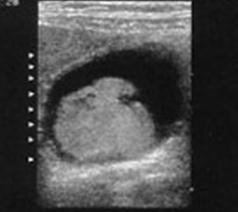
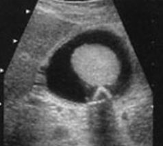
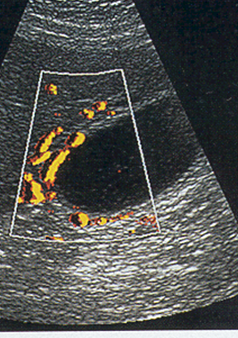
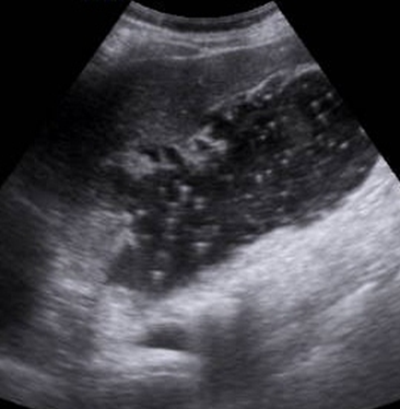
What is this image showing?
Sludge in the GB
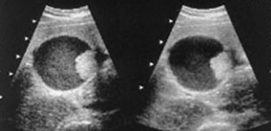
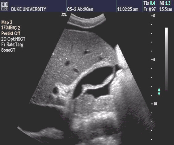
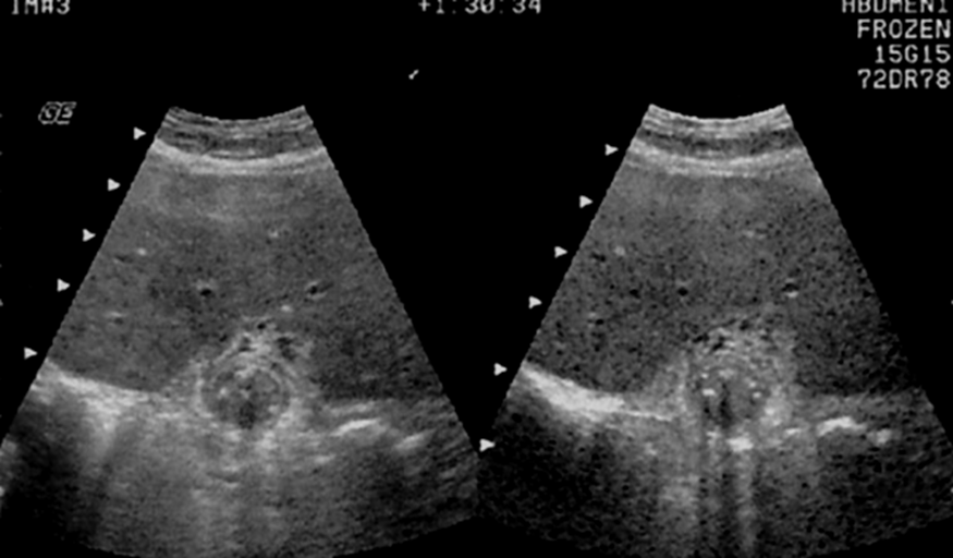
What is this image showing?
Sludge in transverse GB
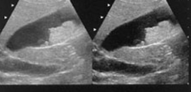
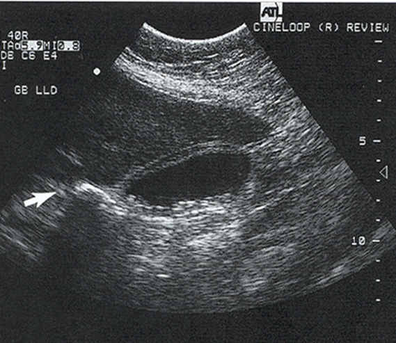
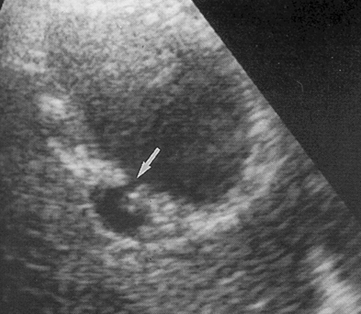
What is this image showing?
Sludge in sagittal GB
What is this image showing?
Sludge in transverse GB
What is this image showing?
Sludge in transverse GB
What are the biliary causes of wall thickness?
Cholecystitis
Adenomyomatosis
Cancer
AIDS
Cholangiopathy
Sclerosing cholangitis
What are the non-biliary causes of wall thickness?
Non-fasting
Diffuse liver disease
Cirrhosis & Hepatitis
Ascites
Pancreatitis
Portal Hypertension
Heart failure
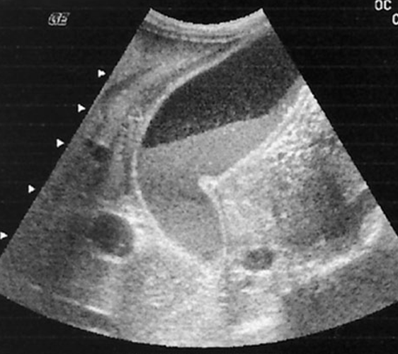
What is this image showing?
Ascites around the GB
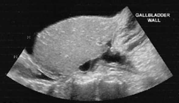
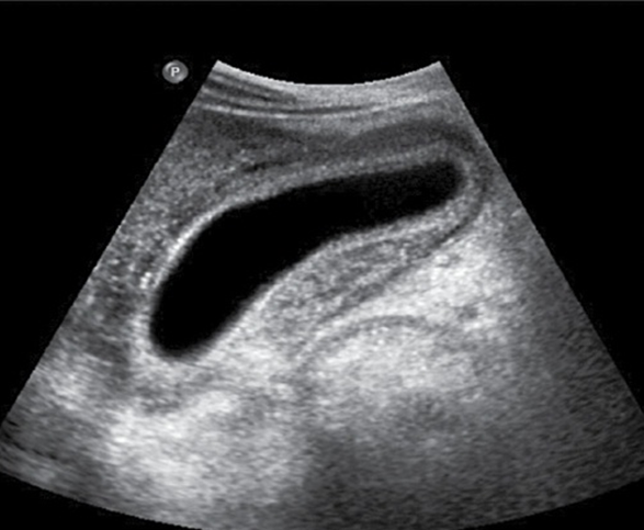
What is this image showing?
Wall Thickness
What are the differential diagnoses for a thickened GB wall?
Cholecystitis
Acute Cholecystitis
Carcinoma
Gangrene
What is Cholecystitis?
Inflammation of the GB
What are the forms of Cholecystitis?
Acute
Chronic
Acalculous
Emphysematous
Gangrenous
What are the symptoms for Acute Cholecystitis?
RUQ pain
+ Murphy’s sign
What is the sonographic appearance of Acute Cholecystitis?
Usually thickened, irregular wall
Distended GB (bigger)
Gallstones may or may not be present
Pericholecystic fluid
May demonstrate color flow in the wall
What is another name for having gallstones throughout the GB?
Calculous Cholecystitis
What is another name for having NO gallstones throughout the GB?
Acalculous Cholecystitis
What is pericholecystic fluid (abscess)?
Bile leaks from the gallbladder and forms a fluid-filled area around it
In relation to Acute Cholecystitis, if localized thickening is found. What other differential diagnosis should you consider?
Abscess
Carcinoma
What is this image showing?
Cholecystitis
What is this image showing?
Acute Cholecystitis
What lab work may possibly be increased with Acute Cholecystitis?
Serum Bilirubin
Alk Phos
Serum Amylase
WBC
To R/O Cholecystitis, what NEEDS to be done?
Patient MUST be fasting, or GB may be just partially contracted
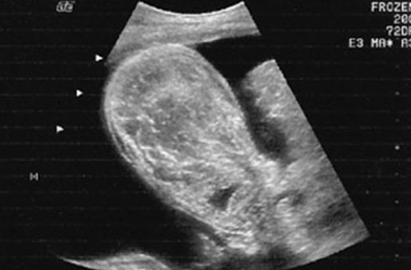
What is this image showing?
Swollen / Edematous GB
What is this image showing?
Swollen / Edematous GB
What is Emphysematous Cholecystitis?
Gas-forming bacteria in GB wall
What is the prognosis for Emphysematous Cholecystitis?
Rare complication
Rapidly progressive & may be fatal (15%)
What lab work may prove Emphysematous Cholecystitis
Abnormal LFTs
What are the US findings for Emphysematous Cholecystitis?
Bright echo in area of GB with ring down or comet tail artifact
May appear as WES
What is Gangrenous Cholecystitis?
Thickened wall
Ulcerations / perforations
In Gangrenous Cholecystitis, ___-___% are gallstones or gravel.
80-95%
What lab work may prove Gangrenous Cholecystitis
Abnormal LFTs
What are the US findings of Gangrenous Cholecystitis?
Echogenic material
Does not shadow
Is not gravity dependent
Does not layer
What is this image showing?
Gangrenous Cholecystitis
What is this image showing?
Gangrenous Cholecystitis
What is this image showing?
Gangrenous Cholecystitis
GB Perforations may occur with…
Gangrenous cholecystitis
Acute cholecystitis
Chronic cholecystitis
When looking at GB Perforations what should you watch for?
Continuity of GB wall
What results from GB perforations?
Pericholecystic fluid collections
What is this image showing?
GB Perforations
What is Acalculous Cholecystitis?
Acute inflammation without Cholelithiasis
+ Murphy’s sign
What lab work may prove Acalculous Cholecystitis
Abnormal LFTs
Increased Amylase
What are the US findings of Acalculous Cholecystitis without gallstones?
GB wall irregular & thickened (> 4 to 5 mm)
Echogenic sludge
Dilated GB
Pericholecystic fluid or sub-serosal edema
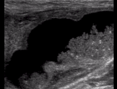
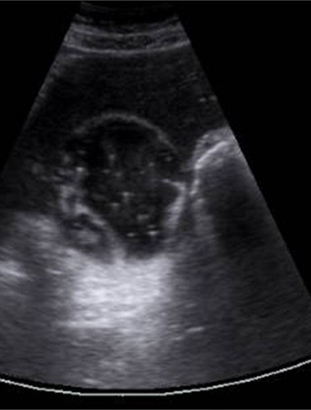
What is this image showing?
Gallbladder