The Central Nervous System

0.0(0)
Studied by 0 people
call kaiCall Kai
Locked
learnLearn
examPractice Test
spaced repetitionSpaced Repetition
heart puzzleMatch
flashcardsFlashcards
GameKnowt Play
Card Sorting

1/63

encourage image

There's no tags or description

Looks like no tags are added yet.

Last updated 4:26 AM on 9/10/26
Name
Mastery
Learn
Test
Matching
Spaced
Call with Kai
Chat

No analytics yet

Send a link to your students to track their progress

64 Terms

1
New cards
Compare the CNS and PNS, including cranial nerves.
The CNS is the brain and spinal cord. The PNS is all nervous tissue outside them and carries signals between the CNS and the body. Cranial nerves arise from the brain/brain stem but are part of the PNS.
2
New cards
Describe nervous-system information flow and its major divisions.
  • Afferent pathways carry sensory action potentials from the periphery to brain/cord

    • Somatic afferents carry information from skeletal muscle

    • Visceral afferents from smooth muscle, cardiac muscle, glands, and organs.

  • Efferent pathways carry commands from the brain/cord to the periphery

    • Somatic motor supplies skeletal muscle

    • Autonomic motor supplies smooth muscle, cardiac muscle, and glands

      • Autonomic divisions are sympathetic (“fight or flight”) and parasympathetic (“rest and digest”)


3
New cards
What is a sensory receptor in an afferent neuron?
A specialized modification of the dendritic/receiving region that detects a stimulus and initiates sensory signaling.
4
New cards
Describe the three functional neuron classes and the role of effectors.
  • Afferent neurons carry sensory information to the CNS

  • Efferent neurons carry CNS commands to effectors, which are muscles or glands that produce the response

  • Interneurons are located mainly within the CNS; they process/integrate information and help formulate appropriate responses.


5
New cards
What is the basic function of the nervous system?
To receive sensory information, integrate/process it, and generate appropriate motor or glandular responses.
6
New cards
Describe CNS protection and name the meninges in order.
CNS protection includes skull/vertebral column, meninges, cerebrospinal fluid, and specialized vascular protection including the blood-brain barrier. From outermost to innermost, meninges are dura mater, arachnoid mater, and pia mater.
7
New cards
Describe the dura mater.

The outermost meninx. It is tough, thick, and difficult to penetrate (like a canvas).
8
New cards
Describe the arachnoid mater.
The middle meninx; it is thin and web-like.
9
New cards
Describe the pia mater.
The innermost meninx; it is delicate and closely adheres to the brain and spinal cord surface.
10
New cards
What is the subarachnoid space?
The space between arachnoid and pia mater that contains cerebrospinal fluid and blood vessels.
11
New cards
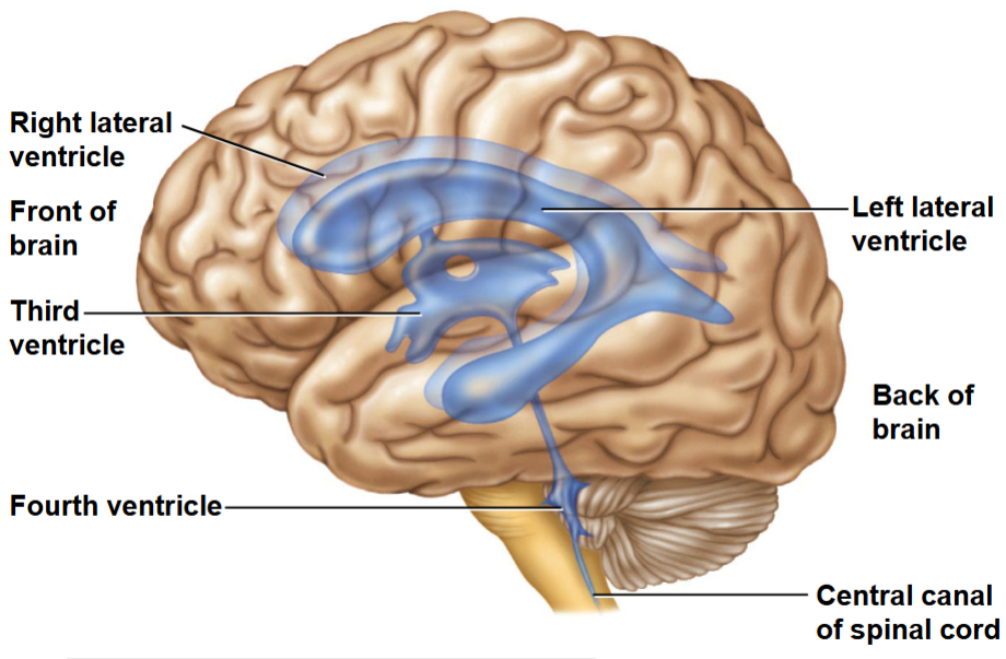
What are the ventricles?
Interconnected cavities within the brain that contain cerebrospinal fluid.
12
New cards
Describe CSF: location, production, functions, and circulation.
  • A clear fluid in the ventricles and subarachnoid space around brain/spinal cord

  • Cushions the CNS, provides buoyancy, stabilizes extracellular conditions, exchanges materials between neural cells and blood, and removes waste.

  • Produced mainly by choroid plexuses in ventricles, flows through the ventricular system and subarachnoid space, and is reabsorbed into venous blood


13
New cards
What causes hydrocephalus?
Impaired CSF flow or absorption causes CSF accumulation and increased pressure in the ventricles.
14
New cards
Why is increased intracranial pressure dangerous?
The skull is rigid, so added volume can compress brain tissue and reduce perfusion.
15
New cards
Describe the blood-brain barrier (BBB): structure, function, and clinical consequence.
  • A selective barrier formed mainly by tight junctions between CNS capillary endothelial cells

  • Limits movement from blood into brain tissue, protecting neurons and stabilizing the chemical environment required for neural signaling

  • The same protection restricts many drugs as well as toxins, complicating treatment of CNS disease


16
New cards
What is a concussion?
A brain injury caused by a blow or rapid acceleration/deceleration that disrupts brain function, often without obvious structural damage on imaging.
17
New cards
Why can coup-contrecoup injury occur?
The brain can strike the inside of the skull at the impact site and then rebound to strike the opposite side.
18
New cards
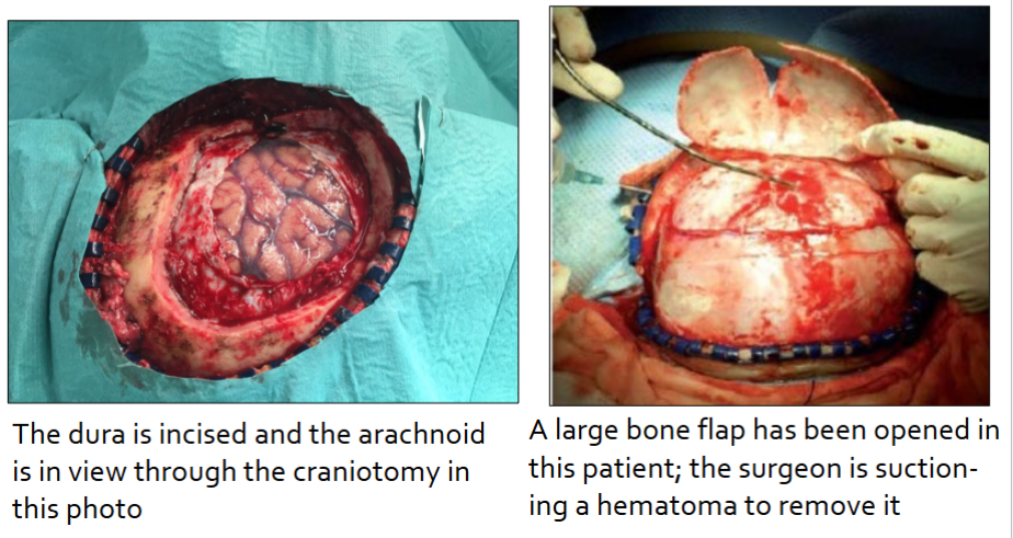
What is a craniotomy?
A surgical procedure in which part of the skull is opened/removed temporarily to access the brain or relieve pressure.
19
New cards
What are the six overall divisions of the brain?
Cerebral cortex, basal nuclei, thalamus, hypothalamus, cerebellum, and brain stem.
20
New cards
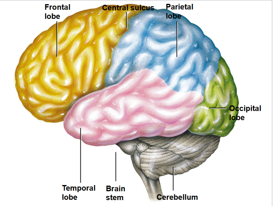
What is the cerebral cortex, and give two broad functions.
  • The outer gray-matter layer of the cerebrum

  • It supports conscious perception, voluntary movement, and integration of sensory information.

  • The four lobes of the cerebral cortex include occipital, temporal, parietal, and frontal

21
New cards
What are basal nuclei, and give two broad functions.
They are deep gray-matter masses within cerebral white matter; they help initiate/select/coordinate movement and suppress unwanted movements.
22
New cards
What is the thalamus, and give two broad functions.
It is a major relay and integrating center for sensory input; it directs attention to important stimuli and helps refine conscious awareness of sensations.
23
New cards
What is the hypothalamus, and give two broad functions.
It lies below the thalamus and integrates homeostatic control; it regulates internal environment, autonomic activity, and pituitary hormone output.
24
New cards
What is the cerebellum, and give two broad functions.
It coordinates the timing and precision of movement and contributes to posture, balance, and motor learning.
25
New cards
What is the brain stem, and give two broad functions.
It links the spinal cord with higher brain centers and contains centers for vital autonomic functions such as cardiovascular and respiratory control.
26
New cards
Where is the frontal lobe, and what is one major function?
It is anterior to the central sulcus; it contains primary motor cortex and is essential for voluntary skeletal-muscle movement.
27
New cards
Where is the parietal lobe, and what is one major function?
It is posterior to the central sulcus and superior to the lateral sulcus; it contains primary somatosensory cortex for body sensation.
28
New cards
Where is the temporal lobe, and what is one major function?
It lies inferior to the lateral sulcus; it contains the auditory cortex for hearing.
29
New cards
Where is the occipital lobe, and what is one major function?
It is at the posterior of the cerebrum; it contains the visual cortex.
30
New cards
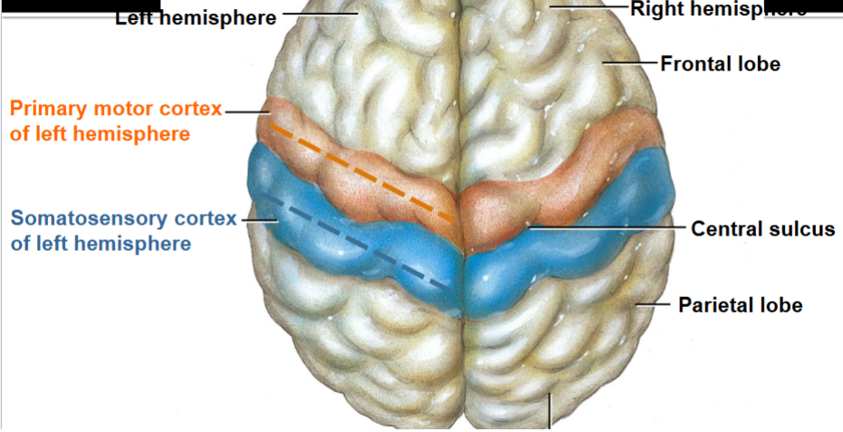
What is the central sulcus?
  • A prominent groove separating the frontal lobe from the parietal lobe

  • Primary motor cortex is located at the caudal most gyrus of the frontal lobe

  • Somatosensory cortex is located at the rostral most gyrus of the parietal lobe


31
New cards
What is the lateral sulcus?
A deep groove that separates the temporal lobe from frontal and parietal lobes.
32
New cards
Compare primary motor and primary somatosensory cortex.
  • Primary motor cortex is in the frontal lobe just anterior to the central sulcus and controls voluntary skeletal-muscle movement, mainly on the opposite body side

  • Primary somatosensory cortex is in the parietal lobe just posterior to the central sulcus. It receives conscious touch, pressure, temperature, pain, and proprioception (awareness of body position/movement), mainly from the opposite body side.

33
New cards
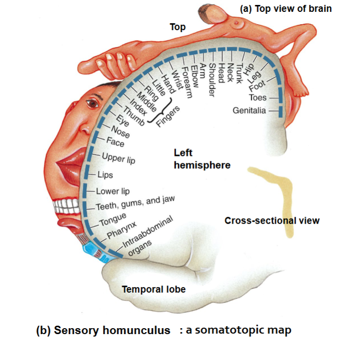
Explain somatotopic maps and the sensory and motor homunculi.
  • An orderly cortical representation in which specific areas correspond to body regions

  • Sensory homunculus shows the relative amount of somatosensory cortex devoted to each region

    • Larger regions = higher receptor density

  • Motor homunculus shows the relative primary motor-cortex area for each region; hands and facial muscles are large because they need fine, precise voluntary control.


34
New cards
Scenario: damage occurs just posterior to the central sulcus in the left hemisphere. What deficit is most likely?
Reduced conscious somatic sensation from the right side of the body, because primary somatosensory cortex lies there.
35
New cards
Scenario: damage occurs just anterior to the central sulcus in the left hemisphere. What deficit is most likely?
Impaired voluntary movement on the right side of the body, because primary motor cortex lies there.
36
New cards
Describe basal nuclei: composition, location, functions, and injury effects.
  • Basal nuclei are several masses of gray matter deep within cerebral white matter

  • Help initiate, select, and coordinate movement, regulate muscle tone and posture, suppress unwanted movement, and coordinate slow, sustained contractions

  • Injury can cause unwanted movement, impaired movement initiation, abnormal muscle tone, or rigidity.


37
New cards
Why is the thalamus called a sensory relay station, and what else does it do?
  • Most sensory information is processed/relayed through the thalamus before reaching cerebral cortex, so it is called a sensory relay station.

  • Also integrates sensory signals, directs attention to salient stimuli, and refines conscious awareness of sensations.


38
New cards
Describe the hypothalamus: location, homeostatic functions, and pituitary relationship.
  • Hypothalamus lies inferior to the thalamus and connects with many brain regions.

  • Integrates internal-condition information and coordinates neural/endocrine responses that keep variables normal, including temperature, food intake, thirst/urine output, autonomic function, and pituitary secretion

  • Controls anterior pituitary secretion and synthesizes oxytocin and ADH/vasopressin, which are released from the posterior pituitary.


39
New cards
What major structures make up the brain stem?

Midbrain, pons, and medulla oblongata. The brain stem connects the spinal cord with higher brain centers.

40
New cards
Name vital functions associated with the brain stem.
Control of cardiovascular and respiratory activity, regulation of sleep-wake/arousal, reflexes, and conduction of ascending/descending pathways.
41
New cards
What is the cerebellum's major role in movement?
It compares intended movement with sensory feedback and helps coordinate timing, force, precision, posture, and balance.
42
New cards
Scenario: a person has poor balance and poorly coordinated, inaccurate voluntary movements. Which division is most likely affected?
The cerebellum.
43
New cards
What is the spinal cord's position and extent?
It extends from the brain stem through the vertebral canal and is protected by the vertebral column, meninges, and CSF.
44
New cards
What are the spinal cord's two major functions?
It serves as the link between brain and PNS and acts as an integrating center for spinal reflexes.
45
New cards
What are spinal nerves?
Mixed nerves that emerge from the spinal cord and carry both sensory and motor fibers.
46
New cards
How many pairs of spinal nerves are there?
31 pairs: 8 cervical, 12 thoracic, 5 lumbar, 5 sacral, and 1 coccygeal.
47
New cards
What are the two enlargements of the spinal cord?
Cervical and lumbar enlargements, which serve limbs and contain many neurons for limb innervation.
48
New cards
<p>Compare grey matter and white matter in the spinal cord.</p>

Compare grey matter and white matter in the spinal cord.

  • Grey matter: the central butterfly-shaped region containing neuron cell bodies, dendrites, interneurons, and unmyelinated portions of axons

  • White matter: outer region composed largely of myelinated axon tracts carrying signals to and from the brain.


49
New cards
What is a tract?
A bundle of axons in the CNS that shares a common origin, destination, and/or function.
50
New cards
Compare ascending and descending spinal tracts.
Ascending tracts carry sensory input toward the brain. Descending tracts carry motor output from the brain toward the spinal cord/periphery.
51
New cards
<p>What is found in each horn?</p>

What is found in each horn?

  • Dorsal horn: cell bodies of interneurons where the incoming afferent neurons terminate

  • Lateral: Cell bodies of autonomic motor neurons, especially in thoracic and upper lumbar spinal cord.

  • Ventral: cell bodies of somatic motor neurons whose axons leave through ventral roots to innervate skeletal muscle.


52
New cards
What cell bodies are located in the dorsal root ganglion?

Cell bodies of sensory (afferent) neurons. The dorsal horn thus carries sensory/afferent information from the periphery.

53
New cards
What is a ventral root?
The root carrying axons of somatic motor/efferent neurons out of the spinal cord.
54
New cards
Scenario: a lesion damages an ascending white-matter tract. What type of deficit should be expected?
Loss or impairment of sensory information reaching the brain from below the lesion.
55
New cards
Scenario: a lesion damages a descending white-matter tract. What type of deficit should be expected?
Impaired motor commands from the brain reaching structures below the lesion.
56
New cards
Scenario: damage to a dorsal root occurs. Is sensory or motor signaling primarily disrupted?
Sensory/afferent signaling, because dorsal roots carry sensory fibers into the spinal cord.
57
New cards
Scenario: damage to a ventral root occurs. Is sensory or motor signaling primarily disrupted?
Motor/efferent signaling, because ventral roots carry motor axons out of the spinal cord.
58
New cards
What is a reflex?
A rapid, predictable, involuntary response to a stimulus mediated through a neural pathway.
59
New cards
Why can a spinal reflex occur before conscious awareness?
The spinal cord can integrate the reflex circuit locally while ascending signals continue to the brain for conscious perception.
60
New cards
What information should be excluded from this deck under the exam guide?
Human-only functions such as memory, cognition, language, learning, new synapses, potentiation, and sleep; also the specified later Section 5.4 subheadings.
61
New cards
Scenario: CSF flow is blocked. What sequence of problems can result?
CSF can accumulate in ventricles (hydrocephalus), increasing intracranial pressure. Because the skull is rigid, pressure can compress brain tissue and reduce perfusion.
62
New cards
Scenario: a medication is effective in blood but has little effect in brain tissue. What CNS protection may explain this?
The blood-brain barrier: tight junctions between CNS capillary endothelial cells restrict many substances, including many drugs, from entering brain tissue.
63
New cards
Trace a somatic sensory signal from the periphery into the spinal cord.
A sensory receptor initiates signaling in an afferent neuron. Its cell body is in a dorsal root ganglion; its central process enters through a dorsal root and synapses in/near the dorsal horn. Ascending tracts then carry sensory information toward the brain.
64
New cards
Trace a somatic motor command from spinal cord to skeletal muscle.
A somatic motor neuron cell body in the ventral horn sends an axon out through a ventral root and spinal nerve to skeletal muscle, the effector. Descending tracts bring the motor command from the brain to spinal motor circuits.