P3-OD-PRIMARY LESIONS BETTER
 Call Kai
 Learn
Learn Practice Test
 Spaced Repetition
 Match
Match Flashcards
 Knowt Play
1/117

There's no tags or description
Looks like no tags are added yet.
Name | Mastery | Learn | Test | Matching | Spaced | Call with Kai | Chat |
|---|
No analytics yet
Send a link to your students to track their progress
118 Terms
Primary lesion
These lesions that arise de novo and are therefore the most characteristic of the disease process
ERYTHEMATOUS MACULES
This type of macule is caused by localized congestion in the vascularized bed. It is also a first degree burn.
✓ Bullae/ blebs
✓ Macules
✓ Nodules
✓ Papules
✓ Pustules
✓ Tumescences
✓ Vesicles
"Baguio May Never Plant Purple Tulip Valleys."
Enumerate the 7 types of primary lesions that may occur in the oral cavity and skin as a disease.
✓ trauma
✓ mastication
✓ maceration (softening and breaking down of skin resulting from exposure to moisture)
✓ movement of tissues
✓ time
Primary lesions usually DO NOT retain their initial appearance because they are altered by _____________?
MACULES
Flat and well-circumscribed colored areas of tissue that vary in size, color, & shape.
pin head to several centimeters
Describe the SIZE of macules.
✓ Red
✓ Brown
✓ White
✓ Black
COLOR of Macules (give at least one)
1. Erythematous
2. Petechiae and Ecchymosis
3. Pigmentary Macules
"Egg, Pie and Egg, Pie Madness"
Three (3) TYPES of Macules
➢ Thermal (hot & cold beverages)
E.g. Extreme cold (frost bite) can cause vasoconstriction → low blood supply → first degree (erythematous) → black discoloration
➢ Chemical (caustic drugs)
E.g. Aspirin - do not place aspirin for too long in your mouth because it can cause a chemical burn (aspirin has acid in it and acid causes burning).
Enumerate the CAUSES of erythematous macules. (in the mouth)
✓ Red area : (tender & painful)
✓ Blanches on pressures (turns white)
✓ Size and shape: (depend on the caustic agent)
✓ Common sites: Buccal and Palatal mucosa
✓ Palatal burns: (ex. aspirin burns_
CLINICAL FEATURES of macules.
if pain is present, prescribe:
✓ Analgesic / Topical application of hydrocortisone emollient base (Kenalog in Orabase)
What is the management plan for Macules:
(Note: The majority of these cases are mild & painless.)
PETECHIAE and ECCHYMOSIS
This type of macule results from hemorrhages into the tissue / damage to the tissue.
When purpura spots are less than 4 mm in diameter, they are called petechiae. Purpura spots larger than 1 cm (centimeter) are called ecchymoses.
petechiae = < 4 mm
ecchymoses = 1 cm <
What is the difference between ecchymosis and petechiae?
Answer: TRUE
color of Petechiae and Ecchymosis = red. (The color red does not disappear on pressure.)
True or False:
Petechiae and Ecchymosis DO NOT blanch (color fades away when pressure is applied).
➢ Physical trauma or blunt traumatic insult to the tissue.
- If examined, it is red in color but when sufficient time has lapsed, it will permit the breakdown of the hemoglobin pigment, and turns bluish in color (bluish brown)
CAUSES of Petechiae and Ecchymosis.
The size varies according to the forces of the physical agents inflicting the damage.
✓ borders are poorly demarcated.
✓ does not blanch under pressure because the RBC is located within the tissue rather than in the vessel.
CLINICAL FEATURES of Petechiae and Ecchymosis.
external trauma on body surface → (blood vessels rupture → bleeding under the skin)
How does a hemorrhage occur?
✓ Self-limiting
✓ Removal of the cause
✓ Avoid trauma (usually persist 4 to 5 days)
✓ Palatal petechiae & ecchymosis patches deserve special attention when they occur as a solitary lesion at the junction of the hard & soft palate.
MANAGEMENT of Petechiae and Ecchymosis.
✓ Palate
✓ Buccal mucosa
✓ Floor of the mouth
COMMON SITES of Petechiae and Ecchymosis.
✓ 4-5 days
Duration of Petechiae and Ecchymosis.
✓ thrombocytopenia
✓ leukemia
✓ hemophilia
- affects clotting factors
Hemophilia A – factor VIII
Hemophilia B – factor IX
Hemophilia C – factor XI
✓ SBE
Note:
A complete blood workout should be done to be able to reach a diagnosis.
- CBC
- CT
– BT
- PT
- PTT
If continuous episodes of petechiae and ecchymosis happen, the patient must have a systemic problem and should be examined for the presence of hemostatic defects such as:
✓ Hemophilia A – factor VIII
✓ Hemophilia B – factor IX
✓ Hemophilia C – factor XI
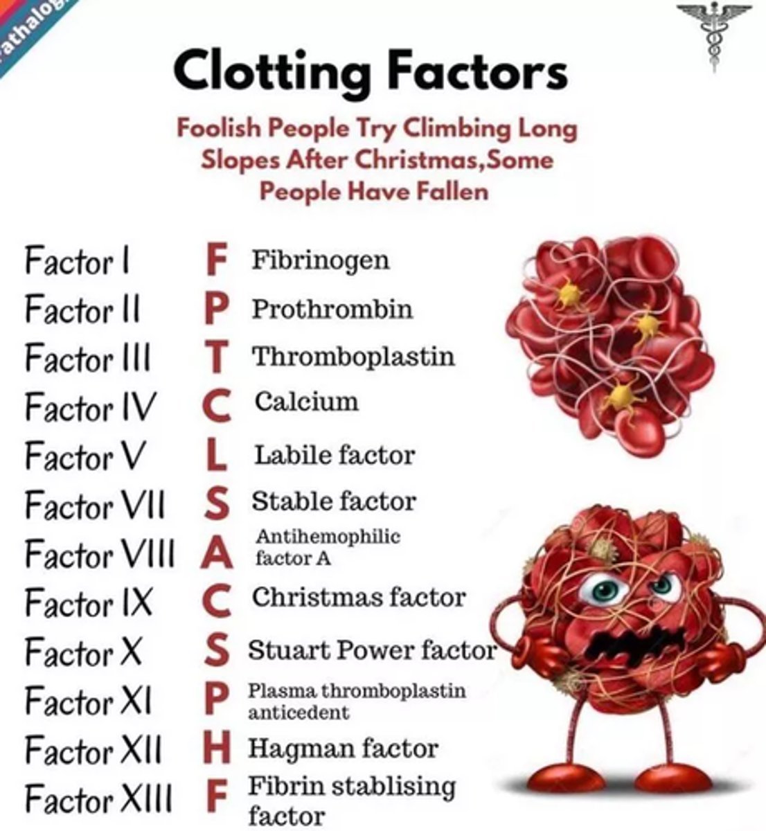
What clotting factors are affected by hemophilia A, B and C?
a. Dengue Fever
b. Idiopathic Thrombocytopenic Purpura
c. Trauma from fellatio
d. Trauma from severe coughing & vomiting
e. Signs of infectious mononucleosis
f. Hemostatic diseases
What are OTHER POSSIBLE DIAGNOSES that can be rolled out based on the presence of petechiae and ecchymosis?
Ans. trauma from fellatio
"..Seen mostly on the posterior palate - the bruise will disappear in a few days after passing the various colors of blue, green, and yellow"
3 multiple choice options
Color and shape:
linear red or bluish bruise
Location:
at the junction of the hard & soft palate
APPEARANCE of trauma from severe coughing & vomiting.
✓ Appears with 6 to 20 petechiae in the soft palate before the patient becomes ill
✓ This petechiae may also occur between the 5th & 12th days of illness. (in the middle of the duration)
✓ It goes along with malaise, enlarged nodes in the neck
Enumerate the SIGNS of infectious mononucleosis.
A macule can be a variety of colors based on the cause.
Ex. Macules can be moles (which are hyperpigmented, or darker, relative to the skin) or vitiligo lesions (which are hypopigmented, depigmented, or lighter, relative to the skin).
Describe the CONCEPT of pigmentary macules.
Ans. Brown to black (extrinsic pigmentation)
COLOR of pigmentary macules.
PHYSIOLOGIC
Also known as "genetic makeup"; this feature/characteristic of pigmentary macules is part of the person already.
Unknown, but has been postulated that macules maybe due to trauma or post inflammation.
ETIOLOGY of pigmentary macules
- Light Brown to Black
- The deeper & heavier the deposit of melanin, the darker it appears
- Light-tanned individuals have normally even coloration, but dark complexion frequently have macules of pigmentation.
CLINICAL FEATURES of pigmentary macules.
Other diagnosis:
a. Ephelis (Freckles)
b. Melanosis
c. Melanoplakia (Black pigmentation)
d. Albinism
e. McCune-Albright syndrome
f. Von Recklinghausen's Disease (Neurofibromatosis 1)
What are OTHER POSSIBLE DIAGNOSES that can be rolled out based on the presence of pigmentary macules?
MELANOSIS
(pigmentary macule)
An abnormal melanin pigmentation of oral mucosa
- usually occurs on the gums
- e.g. cigarette smoking
– stimulates melanocytes
MEALNOPLAKIA (Black pigmentation)
(pigmentary macule)
- People have discernible degree of melanin pigmentation distributed throughout the epidermis of the skin/mucosa
- Usually occurs on the lips
ALBINISM
It happens when melanin formation is impaired by congenital decrease in tyrosinase
McCune-Albright syndrome
- It is associated with mosaicism, meaning that the abnormal genes is present in a fraction, but not all, of the patient cells
- results from a change (mutation) in a gene that occurs by chance in the womb; mutations in the GNAS1 gene
Ans. Both statements are TRUE.
Rationale:
McCune-Albright syndrome is not inherited. Instead, it is caused by a random mutation in the GNAS gene that occurs very early in development.
As a result, some of the body's cells have a normal version of the GNAS gene, while other cells have the mutated version. This phenomenon is called mosaicism.
True or False:
Mc.Cune-Albright syndrome is not inherited and not contagious.
HYPEROCCLUSION
Definition: Whole biting pressure is focused on that area.
Premature contacts
This happens when there is not enough insulation of pulp; exposed dentin.
Sepsis
A serious condition in which the body responds improperly to an infection
Diazepam + Alcohol: Death
Give an example of POTENTIATION.
Antibiotics will have no effect because alcohol will compete and attach to the receptor that the antibiotic is supposed to attach to.
Antibiotics + Alcohol = ?
Antagonism Oplates + Naloxone = Narcotic
Antagonism Oplates + Naloxone = ?
Corticosteroids
Drugs that delay wound healing.
Antihistamine
Effect is for the lungs, causes drowsiness in the brain (Histamine recognizes irritant; causes bronchoconstriction. With antihistamine, it lower down your senses.)
Phenytoin
For gingival enlargement.
fibrous dysplasia of bone (FD)
Description:
- larger bone
- scar-like tissue
- happens with weight bearing bones such as legs causing limping, deformity & fracture.
- no treatment, but surgery may correct fractures and deformities
café-au-lait skin spots
skin; patches of increased or darker skin coloring
precocious puberty (PP)
In the endocrine system; irregular function of growth-regulating glands that produce hormones.
- early puberty like menstrual bleeding before 2 years of age
- boys shows less signs than girls
Thyroid Function - thyroid gland is small in the neck that affects the metabolism.
- majority have thyroid problem, like presence of enlargement or masses (nodules/cyst).
- Hyperthyroidism
➢ Growth Hormone
- too much pit. growth hormone. resulting to coarse facial features like larger hands & feet & arthritis.
- Acromegaly
- Abnormal bone growth in the skull can lead to blindness or deafness due to pinched nerves
- Deformities may be seen in the frontal bone and can cause cosmetic problems
Other effects of McCune-Albright syndrome on the endocrine system:
Cushing Syndrome
- too much pituitary growth hormone resulting to coarse facial features, larger hands & feet & arthritis.
- Tx: medication & surgery
Von Recklinghausen's Disease (Neurofibromatosis 1)
- INHERITED autosomal dominant condition (difference with McCuneAlbright syndrome)
- It has eight (8) category but most common is NF1.
- NO prevalence for gender and race in NF1.
-also manifest café-au-lait spots
multiple fibromatosis that occurs in any part of the body
Von Recklinghausen's Disease: most common sign on the skin is?
lisch nodules
pigmented hemartomas of the iris; translucent brown pigmentation spots in the iris
crowe's sign
inguinal and axillary freckles is usually present in this sign
- bone lesion
- cardiovascular and neurological abnormalities
- mental deficiency
- seizures
- short statute
- scoliosis
MANIFESTATIONS of Von Recklinghausen's Disease (Neurofibromatosis 1).
- tongue and buccal mucosa are affected
- hemifacial disfigurement caused by plexiform neurofibroma of the trigeminal nerve
- presence of impacted, displaced or missing teeth in the mandible and overgrowth of the alveolar ridge
- may have severe periodontitis & calcular deposits.
ORAL MANIFESTATIONS of Von Recklinghausen's Disease (Neurofibromatosis 1).
-enlarged mandibular canal, mandibular foramen, mental foramen → displaced teeth
- Neurofibroma can develop intraosseously resulting to well demarcated unilocular & multilocular radiolucent lesion
RADIOGRAPHIC FINDINGS of Recklinghausen's Disease (Neurofibromatosis 1).
➢ Bilateral Adrenocortical Destruction after TB
➢ Idiopathic Atrophy
➢ Bilateral Tumor Metastasis
➢ Leukemic Infiltration
➢ Amyloidosis of Adrenal Cortex (adrenocortical insufficiency) Addisons disease
PATHOLOGIC causes of Pigmentary Macules
Weakness
- Weight loss
- Nausea
- Vomitting
- Hypotension
- The cheek is the most common site for pigmentation. Highly reliable sign in primary adrenal insufficiency.
Signs & Symptoms of Adrenocortical Insufficiency
- If suspected should not begin treatment, seek medical consultation first.
- Supplemental corticosteroids medication is always required prior to dental treatment to cope up with adrenal insufficiency during stressful treatment.
Management for adrenocortical insufficiency in the dental setting.
a. Addisons disease
b. Peutz Jegher syndrome
c. Systemic Lupus Erythematosus
Other diagnosis aside from Adrenocortical Insufficiency:
Peutz Jegher syndrome
Inherited autosomal dominant may be a causative factor
- similar to ephelides but distribution seen around the lips (peri-oral distribution)
- seen around the lips, buccal mucosa, gingiva, hands, and feet.
abdominal pain, rectal bleeding, and diarrhea
Subjective symptoms of intestinal polyps
Systemic Lupus Erythematosus
This is an autoimmune disease in which the immune system attacks its own tissues - supplemental corticosteroid is given to suppress the immune system.
TRUE: Intestinal polyposis found in the jejenum is a manifestation of the perioral pigmentation.
TRUE or FALSE:
Intestinal polyposis found in the jejenum is a manifestation of the perioral pigmentation.
Butterfly Rash
Common sign of SLE (SYstemic Lupus Erythematosus)
Give prophylactic antibiotic for 7 days
Tx. for SLE
PAPULES
- ELEVATED
- usually associated with stress
- Size = pinhead but measures greater than 0.5cm
- Shape = flat, conical, circular, pointed to umbilicated, has elevated and raised bumped.
Describe PAPULES.
a. pimples
b. lichen planus
EXAMPLES of papules.
➢ Unknown, maybe due to psychological problem like deep emotional strain and remission after a personal crisis personal crisis.
➢ Other causative factors:
- Diabetes Mellitus
- Mechanical Irritation
- Galvanic Currents
- dissimilar metal components (e.g. amalgam + stainless steel resto)
CAUSATIVE FACTORS of papules.
1) Reticular - lacework of intersecting white lines (striae of Wickham – in cheek biting)
2) Papular – looks like pimples
3) Erosive - presence of squamous cells
4) Atrophic - presence of carcinoma
5) Bullous – malignant in nature
BREAP - Bullous, Reticular, Erosive, Atrophic, Papular
CLINICAL FEATURES of papules.
- Buccal Mucosa (85%)
- Gingiva
- Tongue
- Palate
- Floor of the Mouth
- Vermillon Border
SITES where papules are seen.
✓ topical application of cortisone (3x/day)
MANAGEMENT for papules.
This has clinical features of lichen planus.
- drugs like
• beta blockers,
• metallic drugs (gold, salts, arsenical, mercury, • anti-inflammatory (non-steroidal like indomethacin)
• and antibiotics (tetracycline and streptomycin)
describe Lichenoid Drug Reaction
Remove the causative factor
MANAGEMENT for Lichenoid Drug Reactions
➢ macules - flat and have causative factors
➢ papules - elevated and no causative factors
DIFFERENTIAL DIAGNOSIS for macules and papules.
no pathological reactions.
- these are elevated circumscribed solid lesion.
- It is enlarged papules deep seated which involves the submucosa or lower dermis of the skin
describe NODULES
a. Bohn's Nodule
b. Epstein Pearls
c. Tonsil nodules
d. Rheumatoid nodules
EXAMPLES of nodules.
Bohn's Nodules
-seen on buccal ridge; alveolar ridge of new born infants
- multiple white nodules and will be exfoliated by itself.
Remnants of the dental lamina
CAUSE of Bohn's Nodule
Epstein Pearls
seen on palates of newborn infants. (palatine cyst)
cause : arises from the mucosa gland elements
Tonsil nodules
This is seen on the lingual tonsils in lateral base of the tongue-foliate papilla
Rheumatoid nodules
This is common in rheumatoid arthritis and appear in approximately 25% of patients.
VESICLES
circumscribed single or group elevations of the epithelium of the skin or mucous membrane which consist of serum, plasma, and blood.
- 2nd degree burns
e.g. Herpetic gingivostomatitis – from painful vesicles → ulcers
Viral infection - chicken pox (varicella)
CAUSES of VESICLES
PUSTULES
-have pus (usually caused by infection; bacteria)
- considered cutaneous lesion (skin)
- seen as superficial elevations which contains pus, serum, and fluid surrounded by area of erythema.
- like vesicles, but differs in location and infrequent in oral mucosa.
➢ Psoriasis
➢ Impetigo
➢ Superficial Bacterial Disease
- All three are caused by staphylococci & streptococci
- Usually occurs during summer or hot months, humid weather and commonly seen in children.
✓ Tx: antibiotics
EXAMPLES of pustules.
- Pets,
- Dirty Fingernails,
- Beauty Parlor,
- Swimming Pool,
- Turkish Bath
Sources of infection for pustules.
Bullae or Blebs
- larger than vesicles and deep seated and has a roof cavity
- more resistant to rupture
- rubbing of non-vesiculated area may result in a formation to a vesicle
- denudation of the mucosa or epidermis -
Nikolsky sign
- positive to stippling of epithelium.
➢ Autoimmune Mucocutaneous Disease characterized by Intraepithelial Blister
CAUSES of Bullae/ Blebs
➢ Pemphigus
➢ Behcets Syndrome,
➢ Steven Johnson syndrome
➢ Ranula
EXAMPLES of Bullae/ Blebs
✓ Corticosteroid Therapy
- immunosuppressive drug
- will not recognize good and bad bacteria
MANAGEMENT for Bullae/Blebs
TUMENSCENCES
-these lesion maybe reactive or neoplastic in character.
- non-neoplastic lesions, unless confirmed by biopsy.
- Tumor
- is not synonymous with cancer, it simply refers to a a swelling (e.g. dental abscess)
- Cyst (e.g. eruption cyst)
- Epulis (e.g. epulis fissuratum
- surgically remove the lesion and remove denture)
- Exostosis,
- Torus
- Papilloma
- Polyps (e.g. pulp polyp)
EXAMPLES of Tumenscences
This is caused by both hot and cold (frost bite)
1st degree – ERYTHEMATOUS MACULES
- tender (painful on palpation), and painful even if not palpated
2nd degree – VESICLES / BLEB
Describe the concept
Oral Lesion
focal hyperkeratosis, nicotine stomatitis, lichen planus, leukoplakia, cheek biting
Keratosis
abnormal thickening of the outer layer of the epith. Of the mucous membrane or the skin. Ex. Actinic keratosis (solar keratosis)
Papillometoid
refers to nonneoplastic lesion.
• Keratosis - abnormal