Learn: LECOM OMS-1 Histology Labs 4-7 | Quizlet
 Call Kai
 Learn
Learn Practice Test
 Spaced Repetition
 Match
Match Flashcards
 Knowt Play
1/109

There's no tags or description
Looks like no tags are added yet.
Name | Mastery | Learn | Test | Matching | Spaced | Call with Kai | Chat |
|---|
No analytics yet
Send a link to your students to track their progress
110 Terms
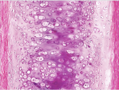
Hyaline Cartilage
What does this image show?
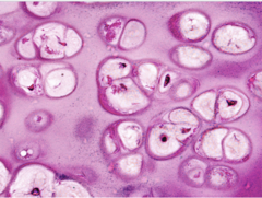
Hyaline Cartilage
What does this image show?
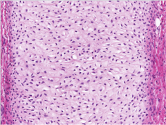
Elastic Cartilage
What does this image show?
Elastic Cartilage: Resorcin-Fuchsin
What does this image show?
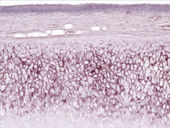
Fibrocartilage
What does this image show?
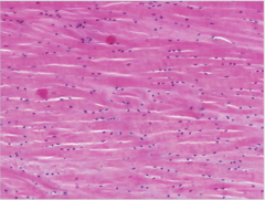
Fibrocartilage
What does this image show?
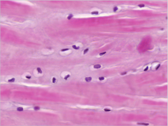
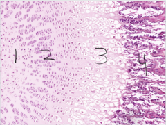
ID the Zones
1. Zone of Reserve Cartilage
2. Zone of Proliferation
3. Zone of Hypertrophy
4. Zone of Ossification
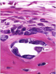
Osteoblast
What does this image show?
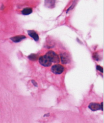
Osteoclast
What does this image show?
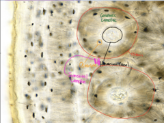
What does this image show?
- Osteon
- Haversian Canal
- Osteocytes
- Concentric Lamellae
- Interstitial Lamellae
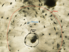
What does this image show?
- Osteon
- Haversian Canal
- Osteocytes
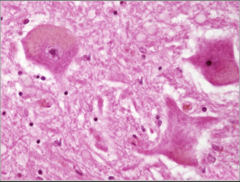
Purkinje Cell in Cerebellum
What does this image show?
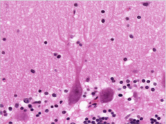
Purkinje Cell (Silver Salt)
What does this image show?
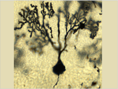
Pyramidal Cell in Cerebrum
What does this image show?
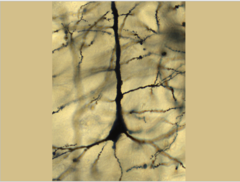
Peripheral Nerve
What does this image show?
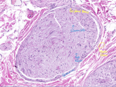
Peripheral Nerve with Node of Ranvier
What does this image show?
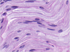
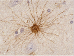
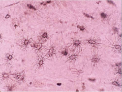
Astrocyte (GTAP)
What does this image show?
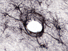
Oligodendrocyte (Silver Salt)
What does this image show?
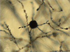
Microglial Cell (Silver Stain)
What does this image show?
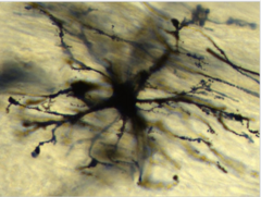
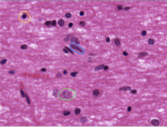
Oligodendrocyte
What does the yellow circle show?
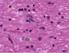
Microglial Cell
What does the blue circle show?
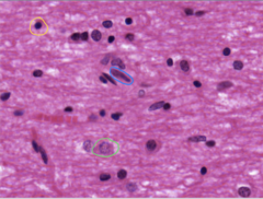
Astrocyte
What does the green circle show?
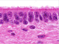
Ependymal Cells (H+E Stain)
What does this image show?
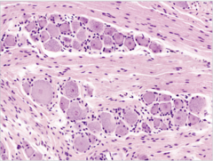
Peripheral Nerve/Ganglion with Pseudounipolar Cells surrounded by Satellite Cells
What does this image show?
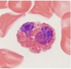
Eosinophil
What does this image show?
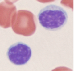
Lymphocyte
What does this image show?
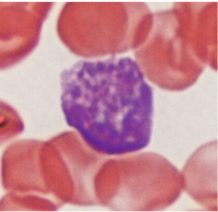
Reticulocyte
What does this image show?
Neutrophil
What does this image show?
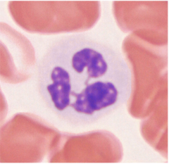
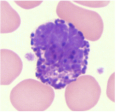
Basophil
What does this image show?
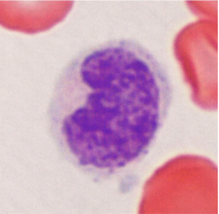
Monocyte
What does this image show?
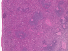
Spleen
What does this image show?
Spleen (Higher magnification)
What does this image show?
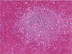
Thymus
What does this image show?
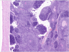
Hassal's Corpuscle in the Thymus (Type VI)
What does this image show?
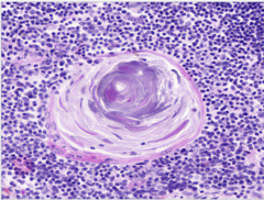
Lymph Node
What does this image show?
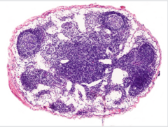
Lymph Node
What does this image show?
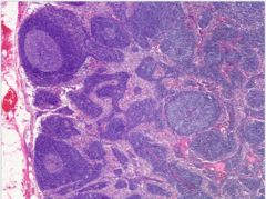
Palatine Tonsil
What does this image show?
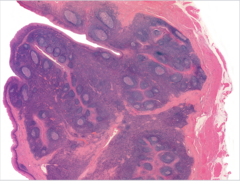
Palatine Tonsil
What does this image show?
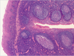
Elastic Cartilage H+E
What does this image show?
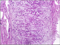
Transitional Epithelium (Urothelium)
What does this image show?
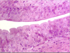
Microglial nuclei
What does this image show?
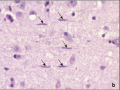
Stratified Columnar Epithelium
What does this image show?
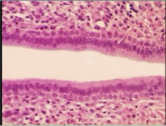
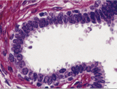
Pseudostratified Columnar Epithelium
What does this image show?
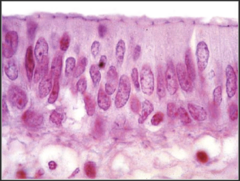
Reticular fibers - Silver impregnation
What does this image show?
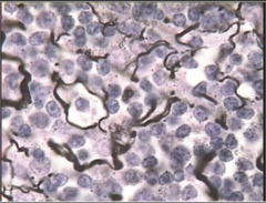
Peripheral Nerve Cross-Section
What does this image show?
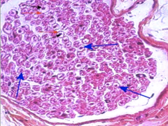
Simple Cuboidal Epithelium
What does this image show?
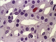
Sensory ganglion - Pseudounipolar neurons and Satellite cells
What does this image show?
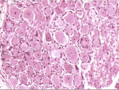
Vessel, endothelium (a form of simple squamous epithelium)
What does this image show?
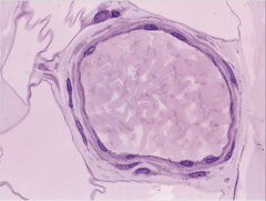
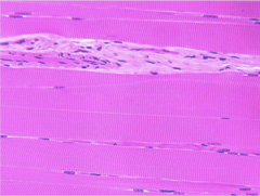
Skeletal muscle, A band, I band
What does this image show?
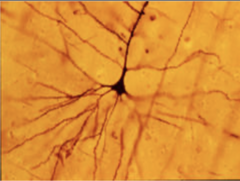
Pyramidal cell in the cerebral cortex
What does this image show?
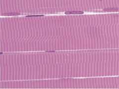
Skeletal muscle, sarcomere, A band, I band, Z line
What does this image show?
Astrocyte
What does this image show?
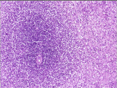
Spleen, Malpighian corpuscle that is a lymphatic follicle perforated by a central artery or arteriole
What does this image show?
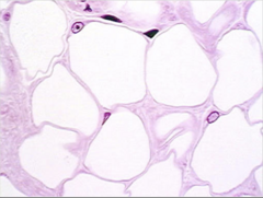
White Adipose Tissue
What does this image show?
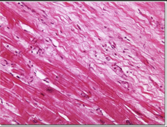
Old myocardial infarction - scar tissue replaced the cardiac muscle on the right. Below is the normal cardiac muscle
What does this image show?
Conducting system in the endocardium - Purkinje fibers. Enlarged fibers, vacuolated cytoplasm, high glycogen content, few myofibrils, cross striation. Left and right we can see the myocardium.
What does this image show?
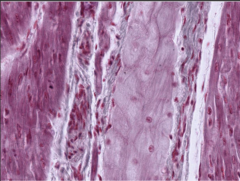
Smooth muscle cross section (left) and longitudinal section (right)
What does this image show?
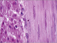
Wharton's Jelly
What does this image show?
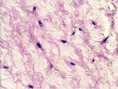
Loose Connective Tissue
What does this image show?
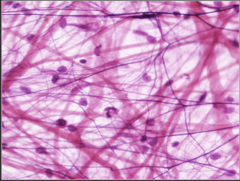
Intramembranous Ossification. Osteoblasts line the bone trabeculae
What does this image show?
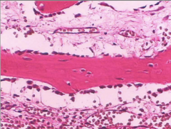
Plasma cells (arrow) and at the bottom there is a lymphocyte
What does this image show?
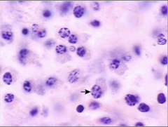
Smooth muscle
What does this image show?
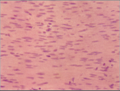
Lymph node, cortex, and paracortex
What does this image show?
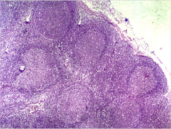
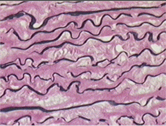
Elastic fibers with Resorcin-Fuchsin
What does this image show?
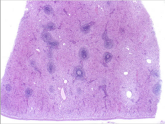
Spleen, Malpighian corpuscles that represent the white pulp surrounded by red pulp
What does this image show?
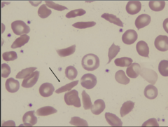
Sickle cells
What does this image show?
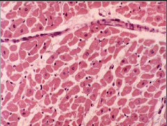
Cardiac muscle cross section. Note the vessels between the cells that appear like white gaps
What does this image show?
Simple columnar epithelium with goblet cells
What does this image show?
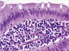
Acute myocardial infarct - most of the nuclei disappeared from the cells
What does this image show?
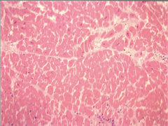
Brown adipose tissue
What does this image show?
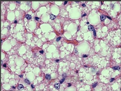
Dense irregular connective tissue - type I. collagen fibers and fibroblasts and submucosa
What does this image show?
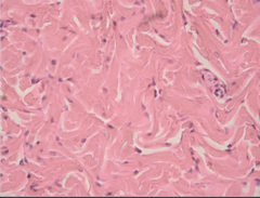
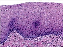
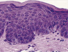
Stratified squamous nonkeratinized epithelium
What does this image show?
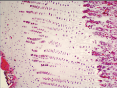
Enchondral ossification, epiphyseal plate. Zone of resting cartilage, zone of proliferation, zone of hypertrophy and zone of ossification.
What does this image show?
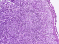
Palatine tonsil, high magnification, covered by stratified squamous nonkeratinized epithelium, lymphatic follicles
What does this image show?
Fibrocartilage, chondrocytes and type I. collagen fibers
What does this image show?
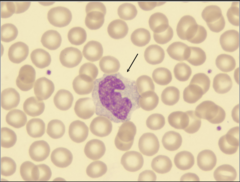
Monocyte
What does this image show?
Dense irregular connective tissue - type I. collagen fibers and fibroblasts and submucosa
What does this image show?
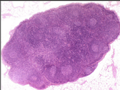
Lymph node
What does this image show?
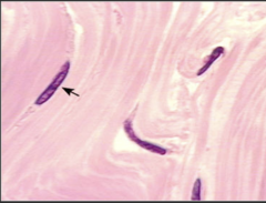
Dense regular connective tissue - type I. collagen fibers and fibroblasts. Tendon.
What does this image show?
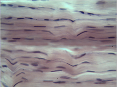
Hyaline cartilage H&E
What does this image show?
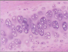
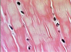
Skeletal muscle, cross section
What does this image show?
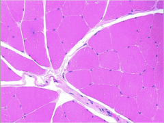
Elastic cartilage, resorcin-fuchsin. Epiglottis.
What does this image show?
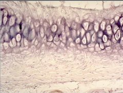
Sebaceous gland - holocrine secretion.
What does this image show?
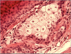
Fibrocartilage, chondrocytes and type I collagen fibers
What does this image show?
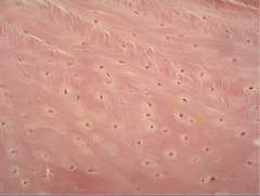
Bone, lamellary systems. Haversian and interstitial lamellae, osteocytes between them
What does this image show?
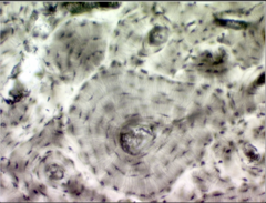
Cardiac muscle, intercalated discs
What does this image show?
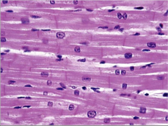
Osteoclasts, resorption bay
What does this image show?
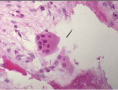
Apocrine secretion in the mammary gland
What does this image show?
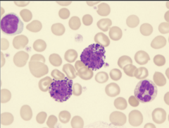
Large lymphocyte, basophils and neutrophil granulocyte
What does this image show?
Osteocytes
What does this image show?
Stratified squamous keratinized epithelium. Skin.
What does this image show?
Nerve longitudinal section. Note the myelin sheath and the node of Ranvier
What does this image show?
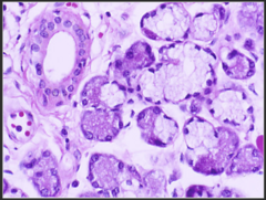
Salivary gland, serous and mucous acini and serous demilunes (artifact). Merocrine secretion.
What does this image show?
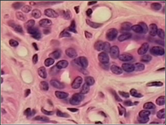
Stratified cuboidal epithelium (sweat gland duct)
What does this image show?
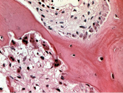
Hyperparathyroidism - numerous osteoclasts. Hypercalcaemia.
What does this image show?
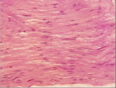
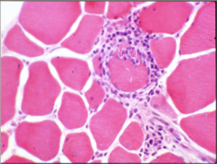
Polymyositis - a muscle fiber is surrounded by lymphocytes and macrophages that break down the fiber - autoimmune disease against the muscle fibers
What does this image show?
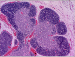
Thymus, lobules, cortex, medulla. Capsule on the surface, vessels
What does this image show?
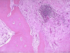
Osteomyelitis - lymphocytic aggregation next to a bone trabecule
What does this image show?
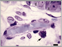
Mastocyte next to a vessel
What does this image show?
Multipolar cells spinal cord ventral horn
What does this image show?