Audition Test 3 - Central Auditory System
 Call Kai
 Learn
Learn Practice Test
 Spaced Repetition
 Match
Match Flashcards
 Knowt Play
1/96

There's no tags or description
Looks like no tags are added yet.
Name | Mastery | Learn | Test | Matching | Spaced | Call with Kai | Chat |
|---|
No analytics yet
Send a link to your students to track their progress
97 Terms
Where are the SGN nuclei located?
cochlea
Where are the CN, SOC, TB, LL nuclei located?
lower brainstem
Where are the IC nuclei located?
midbrain
Where are the MGB nuclei located?
thamalus
Which nuclei is the first to receive the bilateral center?
Superior olivary complex
Do unilateral connections also exist?
yes, cochlear nuclei receives only unilateral projection
What are the 6 major characteristics of higher level nuclei compared to lower level?
Higher level nuclei usually have:
- more complicated structures
- larger number of neurons
- multiple types of neurons (morphological and functional diversity)
- more sophisticated signal processes (involving the interaction between excitation and inhibition, neural circuits)
- control to lower levels through efferent system
- interaction with other systems
What kind of organization is higher level nuclei?
hierarchical
What are four of the cell types? Where are each of them located?
(1) spherical cells and globular cells (bushy cells) in AVCN and PVCN
(2) octopus cells in PVCN
(3) giant cells and pyramidal cells in DCN
What does PSTH stand for?
Peristimulus histograms
What are two reasons that there is a variation in PSTH patterns?
Inhibition and membrane feature
What is one way to verify inhibition in PSTH patterns?
To verify inhibition, neurotransmitter blocker should be used: if the blocker change the response pattern, the change is due to inhibition
Which characteristic has a tonotopic map? Where does it start and go up to (path)
This is inherited from the cochlea and goes up to the CAS, but the frequency region used takes more of a larger size; in the cochlea it's a fixed frequency distribution, but not in the auditory cortex)
What is another name for a principle neuron?
Relay neurons
What is the function of a principle/relay neuron?
- receive input from and send output to outside nuclei
- input from lower level, output to higher level
- relay to synapse
What is the function of the interneurons? What is this important for?
- make synapse inside the nucleus (locally)
- circuits around relay neurons
- this is important for signal processing
Relay neurons can do ____ _____ but internerons cannot provide these connections
long distance
What is responsible for the information pathway from low to higher level nuclei?
Relay neurons
What are two ways and the terminology to describe neural projection as it goes to higher levels?
Relay and bypassing
What is an example of relay? What is an example of by passing?
• Neuronal projection to one nucleus can be relayed by the principle neurons, and extended to a higher level nucleus
• In some nuclei, the projection may not be relayed but "bypass" or "go through" to project to the next nucleus.
- Bypassing example: SOC nuclei. Projection can go from CN to IC, by passing SOC and LL nuclei.
• Relay example: CNs, the projection must be relayed (with no exception)
Where is there the largest amount of SGN? What about the smallest?
largest = cortex
smallest = cochlea
What is the pathway from SGN to nuclei of the auditory cortex where relay is a must?
CN, IC, MGB
What is the pathway from SGN to nuclei of the auditory cortex that is shortest?
CN directly to IC
What is the pathway from SGN to nuclei of the auditory cortex that is the longest?
CN, MNTB, LSO, NLL, IC
How many nuclei are involved in the shortest pathway? What about neurons?
4 nuclei, 5 neurons
How many nuclei are involved in the longest pathway? What about neurons?
7 nuclei, 8 neurons
Endbulbs of Held and Calyx of Held are big synapses, this ensures what? Results in?
ensures a 1 to 1 transmission, results in primary like PSTH
What are 3 major divisions of the cochlea? (map parts)
dorsal, anterior ventral, posterior ventral
What is the general direction for the tonotopic CF going from high to low frequency shifting?
Tonotopic axis: dorsal-ventral direction for high-to-low frequency shifting
What part of the CN is larger: ventral or dorsal?
ventral - VCN
Is the path from the 8th nerve to the CN bilateral or unilateral?
unilateral
where are octopus cells found?
posterior part of the PVCN
where are globular bushy cells found?
posterior part of the AVCN
Where are spherical bushy cells found?
anterior part of the AVCN
What are 2 types of bushy cells?
spherical and globular
What is the special synapse of bushy cells?
endbulbs of held
Bushy cells synpase is for:
AVCN neurons
What do bushy cells synapse with?
spiral ganglia bodies
Where do the bushy cells synapse relay to?
SOC
What is the synapses that SOC relays to?
Calyx of Held
What kind of transmission do bushy cells have?
secure 1:1
What feature is the timing of bushy cells useful for?
sound localization
Bushy cells: Binaural convergent project from ____ to ____
AVCN to SOC for sound localization
Bushy cells response to tone burst with what type of PSTH?
primary
In a PSTH, the response is from single neuron to the same tone burst. Why are they all different responses?
1) inhibition
2) we see different patterns based on different membrane features
What is the dendrite size of octopus cells?
large
Is onset response weak or strong for octopus cells?
strong
How do octopus cells relate to temporal processing?
strong response to transient signal for time coding
Octopus cells are sensitive to what signal?
broad band noise
Octopus cells project to ____ then to ______
VNLL then IC
What is an example of feature detection in relation to octopus cells?
sensitive to the direction of frequency modulation - understand this as the changing of frequency, which can be shown as sliding tones up and down
How are octopus cells sensitive to frequency change in one direction?
- The input fibers are sensitive to different frequencies.
- Excitation sequence determined by the direction of the FM.
Octopus cells receive input from ____ frequency region
wide
What is a major contributor to every level of signal processing in the CAS?
Inhibition
At which level is inhibition seen?
At the level of the SGN, but at the CN. (There is no inhibition in the cochlea)
Describe what is happening in this image where the triangle is the principal neuron?
The circle is the interneuron and the principle is the triangle. When that auditory nerve excites the interneuron, it causes the principal neuron to be inhibited. Therefore this is a mechanism to produce an onset response since the principal neuron will now stop firing after said onset response
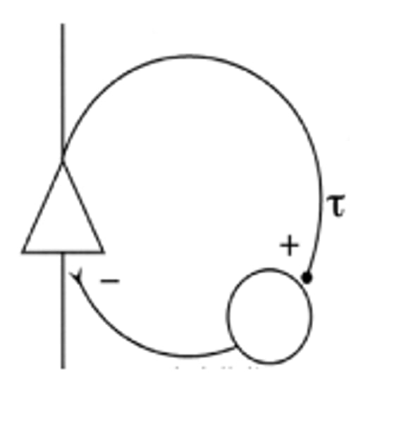
What are the three ways to see the inhibitory response patterns?
PSTH, response area graph, rate/level function graph
Among the different response area graphs, which type shows no inhibition? Where do we see this type?
Type 1; ANF
Describe type II and type III's response area graphs.
Type II: large peak of excitation with little to no inhibition
Type III: large peak with larger area of inhibition
What is the difference between the type II and type III?
Type II: no spontaneous activity (no sound or unheard sound)
Type III: reduction in spontaneous activity with playing of a characteristic frequency tone
What are the two 'aspects' being measured for the rate-level function graph?
Frequency and Intensity
- how the neurons firing rate is affected at different intensities (when it is inhibited)
What are the two patterns for the rate-level function? Which ones are shown by the neurons in the CN in the auditory system?
Monotonic and Non-Monotonic
Monotonic is shown by the CN
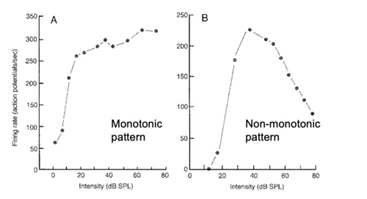
What do both the patterns indicate as it relates to inhibition?
- Non-monotonic suggest more inhibition is evoked at higher sound level, evidence for overlapping between excitation and inhibition
- Therefore, there is no inhibition in the auditory nerve fibers (consistent with the type I response area graph)
What are the two inhibitory neurotransmitters?
Glycine and GABA
How can you verify inhibition by these two neurotransmitters?
Apply a blocker to stop the activity of the glycine and GABA and compare the neural activity with and without this blocker. The difference would be from inhibition.
Where is the SOC located relative to the CN?
At the same level of the CN in the lower brainstem.
What are the three major nuclei in the SOC?
LSO: Lateral Superior Olive
MSO: Medial Superior Olive
MNTB: medial nuclei of trapezoid body
What trait does the SOC help with? How?
Sound source localization, it is the first station where the binaural inputs merge
What are two examples of the binaural cues?
Interaural time difference (ITD) and interaural level difference. (ILD)
Which cue is important for high frequency sounds? Which cue is important for low frequency sounds?
ITD for low frequency; ILD is for high frequency.
Do large or small animals depend more on the interaural level difference? Why?
Small animals since their ears are too close together to depend on timing (too short a time difference)
What is the size of the MSO relative to LSO?
Larger than LSO in humans and larger animals; less developed in small animals
What is the size of the LSO relative to MSO?
Larger than MSO in smaller animals
MSO mainly processes:
binaural in the low frequency cues
LSO mainly processes:
binaural in high frequencies
MSO is sensitive to what difference:
ITD (acoustic delay)
LSO is sensitive to what difference:
ILD (difference in loudness/intensity)
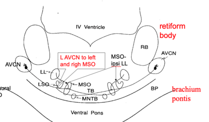
Bilateral innervation for MSO?
yes, AVCN projects directly to MSOs of both sides
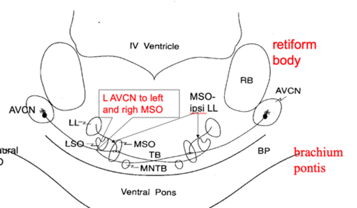
BIlateral innervation for LSO?
No, AVCN projects to ipsilateral LSO
Where does the Calyx of Held synapse occur?
The AVCN input to the MNTB (contralaterally)
What is the inhibitory pathway as it relates to the MNTB?
The output to the ipsilateral LSO and MSO is inhibitory
How many subdivisions does the inferior colliculus have?
Three (the external cortex, the central nuclei (ICC), paracentral nuclei)
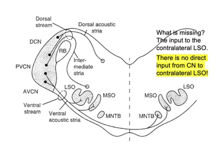
There is no direct input from CN to _____ LSO
contralateral
Which part of IC shows clear tonotopic map?
The central nucleus
Where does the central nucleus get direct monoaural innervation?
VCN and DCN
(Ventral Cochlear Nuclei and Dorsal Cochlear Nuclei)
Where does the central nucleus get indirect binaural innervation?
AVCN- SOC- (LL)- IC
Is the central nucleus contralaterally dominant or ipsilaterally dominant or the same?
Contralaterally dominant (so the central nucleus innervating the opposite side)
For the inferior colliculus, what is the general direction of the frequency on its axis?
Goes from low (dorsal) to high on the ventral side.
How does the general direction of the frequency for the inferior colliculus compare to the cochlear nuclei?
Opposite frequency distribution- the cochlear nuclei is high on the dorsal side and low on the ventral side.
How would you describe the location of the MGB?
Just below the thalamus
What are the three major divisions of the MGB?
Ventral, dorsal and medial
Which part of the MGB shows a clear tonotopic map?
Ventral
What is the MGB and auditory cortex pathway?
Through the internal capsule at the posterior part.
As per Broadmann labelling, which part is the primary auditory cortex? Where is that located?
- Area 41
- Transversely oriented gyri of Heschl.
What area is the secondary auditory cortex located?
Area 42
What is located at area 22? Where is it located? What kind of cortex is this? What area is this called?
- Planum temporale
- located in posterior part of the superior temporal gyrus
- associative cortex
- Wernicke on dominant side
What is the connection called to integrate the two modalities of Wernicke and Broca?
Arcuate fasciculus