Alteration in Perfusion: Peripheral Vascular and Arterial Disease
 Call Kai
 Learn
Learn Practice Test
 Spaced Repetition
 Match
Match Flashcards
 Knowt Play
1/67

There's no tags or description
Looks like no tags are added yet.
Name | Mastery | Learn | Test | Matching | Spaced | Call with Kai | Chat |
|---|
No analytics yet
Send a link to your students to track their progress
68 Terms
Peripheral Artery Disease (PAD)
Involves thickening of the artery walls and progressive narrowing of arteries of upper and lower extremities
Atherosclerosis is leading cause in majority of cases
Symptoms occur when vessels are 60-75% blocked
Peripheral Artery Disease (PAD) risk factors
Risk factors: Tobacco use, Diabetes, HTN, High cholesterol, Age greater than 60
PAD Clinical Manifestations: intermittent claudication
Ischemic muscle pain that is caused by a constant level of exercise
Build up of lactic acid from anaerobic metabolism
Resolves within 10 minutes or less with rest
PAD Clinical Manifestations: paresthesia
Numbness or tingling in the toes or feet from nerve tissue ischemia
Neuropathy causes severe shooting or burning pain
PAD Clinical Manifestations:reduced blood flow to the limbs
Thin, shiny, and taut skin
Loss of hair on the lower legs
Diminished or absent pedal, popliteal, or femoral pulses
Elevation pallor. Pallor of foot with leg elevation
Dependent rubor. Reactive hyperemia (excess of blood) of foot with dependent position
PAD Clinical Manifestations
Pain at rest
Aggravated by limb elevation. Pain relief by gravity
Critical Limb Ischemia (CLI)
Chronic ischemic rest pain lasting more than 2 weeks
Nonhealing arterial leg ulcers or gangrene
PAD Complications
Atrophy of skin and underlying muscles
Delayed healing
Wound infection
Tissue necrosis
Arterial ulcers over bony prominences
Most serious: Nonhealing arterial ulcers and gangrene
May result in amputation
PAD Diagnostics
Doppler ultrasound
Ankle-brachial index (ABI)
Magnetic resonance angiography
Ankle-brachial index (ABI)
Done using a hand-held Doppler
Calculated by dividing the ankle systolic BP (SBP) by the higher of the brachial SBPs
Falsely elevated results can be seen in older patients or those with diabetes
Ex: Ankle systolic is 120 mmHg and the brachial systolic is 140 mmHg, the ABI would be 120/140 = 0.86.
Norm: 1-1.3
ABI of
0.9 or less is generally considered abnormal and may indicate PAD
PAD life style changes
Lifestyle Changes: healthy diet, stop smoking
Walking is most effective exercise for individuals with claudication
30 to 45 minutes daily, 3 times/wk
Patients should consult with their HCP before taking any dietary or herbal supplements especially if they are prescribed: NSAIDs, Anticoagulants
PAD nutrition
Nutrition Therapy: 3% to 5% weight loss yields reduced triglycerides, glucose, A1C, and decreased risk of type 2 diabetes
PAD Collaborative Care
Proper foot care and check feet every day for wounds
Avoid heat, cold, and injury to extremities
Medications
Percutaneous Transluminal Angioplasty (PTA)/stent
Surgery
PAD Collaborative Care Medications
ACE inhibitors: ramipril, captopril, benazepril
Antiplatelet agents
Anticoagulants are not recommended at this point
ACE inhibitors: ramipril, captopril, benazepril
Increases peripheral blood flow
Increases ABI
Increases walking distance
antiplatelets
Aspirin: Low-dose aspirin therapy
Clopidogrel: for aspirin-intolerant patients (effect decreased with omeprazole).
Can be used in combination for high-risk patients
Cilostazol: not with HF patients
Pentoxifylline
PAD Collaborative Care PTA
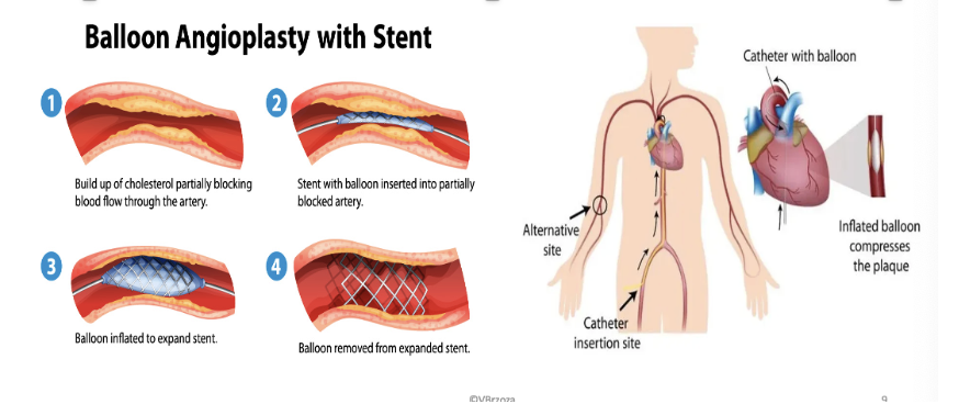
PAD Collaborative Care…Occlusion Emergency: causes
Causes: Thrombus, Embolus, Trauma
PAD Collaborative Care…Occlusion Emergency: symptoms
7 “P”s of limb ischemia (different than book)
Pain
Pulselessness
Pallor
Poikilothermia (inability to regulate temperature)
Paresthesia (numbness, tingling)
Paralysis (loss of muscle function)
Perfusion (capillary refill greater than 3 sec)
PAD Collaborative Care…Occlusion Emergency: emergency treatment
Anticoagulants, Thrombolytics, and/or Surgery (Revascularization, Amputation)
PAD Collaborative Care…Femoral-Popliteal Bypass Femoral-Tibial Bypass

PAD Collaborative Care… Post Op
Maintain circulation and check for a re-blockage of the artery
Assess pulse, color, temp, capillary refill, bruit
Monitor pain, BP
Deep breathing every 1-2 hr
Monitor for graft occlusion
Monitor for wound infection
Monitor for active bleeding, hematoma, thrombosis, embolus, compartment syndrome
Client education
Other Arterial Disorders
Aortic Aneurysms
Thromboangiitis Obliterans (Buerger’s disease)
Raynaud’s Phenomenon
Aortic Aneurysms (AA)
Sudden interruption in arterial blood supply
Permanent, localized, outpouching, or dilation of wall of aorta
If untreated, can result in tissue death
AA Clinical Manifestations
Often asymptomatic
Pulsatile mass in periumbilical area slightly left of midline.
Bruit auscultated over aneurysm
AA Clinical Manifestations assess for
Assess for shortness of breath, coughing, hoarseness, difficulty swallowing
AA Clinical Manifestations may cause
May cause back or chest pain, epigastric discomfort, altered bowel elimination, intermittent claudication.
Sudden excruciating back or chest pain, symptomatic of thoracic rupture
Blue Toe syndrome: patchy mottling of feet
AA Complication…Rupture Emergency: Rupture into retroperitoneal space
Bleeding
Severe back pain
May/may not have back/flank ecchymosis (Grey Turner’s sign)
AA Complication…Rupture Emergency: Rupture into thoracic or abdominal cavity
Massive hemorrhage causes hypovolemic shock, tachycardia, hypotension, pale clammy skin, decreased urine output, altered LOC, abdominal tenderness
Most do not survive long enough to get to the hospital
AA Diagnostics
X-ray of Chest—abnormal widening of thoracic aorta
Ultrasound: Useful in screening for aneurysms, Monitors aneurysm size
CT scan or MRI: Diagnose and assess location and severity
AA Collaborative Care
Vital signs
Assess pain, Oxygen saturation, circulation
Cardiac monitoring
Monitor ABG, electrolytes, CBC, Labs
Monitor Infection, neurological status
Monitor Urine output
Administer O2
IV access
Control BP- Antihypertensive
Surgical repair for AA larger than 5.5 cm
AA Surgery Post Op
ICU monitoring
Arterial line for continuous BP assessment
Hemodynamic monitoring with Pulmonary Artery catheter (shows heart and lung function)
Mechanical ventilation
Peripheral IV lines-maintain hydration with IVF and Blood
Urinary catheter
Nasogastric tube-monitor I/O
Monitor abdominal girth, incision
Continuous ECG Monitoring, VS with Pulse oximetry; monitor temp
Monitor electrolytes (chemistry) and CBC (hematology)
Pain medication
Monitor for complication of embolization or graft occlusion, 7 Ps.
Thromboangiitis Obliterans (Buerger’s disease)
Rare, progressive disease that causes inflammation and blood clots in the small and medium-sized arteries and veins of the hands and feet
Nonatherosclerotic
Inflammatory thrombus eventually causes ischemia
Thromboangiitis Obliterans (Buerger’s disease) Clinical Manifestations
Intermittent claudication of feet, hands, or arms
Rest pain
Ischemic ulcerations
Changes in color and temperature
Paresthesia
Superficial vein thrombosis
Cold sensitivity
Thromboangiitis Obliterans (Buerger’s disease) Collaborative Care
No specific lab or diagnostic tests. Based on history and symptoms and exclusion of other disorders
No smoking tobacco or marijuana; no nicotine replacements
Avoid cold exposure
Walking program
Antibiotics for ulcers
Analgesia for pain
Avoid trauma
IV iloprost—promotes vasodilation
Surgery: Bypass surgery, Amputation
Stem cell therapy (stem cells into a patient's body to promote tissue regeneration and repair)
Raynaud’s Phenomenon
Vasospasm/vasoconstriction of arterioles & arteries
Effects fingers & toes
No loss of pulses
Precipitated by cold, emotional upsets, caffeine, tobacco
Extremity color changes: pallor, cyanosis
Feels like numbness & tingling
Associated with autoimmune disorders (RA, Lupus)
Also, associated with trauma (frostbite)
Raynaud’s Collaborative Care
Avoid temperature extremes; wear appropriate clothing
No tobacco products
Avoid caffeine
No vasoconstrictor drugs
Stress management
Immerse hands in warm water to help decrease vasospasm
Sustained release calcium channel blockers to decrease vasospasm
Vasodilators: Topical nitroglycerin 2% ointment, Prostacyclin infusion
Antibiotics
Analgesia
Venous Disorders
Phlebitis
Venous Thrombosis
Superficial Vein Thrombosis
Venous Thromboembolism (VTE)
Deep vein thrombosis (DVT)
Pulmonary embolism (PE)
Phlebitis
Acute inflammation of the walls of small cannulated veins of the hand or arm (related to IV catheter)
Phlebitis clinical manifestations
Clinical Manifestations: pain, tenderness, warmth, erythema, swelling, and palpable cord
Phlebitis treatments
Remove catheter
Edema…Elevate extremity to promote fluid reabsorption
Pain and inflammation…NSAIDs
Warm, moist heat
Venous Thrombosis
Formation of a thrombus (clot) with vein inflammation
Most common disorder of the veins
Superficial vein thrombosis…saphenous vein
Deep vein thrombosis (DVT)…iliac and/or femoral veins
Venous thromboembolism (VTE) – preferred term
Represents spectrum from DVT to pulmonary embolism (PE)
Venous Thrombosis
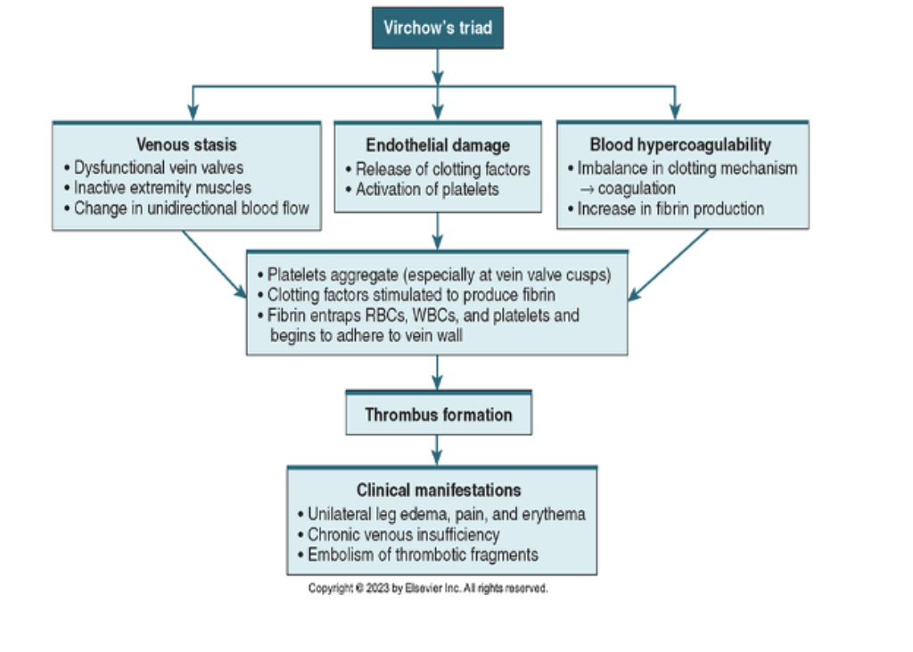
Virchow’s triad - 3 key factors that cause venous thrombosis
Venous stasis (slow blood)
Damage to endothelium (inner lining of vessel)
Hypercoagulability of blood (sticky cells)
Patients at risk for developing VTE usually have predisposing conditions to these 3 factors
VTE SV DVT PE Prevention and Collaborative Care
Early and progressive mobilization. Walk 4 to 6 times/day.
Pneumatic compression devices, TEDs (Not recommended if VTE already exists)
Flex and extend feet, knees and hips every 2 to 4 hours while awake
Passive and active ROM, as indicated
OOB to chair
VTE SV DVT PE Prevention and Collaborative Care: Anticoagulants
Warfarin. Antidote: vitamin K. Lab: INR (Norm 0.8-1.2). Norm while on warfarin is 2 to 3 higher (ie. If normal is 1, the pt on warfarin will have an INR of 2 to 3)
Heparin. Antidote: protamine sulfate Lab: PTT. (Partial Thromboplastin Time norm 30-40): Norm while on heparin is 60-100 secs (1.5 to 2.5 times norm/control).
Enoxaparin. Antidote: protamine sulfate
Superficial Vein Thrombosis: risk factors
Risk factors: pregnancy, obesity, cancer, recent fracture(s), estrogen therapy, recent sclerotherapy, recent surgery or long-distance travel
Superficial Vein Thrombosis clinical manifestations
Palpable, firm, cordlike vein. Varicose Vein.
Itchy, painful, red, and warm
Mild fever, leukocytosis
Often involves varicose veins
Superficial Vein Thrombosis diagnosis
ultrasound
Superficial Vein Thrombosis treatment
Treatment of clot smaller than 5 cm and not near saphenous femoral junction
Oral (or topical) NSAIDs
Compression stockings
Warm compresses
Elevate limb above heart
Mild exercise
Venous Thromboembolism (VTE)…Deep Vein Thrombosis (DVT): Clinical Manifestation
Unilateral edema
Pain, tenderness with palpation
Paresthesia (burning, prickling, tingling, or numb)
Red, warm, Fever greater than 100.4° F (38° C)
Venous Thromboembolism (VTE)…Deep Vein Thrombosis (DVT): complications
PE
VTE DVT Diagnostics
Lab: ACT (Activated Clotting Time), aPTT, INR, bleeding time, Hgb, Hct, platelet count, D-dimer, fibrin monomer complex
Noninvasive venous: venous compression ultrasound, duplex ultrasound
VTE DVT Diagnostics Invasive
Invasive venous: CT venography, MR venography, contrast venography
VTE DVT Collaborative Care medications
Anticoagulation
Heparin
PTT: Norm while on heparin is *60-100 secs (1.5 to 2.5 times norm/control).
Complication: HIT-Life threatening
VTE DVT Collaborative Care
Bed rest initially…do not move clot
No Compression Stockings/boots to affected limb…do not move clot
No massaging, lotion to lower limbs…do not move clot
Daily Measurements of limb
Guaiac Stools…check for additional bleeding
Surgery: Thrombectomy, Inferior Vena Cava (IVC) filter: Greenfield Filter
VTE DVT complication: Heparin Induced Thrombocytopenia (HIT)
Immune response to receiving heparin. Bleeding and simultaneous clotting
Sudden reduction in platelet count (norm = 150,000-450,000)
If platelet count decreases by 50% or goes severely below normal level
Paradoxical increase in venous and arterial thrombosis. Infarcts damage organs
when can Heparin Induced Thrombocytopenia (HIT) occur
Occurs 5-14 days after heparin therapy
Heparin Induced Thrombocytopenia (HIT) treatment
STOP all Heparin
Non-heparin agent if anticoagulation still needed (thrombin inhibitors-argatroban)
VTE Pulmonary Embolism (PE)
Blockage of 1 or more pulmonary arteries by thrombus, fat or air embolus, or tumor tissue
Clot in venous system into pulmonary circulation then lodges in small blood vessel and obstructs alveolar perfusion
Most PEs arise from deep vein thrombosis (DVT)
VTE Pulmonary Embolism (PE) clinical manifestations
Dyspnea most common; mild-moderate hypoxemia
Tachypnea, cough, chest pain, hemoptysis, crackles, wheezing, fever, tachycardia, syncope, pulmonic heart sound
massive PE clinical manifestations
Massive PE: change in mental status, hypotension, feeling of impending doom, cardiorespiratory arrest/death
VTE PE Collaborative Care
Oxygen: via mask or cannula by hypoxemia; FIO2 is titrated based on ABG analysis; mechanical ventilation may be needed
Elevate the HOB slightly for improved breathing
Pulmonary hygiene: prevent atelectasis
VTE PE Collaborative Care medications
Shock: IV fluids, vasopressors
HF: diuretics
Pain: opioids
Anticoagulation – immediate
Fibrinolytic agents—dissolve clot
Tissue plasminogen activator (tPA)
Alteplase (Activase)
VTE PE Collaborative Care surgery
Surgery: Embolectomy, IVC filter
The nurse teaches a patient with peripheral arterial disease. The nurse determines that further teaching is needed if the patient makes which statement?
A. “I should not use heating pads to warm my feet.”
B. “I should cut back on my walks if it causes pain in my legs.”
C. “I will examine my feet every day for any sores or red areas.”
D. “I can quit smoking if I use nicotine gum and a support group.”
A patient in the outpatient clinic has a new diagnosis of chronic peripheral artery disease (PAD). Which of the following medications will the nurse be prepared to include when providing patient teaching about PAD management? Select all that apply.
Captopril
Clopidogrel
Omeprazole
Heparin
Aspirin
The client admitted with deep vein thrombosis is ordered heparin. Two months after discharge, the Healthcare provider orders lab work. Which lab result would be a concern to the nurse?
A. PTT 75 seconds
B. INR is less than 1
C. Platelets 80,000
D. Hgb 15 g/dL
Following an aortic aneurysm repair, the patient suddenly develops severe pain with some numbness and tingling in the right lower extremity. The right pedal pulse is decreased, the capillary refill takes 6 seconds, and the right foot is cool and pale. Which complication should the nurse suspect?
A. Hypothermia
B. A wound infection
C. Bleeding from the graft site
D. An embolization or graft occlusion