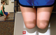
Patella Anatomy and Positioning
 Call Kai
 Learn
Learn Practice Test
 Spaced Repetition
 Match
Match Flashcards
 Knowt Play
1/28

There's no tags or description
Looks like no tags are added yet.
Name | Mastery | Learn | Test | Matching | Spaced | Call with Kai | Chat |
|---|
No analytics yet
Send a link to your students to track their progress
29 Terms
Femur
thigh bone

Patella
Approximately 2in in diameter
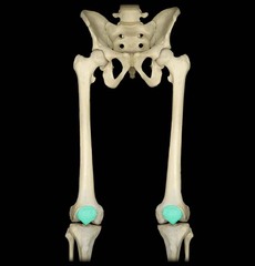
base of patella
Superior Border
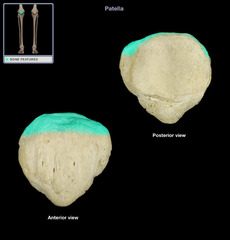
Anterior surface of patella
Rough & Convex

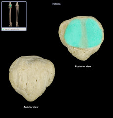
Posterior surface of patella
Smooth & oval shaped, Articulates with femur
Apex of patella
Inferior border

Patellofemoral joint
between patella and patellar surface of femur
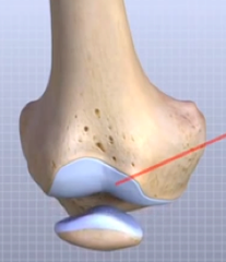
Patellar Ligament
Located anteriorly, Connects to the quadricep femoris tendon, Extends over patella & attaches to tibial tuberosity
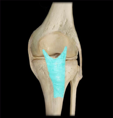
Protects the anterior aspect of the knee joint, Increase the leverage of the quadriceps femoris muscle
What's the role of the Patella?

Patella is loose & movable, Sits more superior
When the leg is extended, how is the patella positioned?
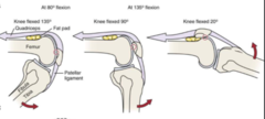
Patella locks into position, Sits more distally
When the leg is flexed, how is the patella positioned?
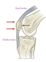
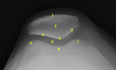
Anterior surface of patella
1?

Patella
2?
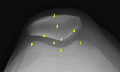
Posterior surface of patella
5?

Patellofemoral joint
6?

Medial condyle of femur
7?

intercondylar sulcus (trochlear groove)
8?

Lateral condyle of femur
9?
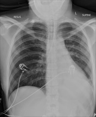
radiopaque objects, clothing removed placed in gown , long hair, oxygen tubing, and pacemaker leads to avoid reduced detail/scatter
during patient preparation, what must be removed?
1. Limit the number of exposures
2. Collimation
3. shielding (led apron)
4. Position correctly
What 4 things help protect you and the patient against radiation?
BEFORE the examination
When must the anatomical marker be placed?
kVp: 70-80 (increase 4-6 for PA)
mAs: Depends
Grid: Depends (bigger than 4in or 10cm?)
SID: Varies
What technical factors are needed for a good Patella (kVp, grid, mA, SID)?
PA
Lateral
Tangential (Merchant, Inferiorsuperior, Settegast, hobbs)
Routine patella projections?
What is the evaluation criteria and procedure for an PA Patella X-ray?
SID: 40in
kVp: 70-80
Grid: depends (bigger than 4in or 10cm?)
CR: perpendicular, directed to midpatella, about midpopliteal crease
Patient position: prone, legs extended, may internally rotate for perpendicular patella
Evaluation criteria: knee joint and patella are shown, no rotation, patella is centered to femur with slight internal rotation of anterior knee

What is the evaluation criteria and procedure for an Lateral (mediolateral) Patella X-ray?
SID: 40in
kVp: 70-80
Grid: depends (bigger than 4in or 10cm?)
CR: perpendicular, directed at mid-patellofemoral joint
Patient position: Flex knee only 5-10°, lateral recumbent position affected side down
Evaluation criteria: patellofemoral joint and femorotibial join demonstrated, true lateral
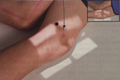
What is the evaluation criteria and procedure for a Merchant Bilateral method patella X-ray?
SID: 48-72in
kVp: 70-80
Grid: No grid (air gap method)
CR: Angled 30˚ caudad, directed to point midway between patellae
Patient position: Patient supine with knees flexed 40˚ over the end of the table, resting on leg support
Evaluation criteria: intercondylar sulcus (trochlear groove) and patella of each distal femur should be visualized, patellofemoral joint space open

What is the evaluation criteria and procedure for a Inferosuperior patella
X-ray?
SID: 40-48in
kVp: 70-80
Grid: No grid
CR: Directed inferosuperiorly at 10-15˚ from lower legs (start at horizontal) to be tangential to patellofemoral joint
Patient position: Place patient in supine position, legs together, with sufficient-sized support placed under knees for 40-45˚ knee flexion (legs relaxed)
Evaluation criteria: intercondylar sulcus (trochlear groove) and patella of each distal femur should be visualized, patellofemoral joint space open
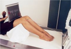
What is the evaluation criteria and procedure for a Settegast Method patella
X-ray?
SID: 40in
kVp: 70-80
Grid: No grid
CR: Directed inferosuperiorly at 15-20˚ from lower legs (start at horizontal) to be tangential to patellofemoral joint
Patient position: prone or seated position, Flex knee to a minimum of 90˚ (Have patient hold onto gauze or tape for prone)
Evaluation criteria: intercondylar sulcus (trochlear groove) and patella of each distal femur should be visualized, patellofemoral joint space open
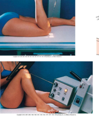
What is the evaluation criteria and procedure for a Hobbs Modification patella X-ray?
SID: 48-50in
kVp: 70-80
Grid: No grid
CR: mid-patellofemoral joint
Patient position: patient seated in a chair, IR under knees resting on a step stool or support (reduce OID), Knees should be flexed with feet placed slightly underneath the chair, perpendicular to IR
Evaluation criteria: intercondylar sulcus (trochlear groove) and patella of each distal femur should be visualized, patellofemoral joint space open