Exam II
 Call Kai
 Learn
Learn Practice Test
 Spaced Repetition
 Match
Match Flashcards
 Knowt Play
1/679

There's no tags or description
Looks like no tags are added yet.
Name | Mastery | Learn | Test | Matching | Spaced | Call with Kai | Chat |
|---|
No analytics yet
Send a link to your students to track their progress
680 Terms
Cranium
skeleton of the head
How many named bones does the cranium have?
22
How is the cranium divided?
Into two parts: the neurocranium and viscerocranium
Neurocranium
Bony case of the brain and its membranous coverings (meninges)
Neurocraniums is formed by:
8 bones: 4 singular frontal, ethmoidal, sphenoidal, occipital, 2 pairs temporal, and parietal
Consequences of a blow to the superciliary arch?
laceration and black eye
Sinuses are what type of cells
Pneumatic cells
Sinuses-function
decrease weight of skull and allow air through nose to be clean
Viscerocranium
Forms the anterior part of the cranium (face)
Viscerocranium consists of:
15 irregular bones: 3 singular mandible, ethmoid, vomer, 6 pairs maxillae, inferior nasal conchae, zygomatic, palatine, nasal, lacrimal
Malar flush
redness of the skin over the zygomatic prominence. Associated with Fever, tuberculosis, and Lupus
Maxillae
Pair of bones fused together that holds your upper teeth, shapes your cheek area, and forms the floor of your eye sockets and the roof of your mouth.
Foramen magnum
large opening at the base of the skull that allows the spinal cord to connect with the brain.
Le Fort I fracture
horizontal fractures passing superior to the maxillary alveolar process (sockets contain upper teeth)
Le Fort II fracture
passes the posterolateral parts of the maxillary sinuses to the bridge of the nose
Le Fort III fracture
horizontal fracture passing through the superior orbital fissures and other bones
All Le Fort Fractures are
horizontal
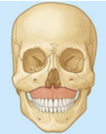
What type of fracture?
Le Fort I
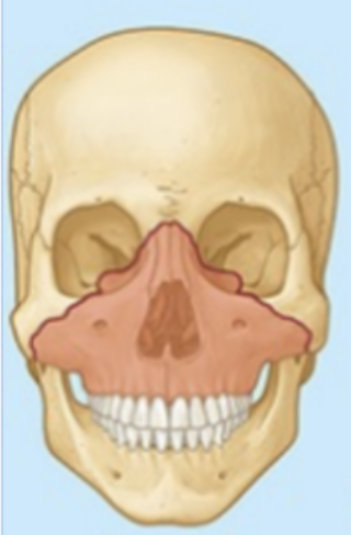
What type of fracture?
Le Fort II
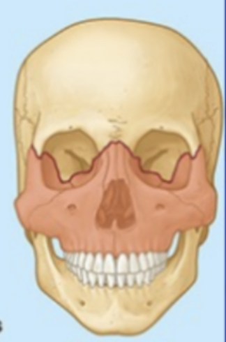
What type of fracture?
Le Fort III
Mandible
houses the lower teeth, forms the chin, and is the only freely movable bone in the face
Mandible function
chewing, shape, and speech
Fractures of the ____ usually occur in pairs, frequently on opposite sides.
mandible
Extraction of the teeth causes
reabsorption of the alveolar bone
Fractures of the coronoid process happens usually
simultaneously on both sides
Fractures of the coronoid process or neck of the mandible occur by:
Fall on chin, punch, trauma, or vehicular trauma
What happens when the mandible shrinks?
It leaves the mental foramen open and the nerves exposed to pain
How does mandible shrinkage occur?
Dentures or crystal meth
Dura
Firmly attached to the bone in the cranial base
Outer layer of meninges (periosteal layer)
Dura- function
Protects brain, stabilizes position, supports blood circulation
How can the dura detach from the head?
blow to the head or fracture of the skull
In a blow to the head the periosteal layer of the dura matter detaches from the
clavaria
A fracture that tears the dura results in
leakage of CSF - will come out from nostrils (rhinorrhea)
The skin of the scalp is normally ____ bearing
hair
What type of tissue is the scalp and what does it cover?
Subcutaneous tissue that covers the neurocranium from the superior nuchal lines on the occipital bone to the supra-orbital margins of the frontal bone
Scalp-function
Laterally, the scalp extends over the temporal fossa to the zygomatic arches
How many layers does the scalp have?
5
Describe first three layers of scalp
First three are connected intimately and move as a unit - skin, connective tissue, aponeurosis (SCA)
List the layers of the scalp from outermost
Skin
Connective tissue (dense)
Aponeurosis
Loose connective tissue
Pericranium
Superficial scalp wound do not gape open due to
the strength of the epicranial aponeurosis
Aponeurosis wounds
Gape widely (lacerate epicranial aponeurosis) - blood or pus spreads easily in the loose areolar layer of the scalp (4th & 5th layers), and may pass anteriorly into the eyelids → black (racoon) eyes
What are meninges?
Membranous coverings of the brain that lie immediately internal to the cranial
Epidural space
space between bone and top of the dura
What are the layers of the meninges?
Dura matter
Subdural space
Arachnoid
Sub arachnoid space
Subarachnoid space
contains CSF
Meninges- function
Protect the brain
Form the supporting framework for arteries, veins, and venous sinuses (vasculature for brain that take blood out)
What is cerebral-spinal fluid (CSF)
clear, colorless liquid that circulates through the brain's ventricles and around the spinal cord
Extradural or epidural hemorrhage originates
in an artery, typically the middle meningeal artery
How does an extradural or epidural hemorrhage occur
blow to the head
What happens when an extra dural or epidural hemorrhages occurs
The patient feels very sleepy, blood accumulates in the extradural hematoma, forces the brain through the foramen magnum (herniation) of midbrain sections → death
Dura matter
tough, thick external fibrous layer
Dura matter innervation
CN V (trigeminal) in the anterior and middle cranial fossae and by the CN X (vagus) in the posterior cranial fossa
Dura matter- function
anchors the brain to the skull and compartmentalizes it to stop the brain from moving too much when you move
May headaches originate in the ____, which is sensitive to pain caused by stretching of the meninges
dura mater
Dural border hematoma (subdural hematoma)- caused by
Caused by extravasated blood between the dura and arachnoid, typically from tearing of a cerebral vein
Arachnoid matter
thin intermediate layer
looks like a spider web
Arachnoid matter- function
acts as a bridge for the layer underneath it. It creates the subarachnoid space which is filled with cerebrospinal fluid (CSF)
Inflammation of the Leptomeninges results from
pathogenic organisms
Subdural hematoma- treatment
burr holes, done to drain blood from the space between the dura mater and the arachnoid mater.
A subarachnoid hemorrhage usually occurs from
an aneurysm or HTN
Pia matter
delicate internal vasculated layer
Pia matter- function
blood vessels to the brain to supply it with oxygen and nutrients. It also helps seal the brain to contain cerebrospinal fluid
What are the leptomeninges
pia matter and arachnoid matter
Dural venous sinuses
Endothelium-lined spaces between the periosteal and meningeal layers of the dura.
Dural venous sinuses- location
From where the dura septa attach along the free edge of the falx cerebri and in relation to formations of the cranial floor
Dural venous sinuses- function
Large veins from the surface of the brain empty into these sinuses and most of the blood from the brain usually drains through them into the internal jugular veins
Examples of dural venous sinuses
Cavernous sinus and Sigmoid Sinus
Infections may pass from the facial veins into the ____ ___ via the superior ophthalmic veins or the vertebral venous plexus into the dural sinuses
cavernous sinus
Triangle of death (or danger triangle)
An area of the face where infection may produce inflammation of the facial vein which can spread to the cavernous sinus, causing infected brain tissue
Fractures of the cranial base could case what artery to tear
internal carotid artery (circle of Willis)
What does a tear of the internal carotid artery through the cavernous sinus cause?
compression of CN VI (abducens: look to the side) and/or structure in the wall of the of the sinus, which includes CNs III, IV, V1 and V2 which affect eye movement and extra ocular muscles
Tentorial notch
Concave gap in the free anteromedial border of the tentorium cerebelli which the brainstem extends from the posterior into the middle cranial fossa
Tentorial notch- function
opening before the cerebellum that allows brain to continue as the spinal cord
An inflamed cerebellum can cause
Parts of the temporal lobe to herniate through the tentorial notch
What happens if the tentorium lacerates the temporal lobe
Damage to the oculomotor nerve causing paralysis of extraocular muscles
Pituitary (master gland) tumors or enlargement may extend superiorly through
sellar diaphragm
Pituitary (master gland) tumors or enlargement can cause _____ disturbance
endocrine
Brain
Delicate structure enclosed in a rigid cranium, surrounded by meninges
List 4 structures of brain
Cerebrum
Cerebellum
Brain stem
Diencephalon
List ways brain can be damaged
blow to the head
compressed by a tumor (benign or malignant)
deprived of oxygen (by leak or clot of blood in one of the cerebral arteries - thrombosis or embolism)
Cerebral concussion
Loss of consciousness after a head injury
Cerebral contusion
Results when the pia is stripped from the surface of the brain (bruising brain tissue), allowing blood to enter the subarachnoid space (blow to head: sports → concussion)
Cerebral lacerations result in
bleeding into the brain, causing compression → herniation
Cerebrum composed of:
Two hemispheres with 4 lobes each
Gyri and sulci
_____ has more neurons than any other part of brain
The cerebellum
Cerebrum- function
memory, reasoning
Cerebellum-function
coordination, balance
Brain stem
contains autonomic centers
Midbrain, pons, medulla obloganta
Brainstem- function
regulates involuntary, life-sustaining functions like breathing, heart rate, and blood pressure
Diencephalon
midbrain
Corpus Collosum
connects right and left hemispheres
Diencephalon-function
brain's primary relay, processing, and integration center. It bridges the nervous and endocrine systems, controlling sensory perception, motor functions, autonomic regulation, circadian rhythms, and homeostasis
Where can CSF be obtained
from the posterior cerebellomedullary cistern through a cisternal puncture
CSF creation pathway
Ependymal cells → lateral ventricles → CSF
CSF is produced initially in the
lateral ventricles
CSF function
acts as a shock-absorbing cushion to protect the central nervous system, delivers vital nutrients, and removes cellular waste
Hydrocephalus
excessive CSF dilates the ventricles, thins cerebral cortex, and separates the bones of the calvaria in infants
Treatments for hydrocephalus
Cisternal Puncture
shunt/valve take fluid from affected ventricle subdermally into abdominal cavity
How to diagnose hydrocephalus
transillumination or MRI
Brain accounts for ____ of body weight but receives about ___ of the cardiac output and ___ of the oxygen consumed by the body at rest
2.5%, 1/6, 1/5