Bone Formation (Exam 1) Diagram | Quizlet
 Call Kai
 Learn
Learn Practice Test
 Spaced Repetition
 Match
Match Flashcards
 Knowt Play
1/83

There's no tags or description
Looks like no tags are added yet.
Name | Mastery | Learn | Test | Matching | Spaced | Call with Kai | Chat |
|---|
No analytics yet
Send a link to your students to track their progress
84 Terms
Name the tissue that lines the medullary cavity (medulla)
Endosteum

Identify the space (orange circle)
medullary cavity, medulla
Periosteum
Identify the tough outer layer of the bone (orange)
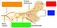
articular cartilage (aka Hyaline cartilage)
Identify the cartilage at the end of the bone (blue).
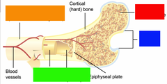
cancellous bone (spongy bone)
Identify the tissue found here (red).

Define anatomical position
Standing facing the front with palms turned to front
Medial
towards the midline
Lateral
away from thje midline
Anterior/ ventral
frontal plane of the body
Posterior/ dorsal
back or rear of the body
Superior
upper portion, towards the head
Inferior
lower portion, away from the head
Cephalad
toward/angled to the head
Caudad
toward/angled to the feet
Proximal
closer to the origin
Distal
further from the origin
Supine
facing up (palms up)
Prone
facing down (palms down)
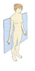
Midsagittal/ median plane
plane that extends from the sagittal suture cutting the body equally into left and right halves
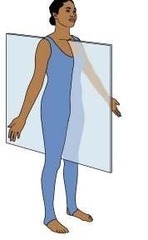
Sagittal plane
any plane that is parallel to the median plane
Coronal plane
plane that extends through the coronal suture cutting the body into anterior and posterior
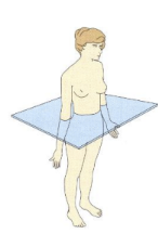
Subcostal plane
transverse plane that extends below the lower border of the ribs
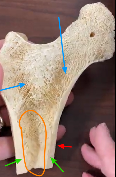
Identify the orange circle.
Medulla/medullary cavity

Identify the blue arrows.
cancellous bone
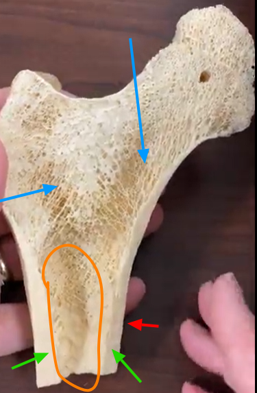
Identify the red arrow
Periosteum
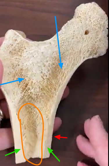
Identify the green arrow.
Cortex
Define long bones and list two long bones.
Bones that are longer than they are wide. ie femur, humerus, tibia, fibula
Define short bones and list 2 examples.
small cubed-shaped bones.
ie tarsal and carpal bones
Define and list 2 flat bones examples
thin, parallel surfaces that assist in protecting internal organs.
ie frontal, occipital, thoracic cage (ribs, sternum)
Define and list two irregular bones.
have complex and varied shapes
Define and list a sesamoid bones.
A bone that forms within a tendon.
ie patella
trabeculae
branching bony plate within the cancellous bone
Endosteum
thin connective tissue that lines the marrow cavity
Medulla
central cavity in the long bone shaft
Lamellae
bone matrix deposited into thing layers. arrange in concentric circles around the osteonic canals.
Osteonic canals
longitudinal tubes (aka haversian canals)
Lacunae
small spaces/cavities in the bone matrix that contain osteocytes.
Osteocytes
bone cells located in the lacunae
canaliculi
tubes in the matrix (location for nutrients transport)
Red bone marrow
produces red blood cells (mainly found in infants)
Yellow blood marrow
replaces red bone marrow in the shaft of the long bones in adults
Nutrient foramen
small canal passing through the periostem and other layers to reach the medulla
Haversian canal
longitudinal canals in compact bone containing blood vessels and nerves.
Volkmann's canal
transverse/horizontal canals that connect Haversian canals and carry blood vessels and nerves.They play a crucial role in the communication between the outer and inner regions of bone.
The cortex comprises of the _______ layer and the ______ layer of the skull
inner and outer
What is the middle spongy bone layer of the the skull between the cortices called?
diploe
What are the five functions of bones?
1.) protection
2.) support and movement
3.) hemopoiesis
4.) mineral storage
5.) muscle attachments
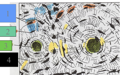
canaliculi
Identify the Blue highlighted area.
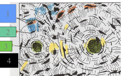
Lacunae
Identify the orange areas.

Lamellae
Identify the yellow areas
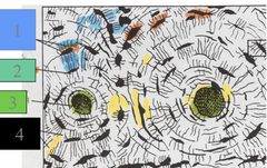
Osteonic canals (Haversion Canals)
Identify the green areas.
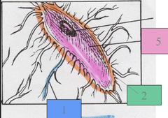
Osteocyte
Identify this cell.
What is the purpose of red bone marrow and where is it found primarily?
Red bone marrow produces red blood cells. Found mainly in the medulla of infants and children.
What is the purpose of yellow bone marrow and where is it found?
Stores fat and is primarily found in the medulla of adults
nutrient foramen
small canal passing through the periosteum and other layers to reach the medulla
Haversian Canals
aka osteonic canals, are small blood vessels in the bone that transports nutrients and oxygen vertically
Volkmann's canal
connect osteonic/ haversian canals, nutrients and blood flow occur horizontally
Ossification
the process of bone formation n(bone forms in 7th-9th week of embryonic life)
Endochondral ossification
bone tissue replaces a cartilage model.
Intramembraneous ossification
fibrous membrane is replaced by bone tissue
Where does the primary center of ossification occur?
Diaphysis
Where does the secondary center of ossification occur?
epiphysis
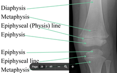
Epiphyseal plate/line
cartilage growth plate between epiphysis and metaphysis in growing bone. After growth stops, it ossifies and becomes the epiphyseal line.
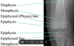
Metaphysis
part of the diaphysis that is adjacent to the epiphyseal line
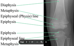
Bone age
is determined by the appearance of the epiphyses

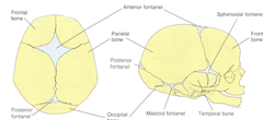
Fontanels
Place of incomplete fusion of cranial bones present at birth
Synarthrosis
bands of fibrous tissue, No movement, ie skull and teeth attachments at the mandible
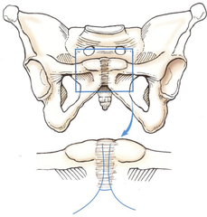
Amphiarthrosis
Joints united by cartilaginous tissue, Limited movement
Diarthrosis
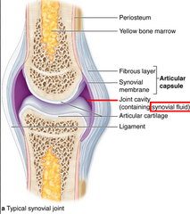
synovial joints with free movement
Name 6 diarthrosis joints.
-saddle
-hinge
-pivot
-ellipsoidal/ condylar
-ball & socket
-gliding
articular cartilage
covers the ends of the bone
articular capsule
fibrous connective tissue that surrounds/encloses a synovial joint.
Synovial Joint cavity
cavity between bones, lined with synovial membrane
Synovial membrane
secretes synovia to lubricate the joint
Ligaments
strong cords of dense tissue that hold the joint tightly together
Bursa
pad of synovial membrane outside the joint cavity where friction occurs
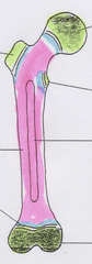
Cancellous tissue
1
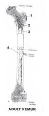
Periosteum
2
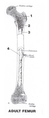
cortex
3
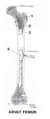
Medulla
4
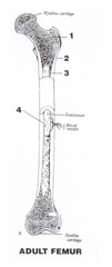
epiphysis
Green
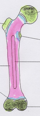
Epiphyseal line
White

Metaphysis
Blue

Diaphysis
Pink