Ophthalmology Lecture: Retinal Conditions
 Call Kai
 Learn
Learn Practice Test
 Spaced Repetition
 Match
Match Flashcards
 Knowt Play
 Podcast
1/13

Earn XP
Description and Tags
Flashcards covering Retinal detachment, Central Retinal Artery Occlusion (CRAO), Central Retinal Vein Thrombosis (CRVT), and Macular Degeneration.
Name | Mastery | Learn | Test | Matching | Spaced | Call with Kai | Chat |
|---|
No analytics yet
Send a link to your students to track their progress
14 Terms
Retinal detachment is defined as the __________ of the retina from the back of the eye.
Separation
clinical feature of retinal detachment is the sudden onset of _________
floaters and flashes
The clinical visual sensation often described by patients with retinal detachment is that a __________ .
curtain came down over the eyes
Fundoscopy for retinal detachment shows a __________ in the vitreous cavity.
large shadow
management of the retinal mangagement
Immediated referral
Central retinal artery occlusion (CRAO) is caused by arterial obstruction from __________ .
Atherosclerotic emboli/thrombi
In patients with CRAO, fundoscopy reveals a classic __________ at the macula.
cherry red spot
the clinical features for central retinal artery occlusion ______
painless
clinical presentation for central retinal artery occlusion (CRAO)
sudden loss of vision in one eye, no light perception
Initial management of CRAO includes direct digital pressure to _________and re-breathing __________ .
massage eye-globe directly , CO2
The medical treatment for CRAO within 30mins of presentation includes administering IV __________ .
acetazolamide
Central retinal vein thrombosis (CRVT) is characterized on fundoscopy by a __________ appearance.
stormy sunset
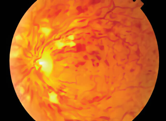
what does this indicate
central retinal vein thrombosis
Visual clinical features of CRVT include sudden loss of central vision and a vision that improves with a __________ .
distorted image