L9, 10, 11, 12, 13 - RNA, Cell Cycle Regulation, Gene Regulation, DNA Repair
 Call Kai
 Learn
Learn Practice Test
 Spaced Repetition
 Match
Match Flashcards
 Knowt Play
1/137

There's no tags or description
Looks like no tags are added yet.
Name | Mastery | Learn | Test | Matching | Spaced | Call with Kai | Chat |
|---|
No analytics yet
Send a link to your students to track their progress
138 Terms
The Transcriptome
The entire set of transcripts in a cell.
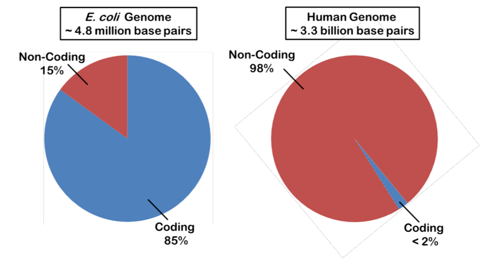
In general, the more complex the organism is, a greater portion of the genome is non-coding.
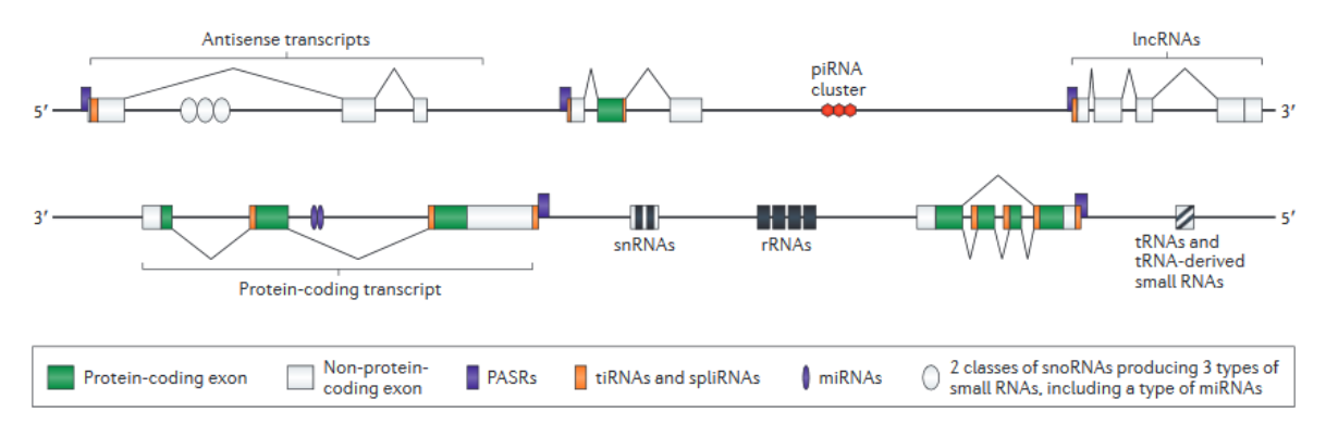
Even though ~98% of the human genome does not code for proteins, most of the genome is transcribed into RNA.
RNA can be characterized as either non-coding or coding (though that distinction itself is oversimplified).
Non-coding RNA
1. Ribosomal RNA: (rRNA) is a major component of ribosomes, which are part of the protein synthesis machinery.
2. Transfer RNA: (tRNA) is a critical component of protein synthesis and acts as a “magic decoder ring” bridging the gap between nucleotide sequence and protein sequence.
3. Other important non-coding RNA that will be discussed later:
microRNA (miRNA)
small interfering RNA (siRNA)
small nuclear RNA (snRNA) or spliceosomal RNA
clustered regularly interspaced short palindromic repeat (CRISPR)
Coding RNA
1. Messenger RNA: (mRNA) is the middleman taking the genes coded in DNA in a readable form to the ribosomes in order to make protein.
Relative Abundance: 3-5% of total RNA in a cell, however these molecules are specific for the proteins they encode, so the abundance of any given mRNA is extremely low! There are 1 – 10 thousand (103-104) different mRNAs in a cell!
Size: highly variable, just like the genes that encode them. (200 nt up to 104 nt)
Base Composition: A, C, G, U with very little modification
Structure: The messages are structured in terms of an Open Reading Frame (ORF) which is the region of a gene or an mRNA that codes for the amino acid sequence of a protein.
Polycistronic mRNA
Polycistronic mRNAs are those that contain more than one ORF and are found in bacterial systems and some protists.

Monocistronic mRNA
Eukaryotic systems use a Monocistronic mRNA that contains only one ORF which codes for one polypeptide.

RNA Biosynthesis in E. coli (Transcription): General Features
1. Requires a DNA template, ribonucleoside triphosphates (ATP, CTP, GTP, and UTP) and an RNA polymerase —> only one RNA P in prokaryotes so we dont have to specify
2. Synthesis of RNA proceeds 5’ - 3’
3. NO Primer – this allows RNA to serve as the primer for DNA synthesis!
4. Possible proofreading, though it is limited and not entirely necessary because the product is not passed on to progeny. Further, it generally has a short ½ life… it is used and destroyed.
5. Template is conserved, it is neither “used up” nor changed
6. Process – overview:
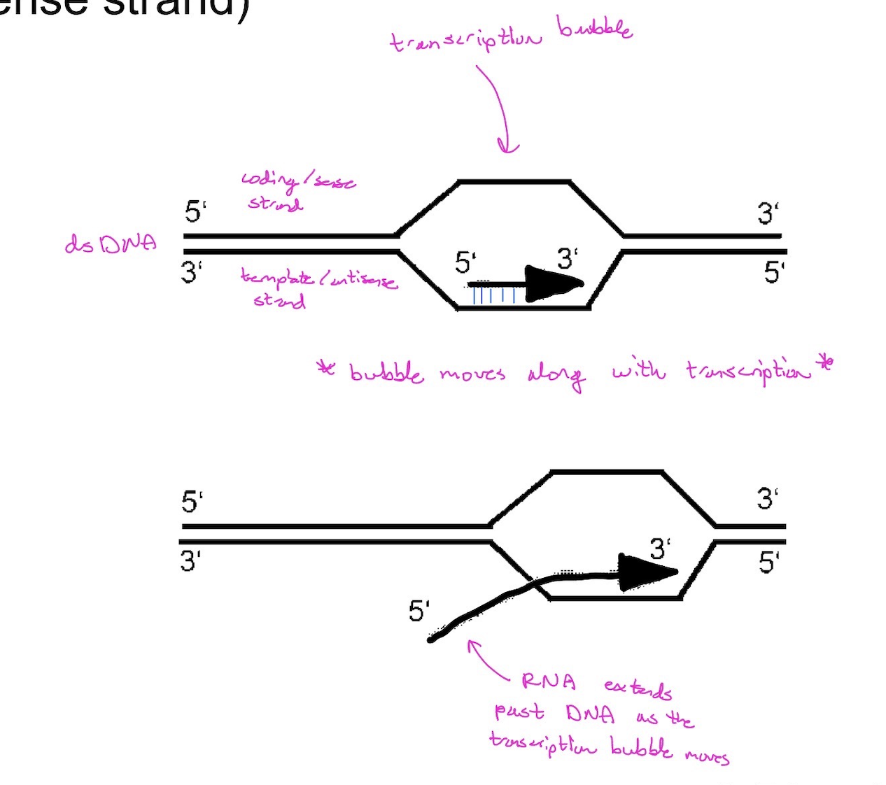
dsDNA opens to form a “bubble”
As RNA is made, it stays paired with the DNA
As the polymerase moves on the DNA duplex recloses, the bubble moves to the right, and the RNA extends out beyond the DNA-polymerase complex.
Product RNA strand is complementary to the template DNA strand (also called the antisense strand, and it reads the “same” as the non-template DNA strand (also called the coding strand or the sense strand)
RNA Biosynthesis in E. coli (Transcription): RNA Polymerase
1. In E. coli, there is only one RNA Polymerase (RNAP)
a. Structure:
Core polymerase (multisubunit) is catalytically active, but does not know where to start transcribing… it’s a big genome
out there.
Holoenzyme is the core polymerase plus Sigma (σ) factor. This additional subunit directs the polymerase to the start site of transcription.
b. Rationale for needing to know where and how often to start transcription: RNA polymerase only copies the bit of information needed for that cell at that moment. Unlike DNA replication which needs to copy the whole genome for passage onto progeny.
2. Reactions of RNA Polymerase
a. Initiation: START
b. Elongation: GO
c. Termination: STOP
d. Initiation: To start at the correct location, the RNA polymerase holoenzyme recognizes a Promoter.

Promoter
is a sequence element (can have variation) that is recognized by the σ factor and directs binding of the RNAP. It is usually upstream of the +1 start site. The +1 start site is represented with a broken arrow. The variation in the promoter elements can change the “strength” of the promoter. [Note: the gene sequence tells the cell what to make, but the promoter tells the cell when to make it and how much to make.]
Inhibition by Antibiotics:
**This is one reason why we care!
Antibiotics can inhibit RNAP by several modes of action which we will group into two classes:
a. Inhibition by DNA-binding
Actinomycin D = dactinomycin = Cosmegen: This drug acts by blocking the unwinding of DNA by RNAP. Used as an anti-tumor drug as it is effective against both bacterial and eukaryotic cells.
doesn’t let RNA P bind to DNA
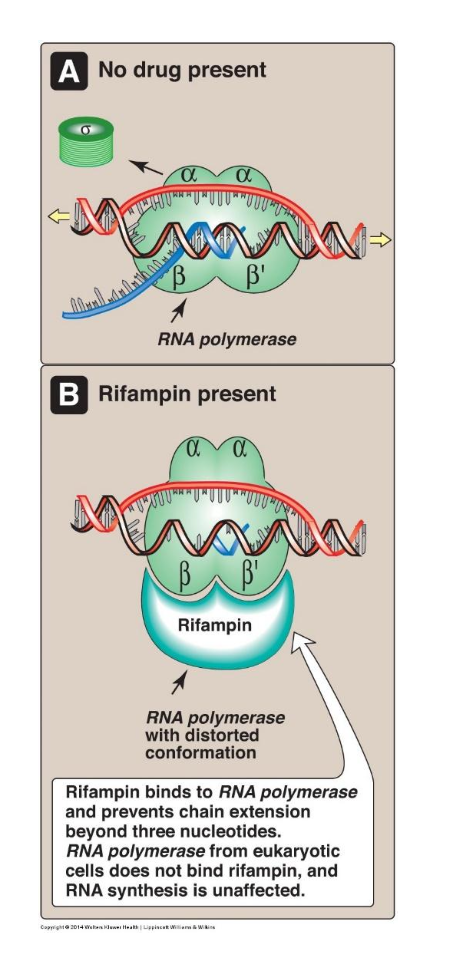
b. Inhibition by enzyme-binding: inhibits RNAP binding and/or enzymatic activity
Rifampicin – used to treat mycobacterial infections (TB, leprosy)
RNA Biosynthesis in Eukaryotes: General Features
1. Shared with Bacteria:
Requires DNA template,
No primer,
5’ → 3’ directional synthesis,
limited proofreading
**NOTE: in prokaryotes —> transcription and translation is coupled
2. Unique to Eukaryotes:
Compartmentalization: transcription (in the nucleus) is separated from translation (in the cytosol)
Chromatin Remodeling: DNA wound tightly around histone proteins must be “relaxed” to be accessible to the transcription machinery. This is mediated by Histone Acetyl-Transferases and Histone Deacetylases.
RNA processing: Primary transcripts get modified into biologically active forms.
RNA Polymerase: There are THREE functionally distinct polymerases in the eukaryotic nucleus. Organelles such as the mitochondria and chloroplasts also have RNAPs, but these more closely resemble bacterial polymerase.
RNA Polymerase II (RNAP-II) & protein encoding mRNAs
The actual product of RNAP-II for protein encoding genes is heteronuclear RNA (hnRNA) not mRNA. It doesn’t become an actual mRNA till it is fully processed (see point 3 below on processing).
RNA Polymerase II (RNAP-II) & protein encoding mRNAs: Gene Structure
a. Incredibly heterogeneous since there are many different genes to make many different proteins!
b. Core Promoters consist of several elements: TATA box, Initiator element, and a Downstream promoter element. (though not all elements are present at all promoters)
TATA box is analogous to the -10 and -35 boxes of bacteria
The initiator element alone is enough to recruit RNAP-II and initiate transcription at the proper site. (though it makes a weak promoter on its own)
Mutation of the TATA results in an 80% decrease in β-globin mRNA. The resulting disease is β-thalassemia.

mutations in gene coding for hemoglobin
**NOTE: more than 200 mutations in HBB gene that makes beta chain of He.
mutations = imbalance of beta chain vs. alpha chain production
there are other causing mutations —> TATA box mutation is one of them
RNA Polymerase II (RNAP-II) & protein encoding mRNAs: Enzyme
RNAP-II is a 10 - 12 subunit complex (some shared with RNAP-I and RNAP-III, some are unique) that employs many accessory factors.
a. BASAL transcription factors (same at every gene to be transcribed)
TFII-A, B, D, E, F, and H
TFII-D (equal to sigma factor for prokaryotes): also called the TATA binding protein or TBP – a bit of a misnomer as subunits of TFII-D are also involved in binding the initiator element and the downstream element!
Function: Binds these elements, recruits the transcription initiation complex and directs RNAP-II to the proper start site.
b. Regulatory transcription factors (since we do not want to transcribe every gene all the time!)
These are specific for particular genes (or sets of genes)
Can have binding sites much further from the initiation site in sequence
***Remember 3-D structure though! They may be quite close in space
Function: Binds these elements and either positively or negatively regulate the initiation of transcription for these genes.

Mushroom Poisoning
i. Within 12-24 hours, ingestion of Amanita phalloides causes severe abdominal cramps, vomiting and diarrhea.
ii. 20% of cases are fatal
iii. Toxin 1α-amanitin is a potent inhibitor of RNAPII
RNA Polymerase II (RNAP-II) & protein encoding mRNAs: Processing
1. This is where the primary transcript (hnRNA) is converted into the mature transcript (mRNA)
2. Consists of 3 processing events:
capping of the 5’ end
polyadenylation of the 3’ end
splicing
3. Important Features or Regions of the mature mRNA: (from the left)
5’ Cap
5’ Untranslated region (UTR)
AUG start codon
ORF (coding region)
STOP codon
3’ UTR**
Poly A tail
**The UTRs (sections of the mRNA that do not code for protein) are still important! For example: In iron metabolism, the Iron Responsive Elements (IREs) are in the 3’ UTR for transferrin and in the 5’ UTR for ferritin. – allow the cell to respond to the local iron levels!

Splicing
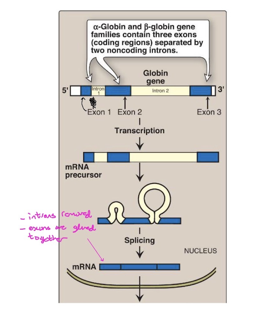
a. Some mRNA encoding genes are “multi-partite” in that they have coding regions that are interrupted by non-coding (or alternately coding) regions.
These regions not being incorporated into the mature mRNA are called INTRONS – intervening sequences – need to be removed
As opposed to EXONS – the coding sequences that are kept and joined.
Removal of introns requires precision. (otherwise the reading frame will be out of register)
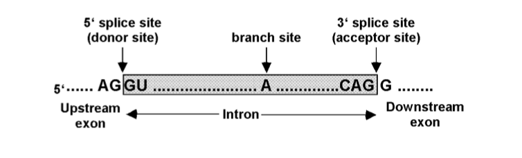
Splicing sites are specified by sequences in the hnRNA
b. Splicing Machinery (Spliceosome) is analogous to ribosomes and contains several hundred proteins and several small nuclear RNAs (snRNA).
Each of the snRNAs is part of a larger complex called a Small Nuclear Ribonucleoprotein Particle or snRNP, (“snurp”).
Each snRNP contains one snRNA and its own set of proteins.
There are 5 snRNPs - and each is characterized by a unique snRNA and has a specific role in the splicing process: snRNPs and other proteins of the spliceosome carry out the endonuclease cleavage and ligation reactions.
*** Medical Significance: snRNPs can be antigens in auto- immune disorders (e.g., systemic lupus erythematosis)
spliceosome
RNA that does the chemistry
ribozyme
RNA component is the catalytically active portion of the complex
splicing sites in hnRNA example
Multiple Introns: ordered splicing
Example: a gene has 3 exons separated by 2 introns can be spliced in exact order, 1 to 2, 2 to 3, without skipping (not 1 to 3)

Multiple Introns: alternative splicing
1. Joining 1 to 3, skipping 2, can give rise to different polypeptide products (may be a tissue-specific mechanism to get similar but not identical proteins)
2. The majority of human multi-exon genes are alternatively spliced.
3. For this example (tropomyosin): One of these products may be in striated muscle and another in smooth muscle... therefore the Ca2+ sensitivity in one muscle may be slightly different than another!

splicing regulation
regulatory sites that enhance or suppress inclusion of exons and introns may be present

Medical Significance: Splicing errors and disease (some examples)
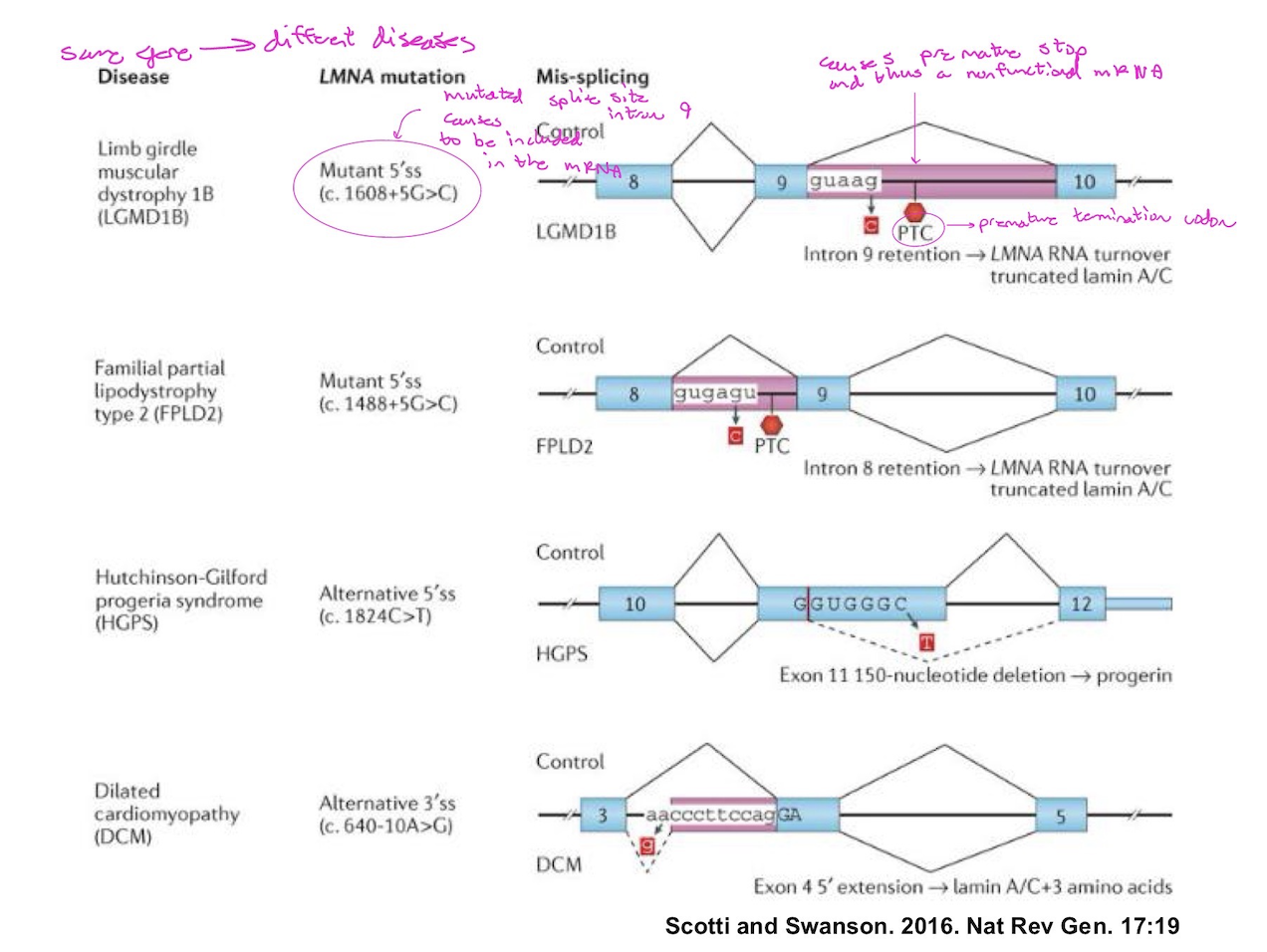

β-thalassemia
1. The resulting mutant β-globin mRNA is shorter and likely less stable
2. The resulting mutant β-globin polypeptide will be shorter; may be out of frame, and likely less stable → NOT Functional!
3. Therefore, there will be an imbalance in production of functional β-chain relative to α-chain → β-thalassemia → anemia

RNA Transport
All mRNAs - from the nucleus where it is transcribed and processed, to the cytoplasm where it can serve as a template for translation.
RNA Silencing
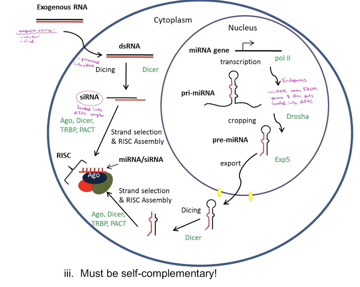
The process by which small non-coding RNAs act as regulators of gene expression. There are two kinds of small non-coding RNAs: short interfering RNA (siRNA) and micro RNA (miRNA). The difference between siRNA and miRNA is based primarily on their source.
siRNA
short interfering RNA
i. exogenous source – from a “foreign challenge” such as a virus.
ii. Viruses can have RNA or DNA genomes.
iii. In the figure you will see how a siRNA can be generated from either a DNA virus or RNA virus.
miRNA
micro RNA
i. endogenous – from the organism’s genome. Micro RNA genes are plentiful and can be found throughout the genome including within some introns of protein encoding genes! (termed mirtrons)
ii. Transcribed from endogenous genome
iii. Must be self-complementary!
RNA Silencing Mechanism
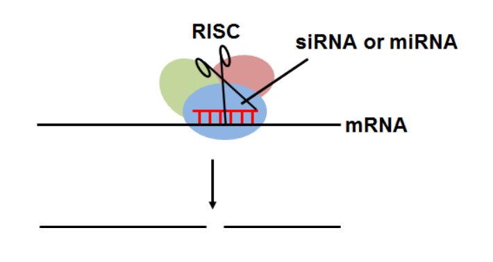
One strand of the 18-24 nt RNA is loaded into the RISC. (RNA Induced Silencing Complex). This “guide strand” targets the complex to an appropriate mRNA and binds to its target region (usually in the 3’ UTR).

RNA Silencing Outcomes
The overall outcome for both mechanisms is the same: a lower level of protein expression for the targeted gene. Hence, this process is called a gene “knock down” (NOT a knock out)
a. Translational interference – Since the complex is bound to the mRNA, translation by ribosomes is physically blocked. (sometimes reversible!) Ribosomes cannot translate through the blockage!
b. mRNA degredation – binding may also mark the mRNA for nuclease cleavage and degredation
c. Nuclease cleavage begins the process of mRNA degredation.
RNA Silencing Biological Context
a. It is estimated that ~1/3 of all genes are regulated by miRNA!
b. Embryology: active miRNAs are involved in maintaining stem cell pluripotency and in determining cell fate upon differentiation. It is also involved in mammalian neurogenesis and certain miRNAs appear to be expressed in “waves” during development.
c. Provides a natural defense against viruses and related “challenges”
RNA Silencing —> Research and Medical Applications
a. Targeted down-regulation of specific genes in specific tissues
b. Determining the existing miRNA profiles in certain normal and diseased/cancerous tissues can aid in the determination of cancers developmental lineage: tissue of origin and differentiation stage.
c. Altering miRNA profiles can be immunogenic in certain tissues
d. Provides an engineered defense against viral infections such as Ebola, HIV, Hepatitis B & C, HSV2
Some are still in animal model research, others are already in clinical trials!
e. FDA approved treatments for hereditary transthyretin amyloidosis (hATTR), acute hepatic porphyria (AHP), primary hyperoxaluria type 1 (PH1), and lowering of low-density lipoprotein cholesterol (LDL-C) in subjects with heterozygous familial hypercholesterolemia (HeFH) or clinical atherosclerotic cardiovascular disease (ASCVD)
CRISPR
clustered regularly interspaced short palindromic repeats
a. small non-coding RNAs in bacteria and most archaea that act as an immune system against viruses
b. The natural function of the CRISPR system can be modified to achieve genome editing
The CRISPR RNA acts as a guide for the Cas9 endonuclease to locate and cleave DNA at sites demarcated by conserved sequences called proto-spacer adjacent motifs (PAMs)
Cas9 introduces double stranded breaks into the target DNA. When the breaks are repaired by cellular machinery the site can be modified or new genetic information can be inserted.
Cell cycle
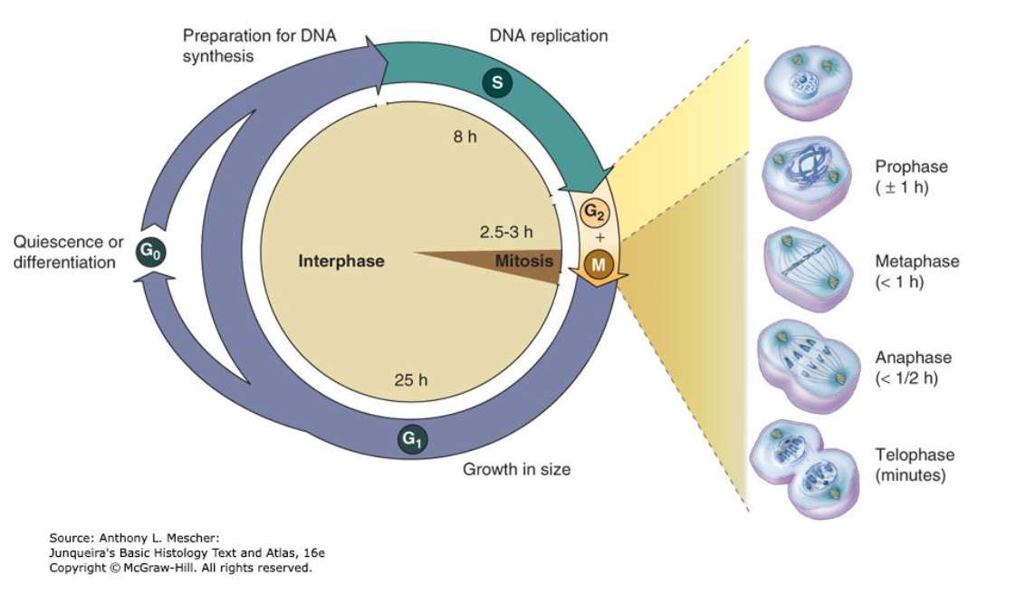
coordination of events surrounding eukaryotic DNA replication and cell division (mitosis).
The length of the cell cycle varies considerably from one cell type to another.
The large difference is the length of interphase.
1. Epithelial cells: ~10 hours
2. Liver cells: one year
3. Skeletal muscles and neuron: lose their ability to divide and replicate in adults
Interphase
Period between successive mitosis. A typical cell spends most of its life in interphase doing whatever it is programmed to do. (G0 for non-dividing cells, indefinitely in G1. G0 is a modified G1 phase.) There is then a period of DNA synthesis (S) in which chromosome duplication occurs followed by a further gap (G2) in time waiting for mitosis to begin. During this gap (G2), essential proteins and cofactors are produced necessary for mitosis to occur.
G1 (Gap 1) phase
interval between mitosis and DNA replication.
a. Cell carrying on its activities, preparing to duplicate its DNA content
b. Cell grows in size and accumulates nutrients
c. Cells accumulate the enzymes and nucleotides required for DNA replication
S (synthesis) phase
DNA replication occurs.
a. The chromatid of each chromosome is replicated – 2 chromatids per chromosome (chromosome X-shaped)
G2 (Gap 2) phase
interval between S phase and next mitosis.
a. Some DNA repair occurs
b. Chromosomes begin to condense in preparation for mitosis
c. Proteins required for mitosis accumulate
d. Short period in preparation for mitosis
G0 phase
Cells that have stopped dividing and left the cell cycle.
These quiescent cells can reenter the G1 phase to resume cell division.
a. Quiescent cells – fibroblasts, liver cells, smooth muscle cells, and
endothelial cells (may reenter cell cycle under certain conditions)
b. Terminally differentiated – skeletal muscle and neurons (cannot reenter the cell cycle)
M phase
Includes all the phases of mitosis: prophase, metaphase, anaphase and telophase.

Regulation of the Cell Cycle
Accomplished by the presence of checkpoints at critical points in the cell cycle. During the checkpoints the completion of critical events is monitored and, if necessary, progression to the next stage of the cell cycle is delayed.
Cell cycle regulators
Two key classes of proteins control the progress of a cell through the cell cycle: cyclins and cyclin-dependent kinases (CDKs).
Cyclins
are a family of cell cycle regulatory proteins that are expressed to regulate specific phases of the cell cycle. Different cyclins are made at the beginning of different phases of the cell cycle and degraded at the end of the phase via the proteasome pathway. Therefore, cyclin concentrations rise and fall throughout the cell cycle due to synthesis and degradation. They are categorized as D, E, A and B cyclins.
Cyclin-dependent kinases (CDKs)
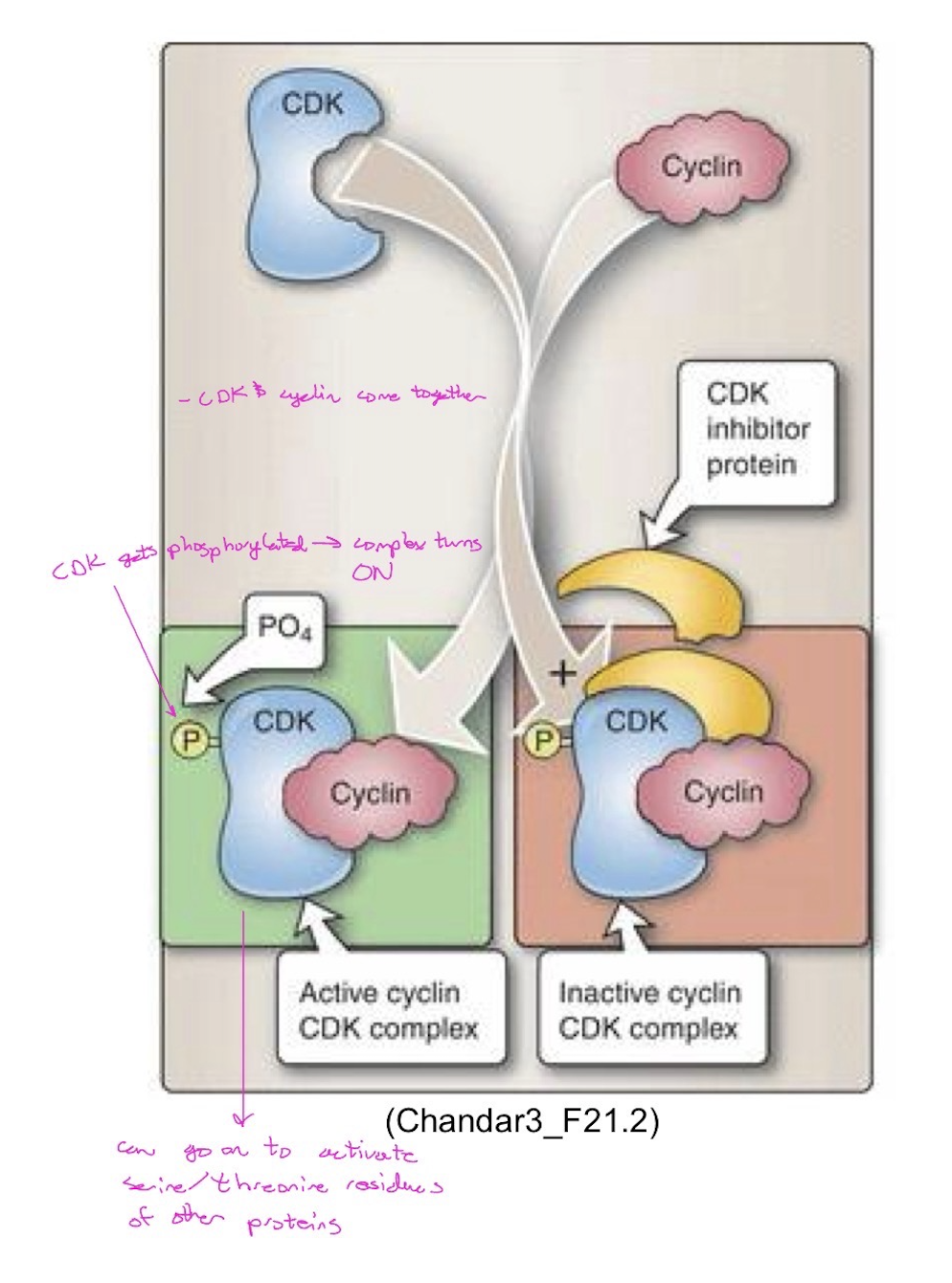
are serine/threonine kinases that are made constantly throughout the cell cycle. CDKs require binding of a specific cyclin to be active and phosphorylate protein substrates. Therefore, CDK enzyme activities fluctuate depending upon available concentrations of cyclins.
Cell Cycle Regulation: Mechanism
a. Different cyclins are present during different phases of the cell cycle.
b. Each cyclin activates one or more specific CDK. When cyclins are paired with their appropriate CDK, the CDK becomes phosphorylated leading to activation of its kinase activity and the cell cycle is permitted to progress.
c. To control cell cycle progression, the amount and type of cyclins varies. Note: the CDK concentration in the cell does not change.
d. When a cyclin-CDK complex is formed, it activates the kinase activity of the CDK.
e. Each activated CDK phosphorylates and activates specific proteins, transcription factors for specific sets of genes, and cytoskeletal subunits – triggering the activities needed for phase specific functions.
f. When the set of activities of that phase are completed, the cyclin controlling that cell cycle phase is removed by ubiquitin–mediated proteasome degradation.
g. The cyclin that promotes activities for the next phase will take over.
Note: cyclin A pairs with both CDK2 and CDK1 to regulate different portions of the cell cycle.
Cyclin-dependent kinase inhibitors (CKIs or CDKIs)
binds to and inhibits the CDK-cyclin complexes, therefore modulating CDK-cyclin complex activity. There are two categories of CKIs: the Cip/Kip family and the INK4 (inhibitors of cyclin-dependent kinase-4) family.
Cip/Kip family: family members include p21 (CDKN1A), p27 (CDKN1B), and p57 (CDKN1C). They have a broad specificity and inhibit multiple cyclin–CDK complexes.
INK4 family: consists of p15 (CDKN2B), p16 (CDKN2A), p18 (CDKN2C), and p19 (CDKN2D). They are specific for the cyclin D–CDK4/6 family of complexes (inhibitors of cyclin-dependent kinase-4).
Clinical Relevance: there are newly available oral cancer drugs that act by targeting and inhibiting CDK4 and CDK6. These drugs inhibits the growth of cancer cells by disrupting the cell cycle. They cause arrest in G1 ultimately leading to senescence and apoptosis.
Tumor Suppressor Genes
a. encode proteins that form a network of checkpoints that help control cell division and suppress inappropriate cell proliferation.
b. the product of these genes applies brakes to cell proliferation and may function as transcription factors, cell cycle inhibitors, signaling molecules, cell surface receptors and regulators of cellular responses to DNA damage.
c. Special classes of tumor suppressor genes:
Gatekeepers: directly involved in cell cycle checkpoints and growth inhibition. Examples include p53, pRb, PTEN.
Caretakers: genes coding for DNA repair apparatus, whose major role is repairing errors/damage in DNA. Mutations in DNA repair genes can lead to mutations in other genes. Examples include BRCA 1and BRCA 2.
Checkpoints and checkpoint regulation
G1 Checkpoint
“START”
a. Decide to “go/not go” into DNA Replication
b. G1 DNA-damage checkpoint:
Is DNA OK—need to repair first?
c. Restriction point (R point):
Activities occurring before this point depend on extracellular resources such as mitogens and growth factors. Once the R point is passed, the cell cycle no longer depends on these extracellular resources.
Considerations: Are cell’s nutrients, size, and environment favorable?
d. This checkpoint is mediated by the interactions between retinoblastoma susceptibility protein (pRb) and essential transcription factors (E2F).
Hyperphosphorylated pRb releases E2F – E2F is a transcription factor that activates genes needed for cell cycle progression. [growth factor stimulation → signaling via MAP/PI3 kinase → phosphorylation and activation of cyclinD/CDK4/6 complex → pRb phosphorylation] —> E2F leads to transcription of Cyclin E and A and other proteins needed for DNA replication
Hypophosphorylated pRb binds to E2F – turns off genes needed for cell cycle progression (blocks cell cycle progression).
![<p>“START”</p><p>a. Decide to “go/not go” into DNA Replication</p><p>b. G<span style="font-family: Arial; line-height: normal; font-size: 9px;">1</span> DNA-damage checkpoint:</p><ul><li><p>Is DNA OK—need to repair first?</p></li></ul><p>c. Restriction point (R point):</p><ul><li><p>Activities occurring before this point depend on extracellular resources such as mitogens and growth factors. Once the R point is passed, the cell cycle no longer depends on these extracellular resources.</p></li><li><p>Considerations: Are cell’s nutrients, size, and environment favorable?</p></li></ul><p>d. This checkpoint is mediated by the interactions between retinoblastoma susceptibility protein (pRb) and essential transcription factors (E2F).</p><ul><li><p>Hyperphosphorylated pRb releases E2F – E2F is a transcription factor that activates genes needed for cell cycle progression. [growth factor stimulation <span style="font-family: Wingdings; line-height: normal; font-size: 14px;">→</span> signaling via MAP/PI3 kinase <span style="font-family: Wingdings; line-height: normal; font-size: 14px;">→</span> phosphorylation and activation of cyclinD/CDK4/6 complex <span style="font-family: Wingdings; line-height: normal; font-size: 14px;">→</span> pRb phosphorylation] —> E2F leads to transcription of Cyclin E and A and other proteins needed for DNA replication </p></li><li><p>Hypophosphorylated pRb binds to E2F – turns off genes needed for cell cycle progression (blocks cell cycle progression).</p></li></ul><p></p>](https://assets.knowt.com/user-attachments/c1e7e30c-60d6-478c-b635-d0150b7377ca.png)
Hyperphosphorylated pRB
transcription factors bind to receptors —> activation of signaling cascades (i.e. MAP kinase and PI3 kinase) —> phosphorylation of and activation of CDK4/6 —> phosphorylates pRB
G1 Checkpoint —> Clinical Relevance
Hereditary retinoblastoma is an inherited eye malignancy caused by a mutation in the pRb gene. The mutant pRb protein is unable to stop the cell cycle in G1 allowing unregulated progression through the remainder of the cell cycle.
Tumor protein p53 (p53)
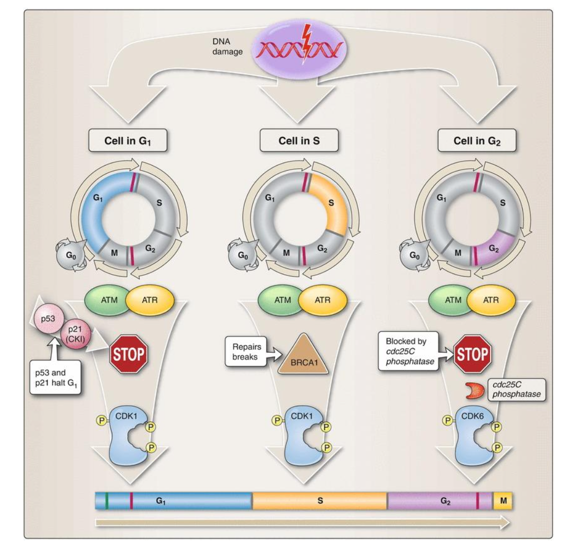
i. Is phosphorylated (activated) when there is DNA damage.
ii. Activation induces transcription of p21 (inhibitory protein — CIP-KIP family go CDK inhibitors) – stops cell cycle progression.
iii. Cell tries to repair DNA damage. If damage not repaired → programmed cell death (apoptosis).
p53: Clinical Relevance
More than 50% of human tumors contain a mutation or deletion of the p53 gene and those who inherit only one functional copy of the p53 gene will most likely develop tumors in early adulthood. Li-Fraumeni syndrome occurs when a mutation in p53 is inherited and is characterized by multiple types of tumors. Some tumors with p53 mutation are resistant to radiotherapy because apoptosis will not be triggered in the absence of functional p53.

HPV/p53: Clinical Relevance
Human papillomavirus (HPV) strains 16 and 18 are associated with cervical cancer. In cervical cells infected with these viral strains of HPV:
i. Viral protein E6 – binds to p53 and targets it for degradation via the proteasome pathway.
ii. Viral protein E7 – binds to pRb and prevents it from inhibiting proliferation.
iii. Both p53 and pRb are inactivated leading to possible upregulated cell cycle progression and malignancy.

S DNA-damage checkpoint
a. Monitors the quality of replicating DNA.
b. Slows the rate of DNA synthesis if DNA damage occurs during S phase.
c. BRCA1 plays a role in the repair of double-strand DNA breaks.
G2 checkpoint
a. Is replication complete? S phase needs to be completed and DNA completely duplicated before mitosis start in order to maintain the integrity of the genome.
CDK1 is phosphorylated in tyrosine residues leading to inhibition of its activity during G1, S, and into G2.
cdc25C phosphatase removes the inhibitory phosphorylations from CDK1 allowing the cell to progress through G2 and into M phase.
Dephosphorylated CDK1 can bind to cyclin B and the activated CDK1-cyclin B complex phosphorylates key components of subcellular structures (e.g., microtubules) to activate mitosis.
b. Is the cell ready to divide?

Metaphase checkpoints
a. Spindle-assembly checkpoint:
Are all chromosomes attached to the mitotic spindle?
If OK, cells will progress into anaphase.
b. Chromosome-segregation checkpoint:
Have all the chromosomes been correctly separated?
c. Now cells divide if everything is OK.

Five stages are defined for Prophase of meiosis I
a. Leptotene: chromosomes become visible as they begin to condense
b. Zygotene: homologous chromosomes align opposite each other, a process known as synapsis
c. Pachytene: Each pair of homologous chromosomes become tightly coiled ******Where crossing over occurs!!
Bivalent = two homologous chromosomes in the unit
Tetrad = four chromatids in the unit
d. Diplotene: The homologous recombinant chromosomes begin to separate but remain attached at the points where crossing over occurred --- these are called chiasmata.
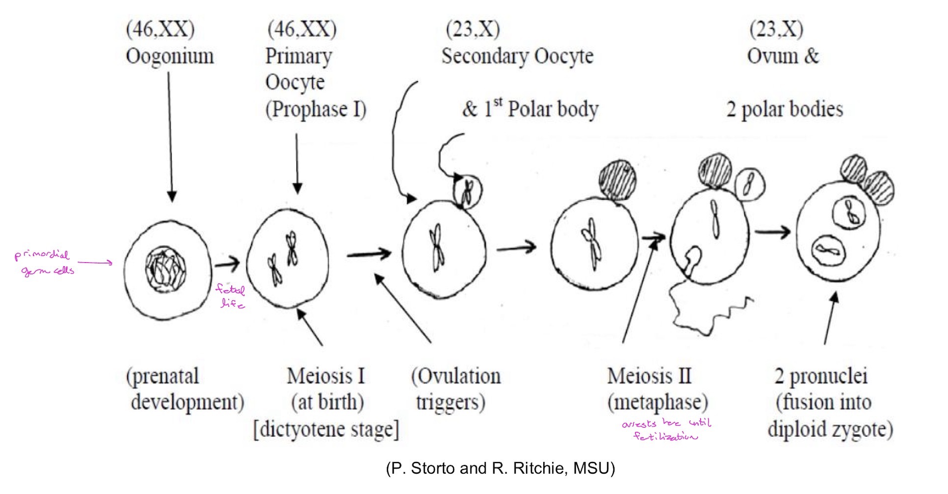
In female gametogenesis (egg formation), there is a special step of diplotene, called dictyotene, at which meiosis I is arrested at the 9th month of gestation for 10-50 years (until ovulation) (see below).
Crossing over is a normal and probably necessary step in meiosis. Each pair of homologs needs to undergo at least one crossover event during each meiosis. Absent (achiasmata) or reduced recombination has been found in 45% of females who yielded Trisomy 21 offspring but is very rare in meiosis of females with normal offspring
e. Diakinesis: separation of the homologous chromosome pairs proceeds as the chromosomes become maximally condensed for metaphase I.

crossing over
homologous recombination (exchange of homologous regions of DNA between non-sister chromatids) occurs, resulting in chromosomes with new combination of alleles (expanding genetic diversity, which may improve the fitness of the organism)

At the end of prophase of meiosis I, a chromosome pair that has undergone one crossover event will consist of:
Two parental chromatids, one each of egg and sperm origin;
Two recombinant chromatids, each containing both egg-derived and sperm-derived alleles in a complementary fashion.

Spermatogenesis
i. Human males produce ~1000 sperms/second (~100-200 million per ejaculate; ~30 billion/year)
ii. The spermatogonia develop from the primodial germ cells by a series of ~200 mitoses.
iii. Due to the repeated replication of the chromosomes, there is an increased risk for accumulating minor chromosomal deletions or duplications over time.
iv. One example: paternal age associated with mutations in FGFR3 (fibroblast growth factor receptor 3), leading to achondroplasia genetic short stature).

Oogenesis (largely confined to prenatal development)
i. Oogonia are derived from the primodial germ cells via ~30 mitoses.
ii. Women are born with all of the primary oocytes, arrested at dictyotene of Prophase I), they will ever have (~2 million). Most of these willdegenerate; those that do not will remain in that stage for decades.
iii. At puberty, ~400,000 primary oocytes left; only ~400 of which will eventually mature.
iv. Ovulation occurs once every ~28 days each month (~1000 primary oocytes will attempt to mature; most of them will die). Females ovulate ~400 time during their lifetime.
Segregation (Mendel’s 2nd Law)
The two members of a single gene pair (alleles) are never found in the same gamete; they always segregate and pass to different gametes.

Independent assortment (Mendel’s 3rd Law)
Members of different gene pair assort to gametes independently of one another; the segregation of one pair of homologs is independent of and does not affect the segregation of other pairs of homologs. 223 (more than 8 million!) combinations are generated by independent assortment of the 23 chromosomes!
i.e. this is why chromosome 1 doesn’t have to line up with its respective chromosome 20

Gene expression is regulated at…
multiple levels
Transcriptional regulation
transcription initiation (main site of control)
assembly of the RNA polymerase complex (on which we will focus most of our attention and provide the most number of examples) but other events can also be controlled
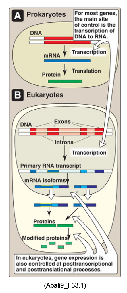
Post-transcriptional regulation (at mRNA level)
RNA elongation, processing, transport, alternative splicing, stability, efficiency of translation
Post-translational regulation (at protein level)
protein translation, processing, transport, stability, targeting
Levels of transcriptional regulation (eukaryotes)
There are two levels of transcriptional regulation:
1. at DNA level
2. at chromatin level

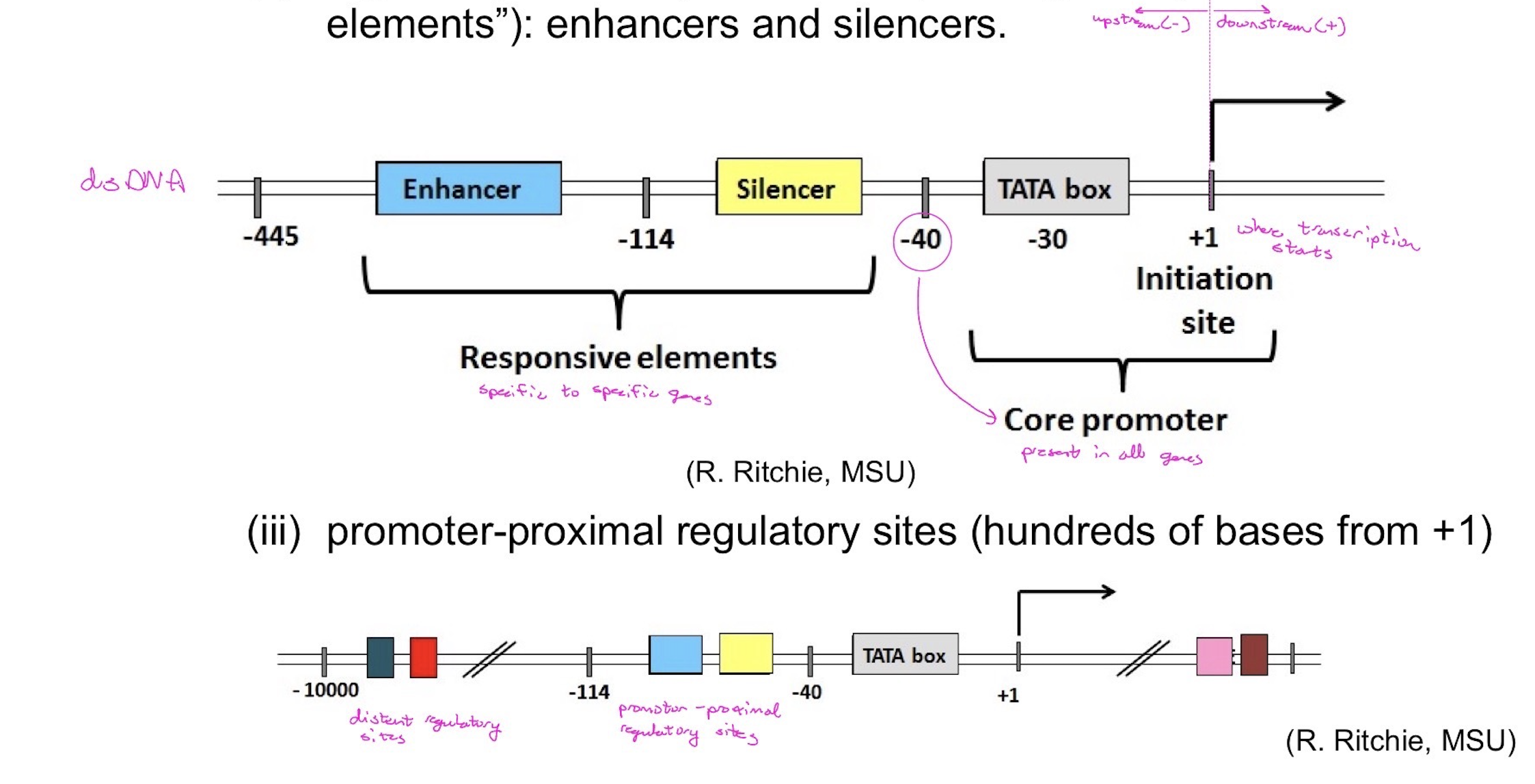
Eukaryotic promoter elements — RNA pol II
(i) core promoter: TATA box, initiator site
(ii) regulatory DNA sequences for specific genes (“responsive elements”): enhancers and silencers.
(iii) promoter-proximal regulatory sites (hundreds of bases from +1)
(iv) distant regulatory sites (thousands or tens of thousands of bases from +1)
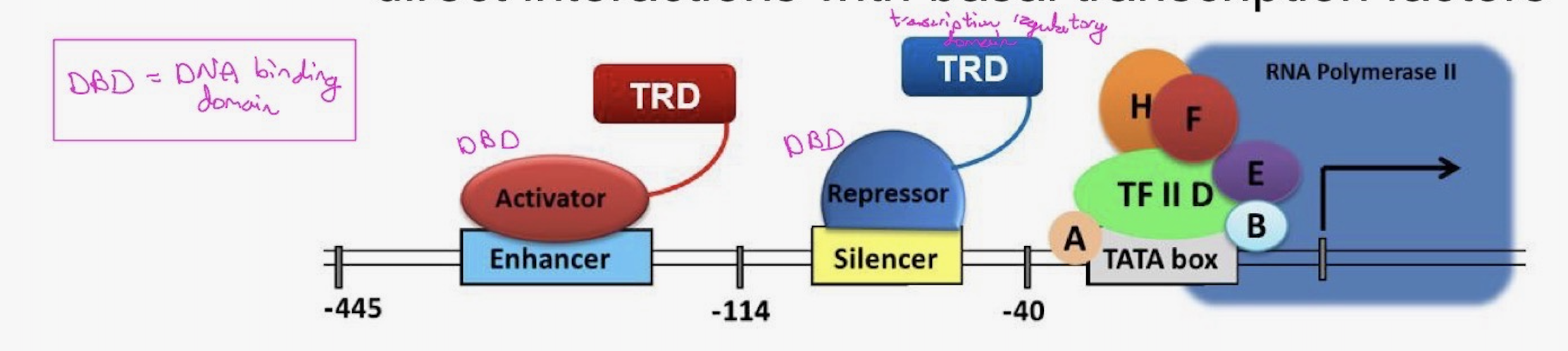
Transcriptional regulatory proteins
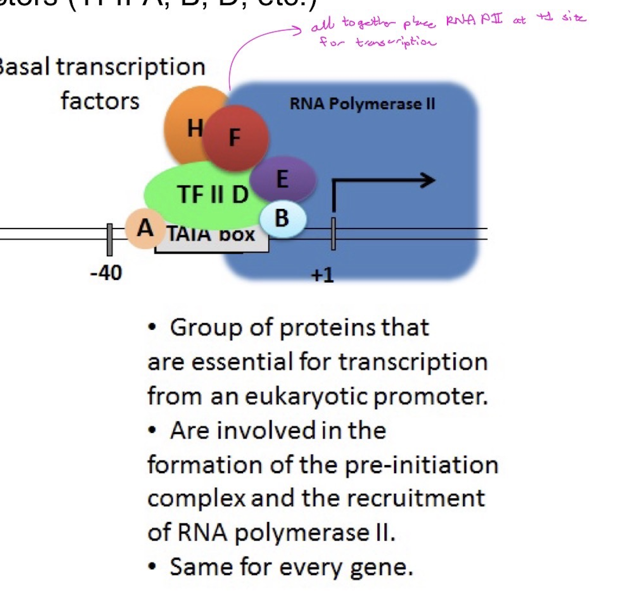
(i) basal (general) transcription factors: TFII-A, TFII-B, TFII-D, etc
(ii) specific transcriptional factors - OVER and ABOVE the general (basal) transcription factors (TFII-A, B, D, etc.)
(iii) Transcription factors bind to promoter elements
basal transcription factors
same for every gene
essential for transcription
involved in formation of pre-initiation site

specific transcription factors
specific for certain genes
binds to DNA at specific sites (i.e. enhancers/silencers)
called activators and deactivators
Transcription factors bind to promoter elements

Regulatory proteins often have…
two distinct domains
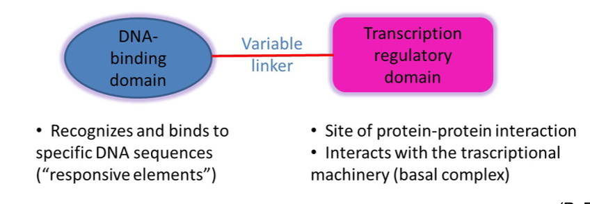
What might TRD do?
direct interactions with basal transcription factors
recruit co-activators that modify chromatin
covalent modification of histone proteins (acetylation; methylation; phosphorylation, etc.)
ATP Dependent Remodeling Enzymes (co-activators that modify chromatin)
ATP-dependent “remodeling” enzymes that induce “sliding” of nucleosomes along DNA, increasing the gaps for RNA pol II, TFII-D, etc. to gain access to DNA
activator recruits protein —> protein then slides nucleosome off DNA —> opens up space in DNA for GTF

What “turns on” transcriptional activators?
steroid hormone receptors

Steroid Hormone Receptors
Steroid hormones are hydrophobic and can cross the plasma membrane to bind to specific receptors in cytosol.
The receptor has:
hormone or ligand-binding domain;
DNA-binding domain (DBD); and
transcription regulatory domain (TRD).
Upon binding to the steroid hormone, the hormone-receptor complex translocates into the nucleus. Using its DBD, the hormone-receptor complex binds to hormone-responsive elements on the DNA. Via its TRD, the hormone-receptor complex recruits the machinery to activate hormone-responsive genes.
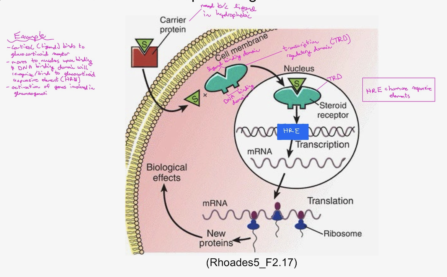
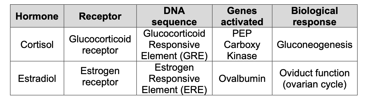
Examples of the receptor as a transcription factor pathway of signal transduction + clinical relevance
Clinical Relevance: Tamoxifen is an antagonist (competitive inhibitor) that targets the estrogen receptor; it is used against breast cancer because some breast tumors rely on estrogen-mediated pathways for proliferation.

Sterol Responsive Element-binding Protein (SREBP)
transmembrane protein
in ER —> held in membrane by cholesterol
The signaling pathways below show similarities to the receptor as a transcription factor pathway of signal transduction (e.g. use similar elements):
(ii) cholesterol metabolism: SREBP
Sterol Responsive Element-binding Protein (SREBP) is a trans-membrane protein at the Endoplasmic reticulum (ER) membrane. Low cholesterol concentration triggers SREBP precursor movement to the Golgi where it signals two proteases, which cleaves the cytoplasmic domain, releasing it to be translocated into the nucleus.
This cytoplasmic domain has:
DBD: binds to sterol regulatory element
TRD: activates genes in cholesterol metabolism
(iii) NF-κB
Nuclear Factor (κ-chain) is anchored in the cytoplasm by Iκ-B. Bacterial lipopolysaccharide (LPS) or viral infection (induction of a cytokine such as interleukin 1β), results in ubiquitin-tagging and degradation of Iκ-B. This releases NF-κB to be translocated into the nucleus and activate genes for inflammatory response.

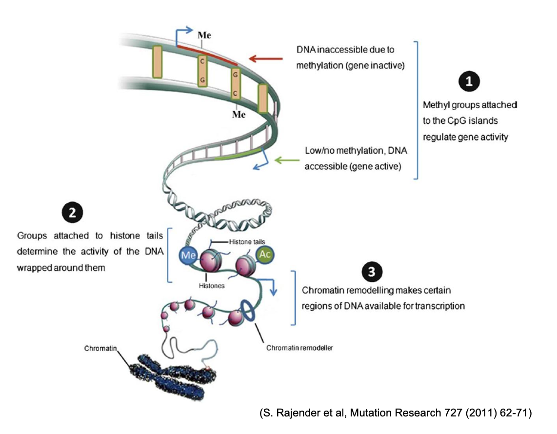
Transcriptional Regulation at the Chromatin Level: Epigenetic Modifications
chemical modifications to the chromatin that regulate gene expression, without changing the primary DNA sequence, and are heritable. Includes DNA methylation and covalent histone modification.
DNA methylation
methylation = gene repression
What gets methylated?
DNA methylation, in the context of gene regulation, is the covalent attachment of a methyl group to the C5 position of the cytosine pyrimidine ring of a CpG dinucleotide in the DNA. CpG dinucleotides consist of a cytosine bound to a guanine by a phosphodiester bond, rather than CG base-pairing.
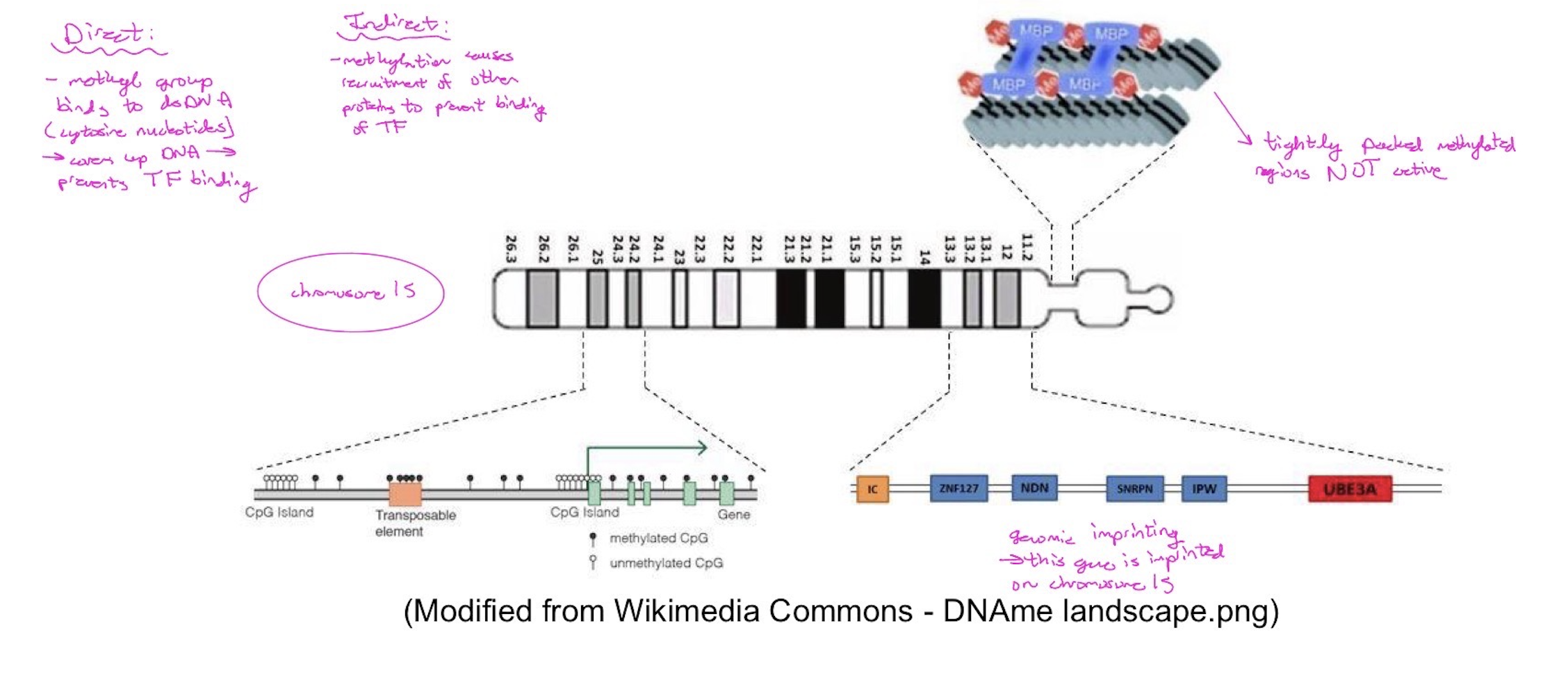
DNA methylation is important for normal embryonic development and for normal functioning of adult organism. DNA methylation covers most of the genome, but it is found predominantly in repetitive regions of the genome, such as satellite DNA and transposable elements [including long interspersed elements (LINES) and short interspersed elements (SINES)]. DNA methylation is also found, to a lesser extent, at imprinted genes.
Heterochromatin (not expressed) is heavily methylated, resulting in tight packing.
Euchromatin (expressed) contains much less methylation and is more loosely packed.
![<p>methylation = gene repression </p><p>What gets methylated?</p><p>DNA methylation, in the context of gene regulation, is the covalent attachment of a methyl group to the C5 position of the cytosine pyrimidine ring of a CpG dinucleotide in the DNA. CpG dinucleotides consist of a cytosine bound to a guanine by a phosphodiester bond, rather than CG base-pairing.</p><p>DNA methylation is important for normal embryonic development and for normal functioning of adult organism. DNA methylation covers most of the genome, but it is found predominantly in repetitive regions of the genome, such as satellite DNA and transposable elements [including long interspersed elements (LINES) and short interspersed elements (SINES)]. DNA methylation is also found, to a lesser extent, at imprinted genes.</p><ul><li><p>Heterochromatin (not expressed) is heavily methylated, resulting in tight packing.</p></li><li><p>Euchromatin (expressed) contains much less methylation and is more loosely packed.</p></li></ul><p></p>](https://assets.knowt.com/user-attachments/4b86c288-6a1b-4ac6-a99b-9d4ecf8ebcea.png)
Functions of DNA methylation
(1) Methylation that happens on ALL chromosomes for regulation of gene expression:
• Transcriptional gene silencing
Directly by inhibiting binding of specific transcription factors
Indirectly by recruiting proteins with associated repressive chromatin remodeling activities
• Regulation of chromatin structure --- euchromatin (transcriptionally active – less methylated) vs heterochromatin (transcriptionally inactive – heavily methylated)
Genome stability --- suppression of repetitive elements
silencing of repetitive and centromeric DNA
suppression of homologous recombination between repeats —> helps prevent recombination of non homologous chromosomes (we don’t want translocations)
transposon silencing —> jumping genes can move around the genome and cause issues
(2) Methylation that happens only on selected chromosomes (e.g. chromosome 15) for genomic imprinting:
Genomic imprinting
(3) Methylation that happens only on the X-chromosomes of females (inactivation of the X-chromosome to maintain gene dosage):
X-chromosome inactivation (females)
DNA methylation: clinical relevance
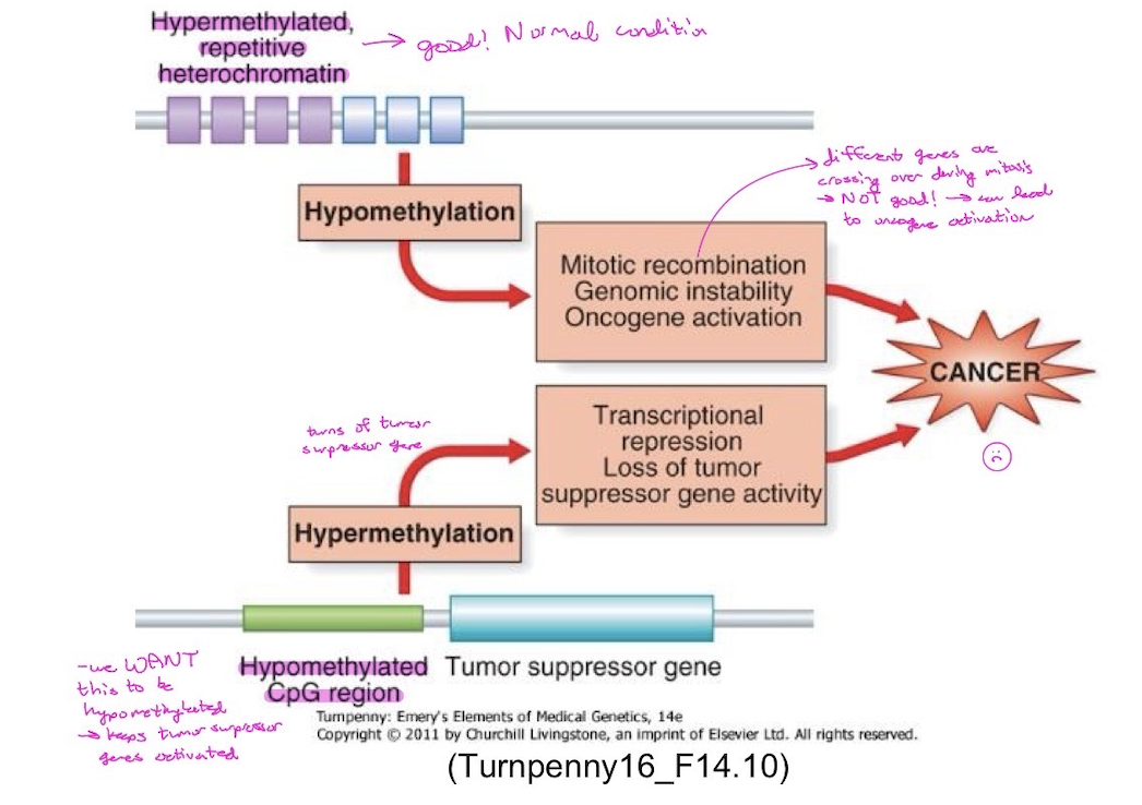
Epigenetic alterations contribute to cancer development.
• Hypomethylation
Genomic instability
Loss of imprinting (LOI)
Activation of oncogenes
Reactivation of transposons
• Hypermethylation
CpG islands may be aberrantly methylated in cancer cells leading to gene silencing (usually of a tumor suppressor gene).
donor of methyl group
S-adenosyl-methionine (SAM)

DNA methyltransferases (DNMT)
class of enzymes that carry out the DNA methylation reaction. DNA methyltransferases transfer methyl groups from SAM to the cytosine in the CpG dinucleotides, and they establish and maintain DNA methylation patterns in the genome.
DNMT1: this enzyme is responsible for maintenance of DNA methylation patterns. After DNA replication, DNMT1 adds methyl groups to the newly synthesized DNA strand based on the presence of methylation in the CpG dinucleotide in the complementary template DNA strand. —> so making sure methyl groups on parent strand get added to new strand
DNMT3A and DNMT3B: these enzymes catalyze the de novo DNA methylation. De novo DNA methylation patterns are established early in embryogenesis, around the time of implantation.
DNMTs: clinical relevance
DNMT3A is mutated in about 20% of acute myeloid leukemia (AML) cases.
Mutations in DNMT3B → Immunodeficiency, Centromeric region instability, and Facial anomalies syndrome (ICF) → DNA hypomethylation and abnormalities localized mostly to the centromere-adjacent heterochromatin of chromosomes 1 and 16 in mitogen-stimulated lymphocytes.
Genomic imprinting
instead of both copies of genes being expressed —> only one is
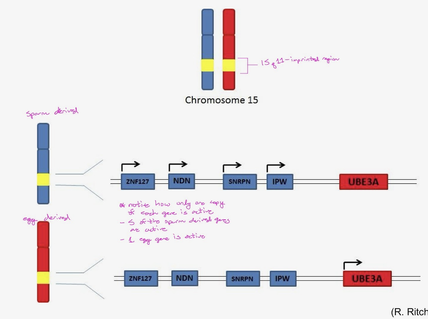
although you inherit duplicate copies of your genes, the egg-derived copy of some genes (or the sperm-derived copy of other genes) may be required/utilized in unique manners during specific stages of development. Imprinting refers to modification of a gene (e.g. methylation pattern) as it is transmitted through the sperm or the egg.
Normally, for imprinted genes, only one copy of the gene is active. Improper imprinting can result in two active copies or two inactive copies, which can lead to abnormal developmental processes.
The regions of the genome in which imprinting happen are known as differentially methylated regions (DMRs). Imprinting control regions (ICRs) are present within DMRs and function to control gene expression across imprinted domains.
Epigenetic reprograming
process through which most genomic DNA patterns are erased (usually by demethylation) and reestablished. There are two epigenetic reprogramming events during embryonic development, one that occurs during pre- implantation development and affect all cells, and a second one that will only occur on primordial germ cell (embryonic cell that will form germ cells).
Epigenetic reprogramming in the zygote
most DNA methylation is removed or erased following fertilization during pre-implantation development, when extensive demethylation of the genome takes place (does NOT affect genomic imprints). This process functions to reestablish totipotency.
so we want a cell to be able to differentiate into any kind of cell
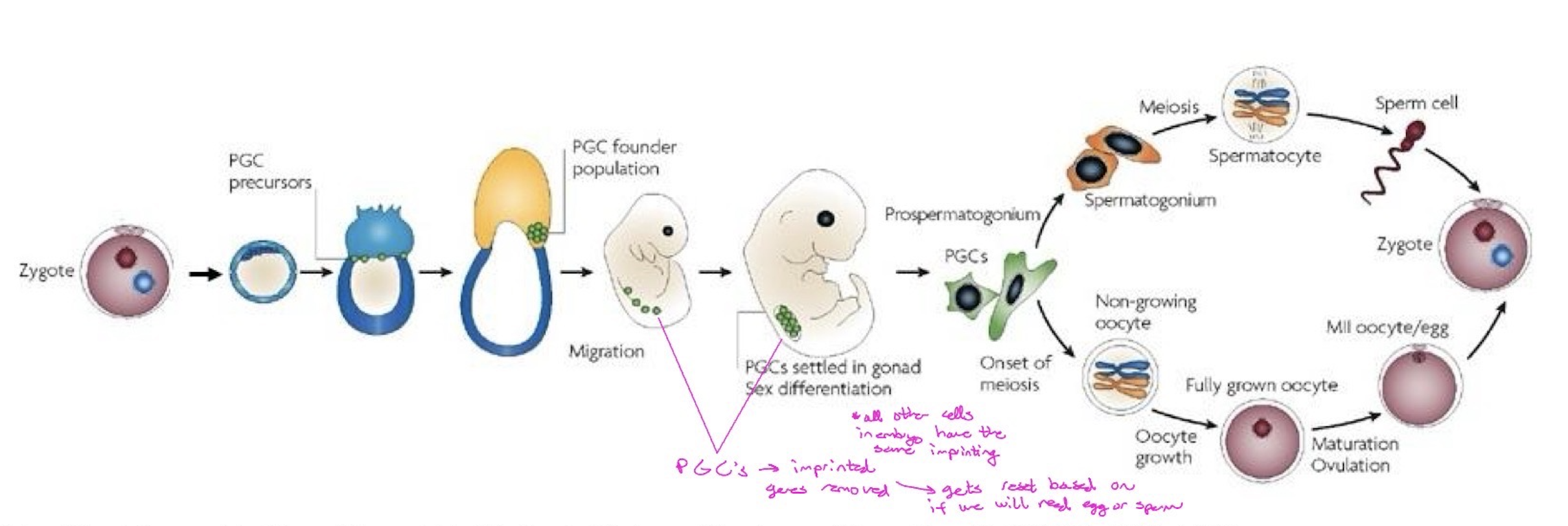
Epigenetic reprogramming in primordial germ cells (PGCs)
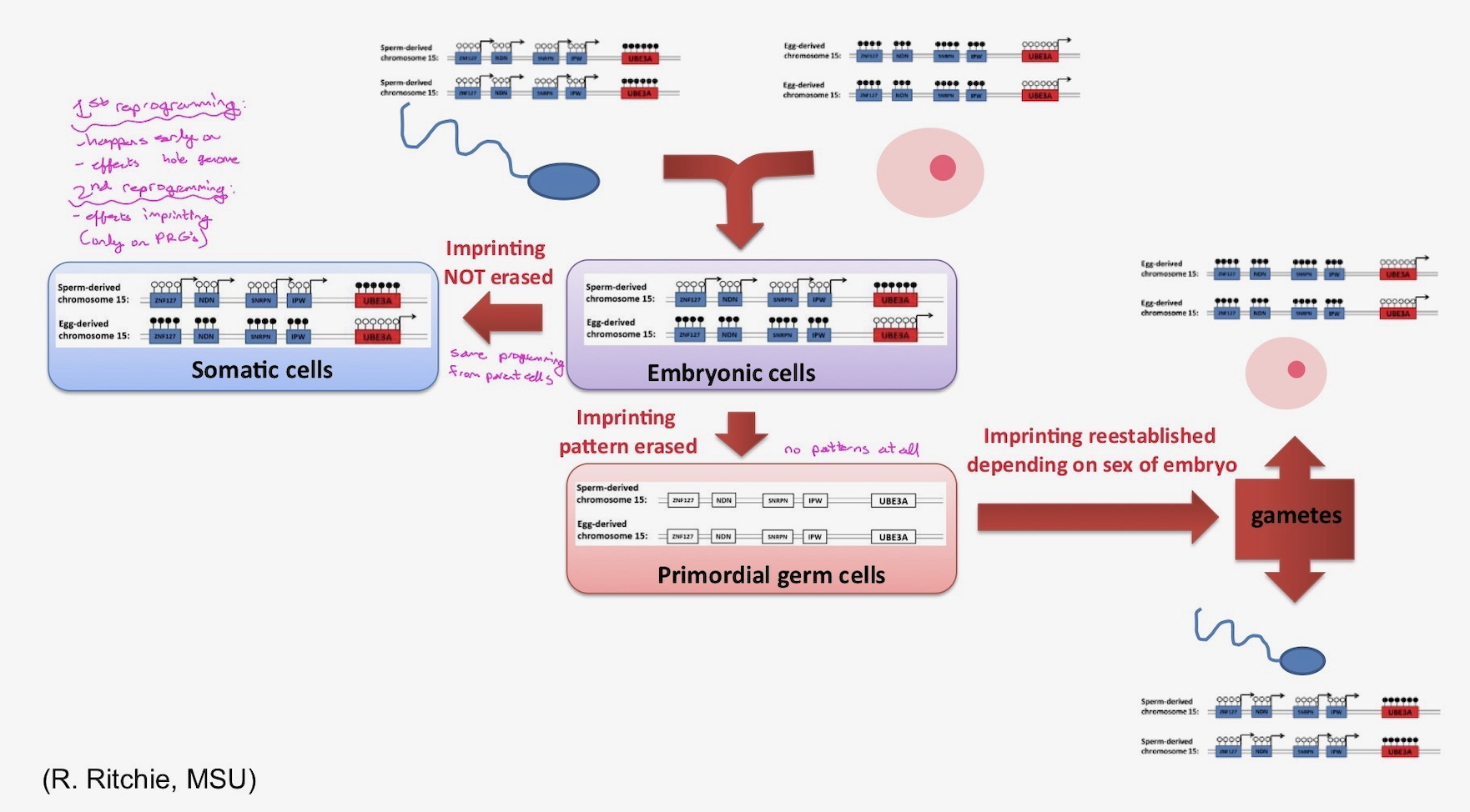
happens during post-implantation development and erases the methylation marks of imprinted genes in the primordial germ cell genomes. This erasure is important to make sure that the epigenetic marks in the primordial germ cells can be reset and reflect the sex of the developing embryo.
Remember that germ cells need to be imprinted with proper methylation patterns according to the sex of the developing embryo.
Somatic cells do not undergo this second epigenetic reprogramming. During post-implantation development imprinted marks must be maintained in somatic cells because correct expression of the imprinted genes is essential for normal somatic development.
Note: The figure below is pertaining to imprinted genes only and it illustrates how the imprinting patterns are erased during the epigenetic reprogramming in primordial germ cells and reset depending on the sex of the embryo. It also illustrates how the imprinting patterns are not erased in somatic cells since they do not undergo this second epigenetic reprograming.
Imprinting: clinical relevance
Prader-Willi syndrome and Angelman syndrome are two different syndromes, which are both linked to the same imprinted region of chromosome 15. In this imprinted region some of the genes are silenced in the egg-derived chromosome and some are silenced in the sperm-derived chromosome. Therefore, a defect on chromosome 15 will lead to loss of different gene activities, depending on whether the chromosome came from the egg or the sperm.
In Prader-Willi syndrome, gene activity that normally comes from the sperm is missing.
On the other hand, on Angelman syndrome gene activity that normally comes from the egg is missing.
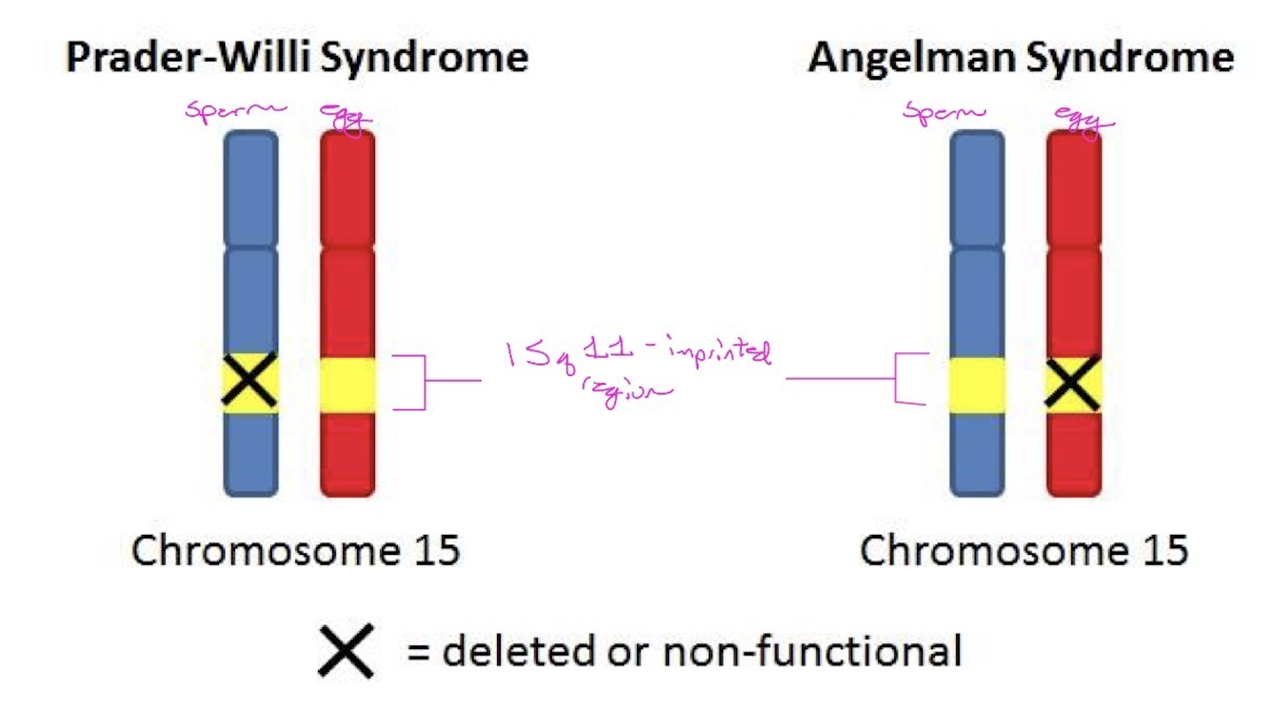

If you look at a female’s chromosome 15 what would be the status of the following genes in her sperm-derived chromosome 15 in her somatic cells? How about in the female’s egg cells?

Covalent histone modification
Histones are globular proteins with an N-terminal tail. Amino acid residues in the histone N-terminal tails can be modified in a variety of ways, which results in changes in the chromatin structure. N-terminal tails of histones can be post-translationally modified (see below) which will change chromatin structure.
Functions of covalent histone modification
• Establishment of chromatin structure (usually by affecting the interaction of histones with DNA or by affecting the contact between different histones in adjacent nucleosomes)
• Regulating the binding of non-histone proteins (which may carry enzymatic activities, such as remodeling ATPases that further modify chromatin).
modified histones can function as a binding site
histone modification can function to disrupt an interaction between a histone and a binding factor.

Post-translational modification of the core histone N-terminal tails include:
acetylation, methylation, phosphorylation, ubiquitylation and sumoylation
Covalent histone modification - Acetylation:
• Residue: addition of acetyl groups to lysine residues (K-ac) in H3 and H4
• Enzyme: histone acetyl transferases (HATs)
• Functions:
transcriptional activation
Opens up chromatin by reducing positive charges of histones, therefore weakening interaction with PO4 on
DNA backbone and reducing the packing of nucleosomes
Function as a binding site for ATP-dependent “remodeling” enzyme, which induces sliding of nucleosomes to increase gaps for the basic transcriptional machinery (RNA pol II, TFII-D, etc) to gain access to DNA sequence
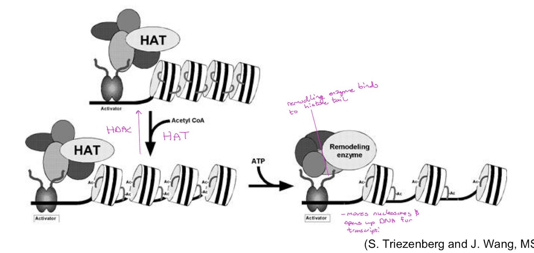
Histone deacetylases (HDACs)
Histone deacetylases (HDACs) remove acetyl groups from histone tails causing chromatin to be more compact and leading to transcriptional repression.
Thus, DNA methylation, along with histone modifications, play a role in packing and unpacking of chromatin → regulation of gene expression