IEM Boards Conditions (All)
 Call Kai
 Learn
Learn Practice Test
 Spaced Repetition
 Match
Match Flashcards
 Knowt Play
1/28

There's no tags or description
Looks like no tags are added yet.
Name | Mastery | Learn | Test | Matching | Spaced | Call with Kai | Chat |
|---|
No analytics yet
Send a link to your students to track their progress
29 Terms
Amino Acid Homeostasis
There has to be a balance of our intake (sources) and output (products) of A.As.
Sources
Dietary protein: intake via intestine provides essential and non essential A. As
Tissue Catabolism: periods of fasting or metabolic stress trigger breakdown of tissue; largest protein repository is muscle
Synthesis: metabolic pathways for non-essential AAs
Products
Anabolism: incorporation into complex proteins
Conversion: into Non-essential or metabolic intermediates (Keto or gluccogenesis)
Excretion: in urine through conversion of nitrogenous waste to urea
5 Major Aminoacidopathies
When there are defects in the enzymes of the metabolism of Amino Acids, they result in a group of disorders call Aminoacidopathies: (the most common and well understood of the group)
Phenylketonuria (PKU)
Tyrosinemia
Maples Syrup Urine Disease (MSUD)
Homocystinuria
Non-Ketonic Hyperglycinemia (NKH) —> not needed for boards
Phenylketonuria (PKU)
Found in individuals with Phenylpyruivic in their urine; had mental retardation
Inheritance:
Autosomal recessive
Incidence: 1/10-25,000 (Higher in turkey, Ireland, Japan, Africa)
Genetic Basis: Always a mutation in Phenylalanine Hydroxylase (PAH) Deficiency
Most PKU are compound heterozygotes with >500 mutant alleles (no one allele has overwhelming frequency)
Metoblic Pathway of PKU
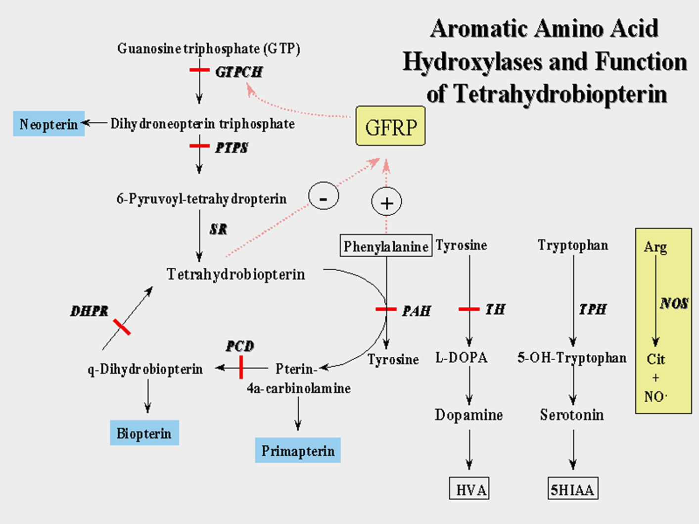
Conversion of Phenylalanine to Tyrosine via PAH enzyme
we get all out Phenylalanine from Diet: we do no syntheses ,it is an ESSENTIAL AMINO ACID
PAH has a cofactor: Tetrahydrobiopterin, necessary for peak PAH activity
Insufficient tetrahydrobiopterin (THBP) can also cause accumulation of Phenylalanine as well as other intermediates in the THBP synthesis (and also the recycling) pathway
A DEfeicny of TYROSINE
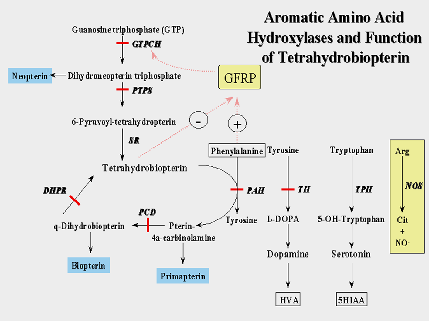
Tetrahydrobiopterin (THBP)
A cofactor that is important in maximizing the activity of some Amino Acid enzymes
Phenylalanine —> Tyrosine (affecting the PAH enzyme)
Tyrosine —> L-DOPA {—> Dopamine} (affecting the Tyrosine Hydroxylase enzyme)
Tryptophan —> 4-OH-Tyrpothan {—> Serotine} (Affecting the Tryptophan Hydroxylase enzyme)
Arginine —> Nitric Oxide (Affecting Nitric Oxide Synthase)
A defiecny of THBP = A defeicny in all these pathways (Overlap with PKU for high Phenylalaine, but also low Dopamine, Seratoin and Nitoric Oxid)
Untreated Classic PKU
At Birth: asymptomatic; moms enzyme activity avoids accumulation of phenylalanine in the fetus
Neonatal period: take-in of phenylamine via diet; accumulation begins; demonstrate symptoms
No fatal neonatal encephalopathy (unlike other metabolic disorders)
Fair skin and hair pigmentation: PHE inhibition of tyrosine hydroxylase = Melanin deficiency
Urine w musty order: Phenylpyruvic acid in urine
Infancy onward: intellectual disability and delays manifest
irreversible ID: Severe in 50%, moderate-mild in 50%
Autism, microcephaly, behavior issue
Large % with epilepsy, eczema
Diagnosing PKU
Newborn Screening: Tandem Mass Spectrometry on heel-prick newborn blood sample, give PHE levels
Normal PHE: <120 umol/L (<6 mg/dL)
Classic KU >1,200 umol/L (>20 mg/dL)
Hyperpheylalaniemia 120-1200 umol/L (2-20 mgdL)
False negative (<1%): sample collection to early, mishandling or lab error
Fals positive (frequent): liver disease/immaturity (PAH active mainly in liver), high protein load, sample collection or lab error
Confirmatory Biochemical testing:
Plasma amino acid analysis: we should see Elevated PHE AND elevated PHE/TYR ratio (due to the PHE→TYR BLOCK)
(RULE OUT LIVER DIEASE OVERLAP: WE WOULD SEE PHE AND OTHER AAs)
THBP testing
Genotyping
PAH gene sequencing: genotype / phenotype correlation incomplete but can predict some treatment response
Treating PKU
Anyone with PHE >360-600 umol/L ( >6-10 mg/dL)
Dietary PHE restriction: Limit dietary protein intake to keep PHE <360umol/L (,6mg/dL) - Classic PKU <1500-250 mg/day PHE intake (not all Hyperpheylalaniemia pts need dietary PHE restriction)
Dietary PHE-free protein formulas: PHE-free protein formulas to avoid amino acid deficiency (TYR)
Outcome with treatment if started by 3 weeks old)
Normal IQ
Increased risk for subtle Neurocognitive deficits and signs
Effect of treatment withdrawal (why it is LIFELONG TX)
increased ADD/ADHA, depression, anxiety, agoraphobia
Progressive demyelination on MRI
Decreased IQ scores (5-30 points)
Maternal PKU Embryopathy
Elevated maternal PHE highly teratogenic (Females with PKU need even MORE dietary control)
PHE level in developing fetus is 2x maternal level: (75-90% teratogenicity vs 50% with thalidomide)
Goal: Maternal PHE levels <360 umol/l (<6 mg/dL)
Higher PHE levels correlate with risk for
Microcephaly, Mental + Growth retardation, Congenital heart defects
Offer prenatal ultrasound and echocardiography to look for these issues
Newer PKU Treatments
Sapropterin Powder
Phenylalanine Ammonium Lyase SQ injection
Large Neutral Amino Acids Tablets/GMP
PKU Carrier Detection
If mutations are detected in parents
Molecular testing of PAH gene in affected individual can facilatate carrier testing: if no mutation identifiable linkage analysis (dup/del anayslsi needed)
Enzyme analysis not practical as PAH is essentially only exressed in liver (need a liver biopsy which is NOT typically done)
Prenatal implantation diagnosis: Available when molecular testing informative
Tyrosinemia Type I
Also called “Hepatorenal” Tyrosinemia due to progressive liver and kidney failure if untreated : type II and III dont present the same way
Inheritance: Autosomal Recessive
Incidence: 1/120,000 (higher in Quebeec)
Genetic defect in: Fumarylacetoacetate Hydrolase (FAH) Deficney
Tyrosinemia Type I Metabolic Pathway
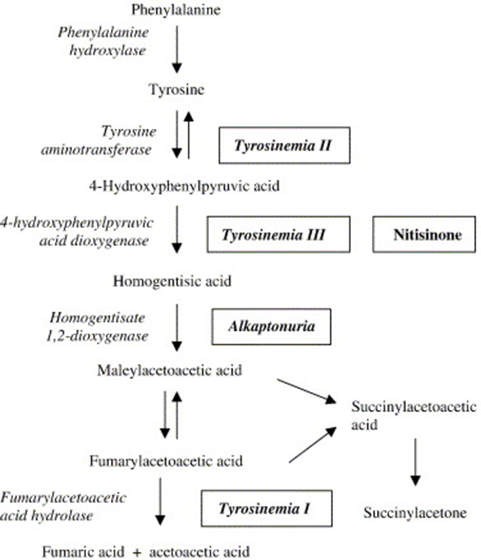
Phenylalanine → Tyrosine via Phenylalanine Hydroxylase (PAH, PKU remember?)
Tyrosine → Fumaric Acid + Acetoacetic Acid via 5 step reaction
Step 1 Enzyme Block: Tyrosinemia II (Eye irritation, skin changes
Step 2 Enzyme Block: Tyrosinemia III (cognitive disability in some but not all)
Step 3 Enzyme Bock: Alkaptonuria (Black pee disorder first IBEM identified)
Step 4: Fumarylacetoacetic Acid Hydrolase (FAH) Block : Tyrosinemia I
High levels of Fumarylacetoaecetic acid accumalte -→ conversion to Succinylacetosectic acid which would normally not accumulate: both Cause TOXICITY AT HIGH LEVELS
Untreated Tyrosinemia I
At birth: Asymptomatic
Infancy: Tyrosine, Fumarylacetoaecetic acid (FAA), Succinylacetone (SAT) accumulate
Cabbage-like odor in urine
Liver dysfunction progression (from FAA)
Renal dysfunction progressing to failure
Rickets, poor growth
Neurologic “crisies”; episodic abdominal +neuropathic pain, weakness, repository failure (from SAT inhibition of porphyrin pathways)
Adolescence: Death typically by age 10 due to
liver failure,
respiratory fialure in neurologic crisis,
hepatocellular cancer
Diagnosing Tyrosinemia I
Newborn Screening: on heel-prick blood sample
Elevated Tyrosine and SAT on tandem Mass spect.
some false negative: may take time for sufficient elevations to elevations
Often false positive: TYR elevation also with liver disease, transient benign or the other forms of tyrosinemia
Confirmatory Biochemical Testing: Plasma amino acid profile
High TYR in plasma A.As: non-specific to Type I
High SAT in blood or urine organic acids: Specific to Type I
Rarely skin fibroblast FAH enzyme activity: PBG in blood (Quebec)
Genotyping
FAH gene sequencing with del/dup analysis (>95%): cancer be targeted in some populations (French Canadians)
Genotype: Phenotype correlation no consistent
Treating Tyrosinemia I
Treatment
Dietary restriction of PHE and TYR
Dietary supplementation: PHE and TYR-free protein formulas
Medications : Orfadine - inhibits enzyme prior to FAH (p-HPPD) reducing FAA and SAT
Liver Transplantation: reserved for liver failure or hepatocellular carcinoma despite treatment
Outcome with treatment:
with NBS: 90% survival into adulthood with normal growth and function
Screening for Tyrosinemia I
Carrier Screening
Moelcualr tesing for FAH gene if mutation idefineted
Linkage analysis if mutation unidentifiable
Prenatal/Preimplantation diaognsis
availbe if moclaur testing infromative
if molecualar testing uninformative: SAT acumination can be Measured in the Amniotic fluid when molecular basis in family unknown but not 100% accurate
Branch Chain Keto-Acid Dehydrogenase Deficiency (Maple Syrup Urine Disease)
Urine smells like Maple Syrup / Sweet
Inheritance Recessive
Incidence 1:185,000 (increased in Mennonite pop.)
Genetic Basis
Mutations in genes coding Branch Chain Keto-Acid Dehydrogenase complex subunits: multiple subunits, multiple genes that can cause MSUD)
4 subunits in BCKD complex: E1a, E1b, E2, E3
3 MSUD genes:
BCKDHA (E1a)
BCKDHB (E1b)
DBT (E2)
***DLD (E3) mutations cause different disease***
Branch Chain Amino Acid (BCAA) Metabolism
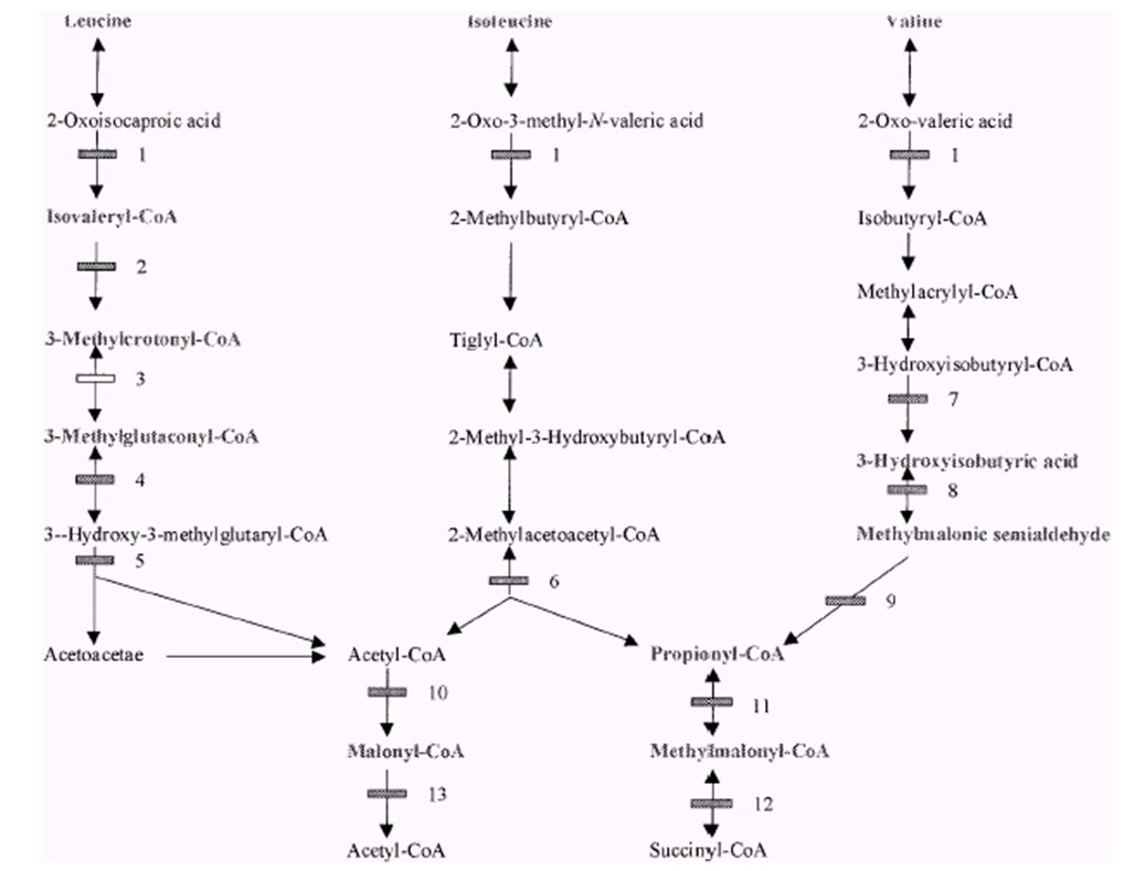
The Branch Chain Amino Acids: Leucine, Isoleucine, Valine
These three are converted via multiple steps to energy intermediates Acctly-CoA and Succinyl-CoA
First Step is via BCAA: When blocked, accumulation of the three branch chain amino acids + their associated keto-acid forms (which are what BCAA actually works with)
Untreated Classic MSUD
At Birth Asymptomatic
Within 12-24 hrs after birth: Maple syrup urine and ear wax (from Isoleucine)
Dietary intake OR Catabolism in the newborn
Within 5 Days
progressive irritability and decreased feeding
progressive encephalopathy/cerebral edema (from leucine: neurotoxic)
Within 7-10 days after birth
massive cerebral edema, coma respiratory failure
death within weeks
Some milder “intermediate” or “intermittent” forms
may only experience “decompensation” during catabolic stress
MSUD unlike PKU and Tyrosaenmia I: No threat of neo-natal death + symptom onset longer vs. rapid onset and death in MSUD
Diagnosing MSUD
Newborn Screening: Heel prick blood tandem Mass spec. analysis
Elevated Leucine and BCAA ratios
rarely false negative: early sample collection (before 24hrs), collection or lab error
Occasional false positives: other disorders appear positive via elevated LEU (hydroxyprolinemia)
Confirmatory Biochemical Testing
Plasma AA analysis:
Elevated BCAA’s; Leucine, Isoleucine, Valine
Allo-Isoleucine presence (only elevated in MSUD)
Urine organic acid analysis
BCAAs present
Enzyme activity: BCKD enzyme activity measurable n fibroblasts
Genotyping
BCKD subunit gene sequencing with del/dup analysis: E3 subunit mutations cause distinct syndrome due to its activity in Mutiple metabolic pathways: genotype/phenotype correlation not consistent
If successful, allows for prenatal/preimplantation genetic diagnosis
Treating MSUD
Preventing Acute decompensation (rapid buildup of BCAAs)
Decrease catabolism and enhance elimination of Leucine
Maximize BCAA-free calories (avoid catabolism)
Intravenous infusion/metabolic formula (Dextrose, lipid and BCCA-free nutrient)
Supplemental Isoleucine and Valine solutions (avoid deficiency with prolonged treatment - just the precise amount they need, no more)
Thiamine supplementation (BCKD coenzyme, increases activity)
Hemodialysis in acute severe illness
Long-term Management
Avoid decompensation: fever, fasting, high protein
Optimize BCAA-levels: metabolic formula and sing AAs
Thiamin: if thiamine responsive
Liver transplant for severe poorly responsive forms
Outcome
Normal (20 points lower): BUT ONLY IN THE FIRST 10 years of LIFE
After 10days: Severe, irreversible brain damage and/or death
MSUD Carrier Detection
Carrier Screening: can be done if BCKD genes have been found in affected individuals
linkage analysis if mutations unidentifiable
Prenatal/Preimplantation diagnosis
Available when molecular testing informative
Can obtain BCKD enzyme activity on amniocentesis or CVS
Homocystinuria
Deficiency of Cystathionine Beta-Synthase (CBS)
Inheritance: Autosomal Recessive
Incidence 1:3350,000 (higher in Qatar, Norway)
Genetic basis: Mutation in Cystathionine Beta-Synthase gene
Onset typically after 3 years old
No neonatal encephalopathy
Many cased missed on NBS
Homocystinuria Metabolic Pathway
Methionine (taken in via diet) → Homocysteine via multiple steps
Homocysteine → Cysteine via Cystathionine Beta-Synthase (CBS) + B6 (coenzyme)
Block of CBS = Elevated Homocysteine and decreased Cysteine levels
Homocysteine converted back to Methionine (Methionine synthase + B12 coenzyme)
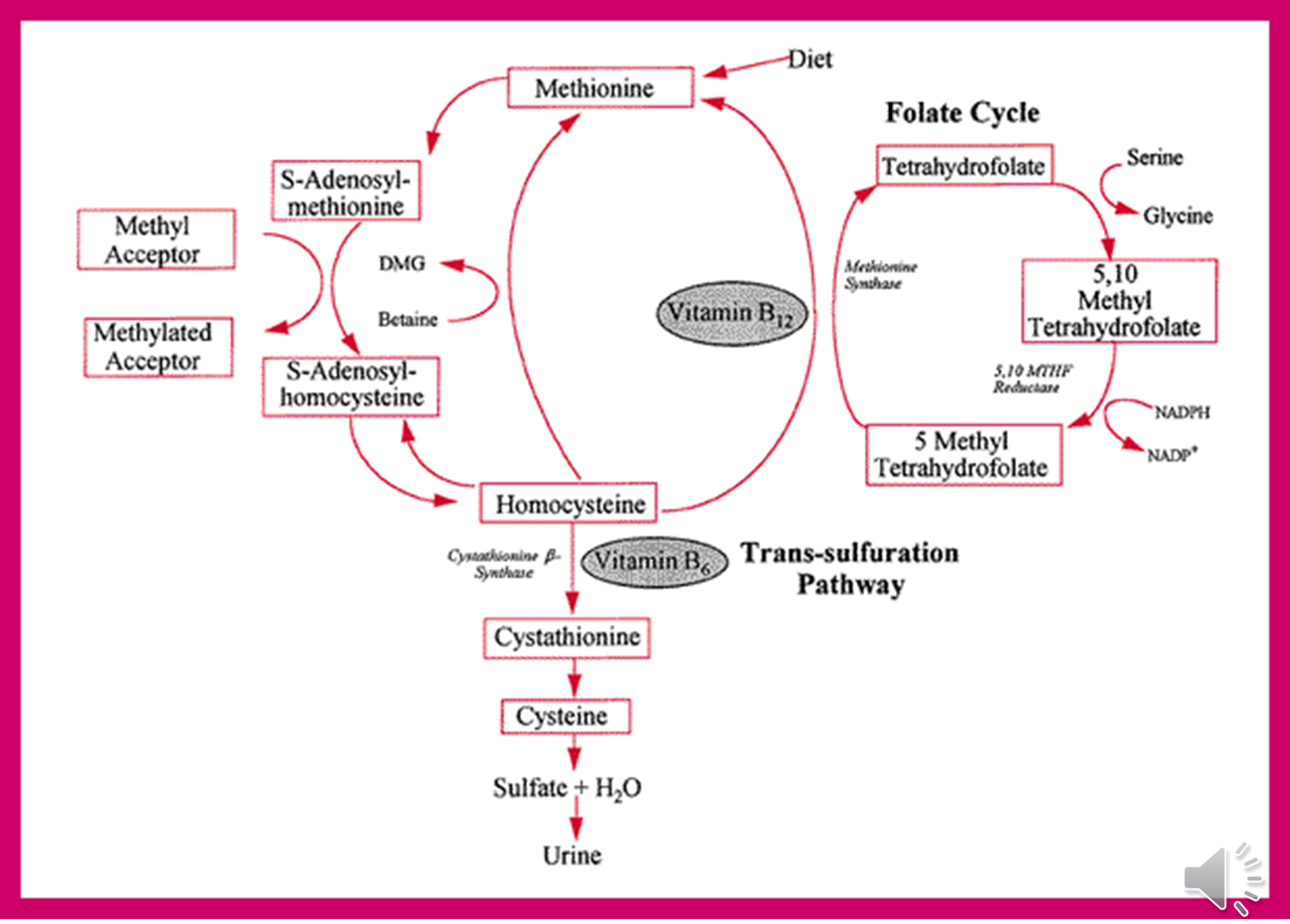
Untreated Homocystinuria
Marfanoid Skeletal changes
Tall and thin, long arms legs fingers
Chest pectus
High-arched palate
Crowded teeth
Ectopia Lentis (dislocation of lens)
Can be first clinical manifestation (typically 3-5 years): different displacement DOWN (vs UP in Marfan’s)
Neuropsycatric Disease
Spontaneous Veinous/Arterial Thromboembolism
Up to 25% by age 15: casue stroke, death pulmonary embolism
Diagnosing Homocystinuria
Newborn screening: via tandem Mass Spect.
Elevated Methionine levels
false negative: accumulation of MET not high in the first few days
false positives: multiple conditions cause elevated MET: liver disease, hypermethioninemia (MET elevated, but not homocysteine)
Biochemical Confirmation
Plasma AA analysis
elevated homocysteine + MET
low/absent Cysteine
Urine: elevated homocysteine (NOT AS SPECIFIC as plasma AA)
CBS enzyme activity: Skin fibroblasts and hepatocytes
Genetic Confirmation
CBS gene sequencing and dup/del analysis
Many mutant alleles, mostly compound heterozygotes
Homocystinuria Treatment
Diet
Restrict methionine and protein
MET-free metabolic formula
Medication
Vitamin B6 (also called Pyridoxine)
BBS cofactor challenge (50% B6-responive)
Supplement folate and B12 (allow for conversion of homocysteine back to methionine)
Betaine
offers alternative route for homocysteine remethylation: a methyl group donor which lowers homocysteine levels by remethylating homocysteine to methionine
used in patients not dramatically B6-repsonive
Outcome
near normal IQ if treated early and effectively
Marfan-like complicaitons significantly reduced
Homocystinuria Carrier Detection
Carrier Testing
If mutation found in affect Individuals
Linkage analysis if mutation not found on affected individuals
Prenatal/Preimplantation Diagnosis
CBS enzyme activity assay in amniocytes (not possible on CVS)
Amniotic fluid homocysteine levels