exam 2 - integumentary & burns
 Call Kai
 Learn
Learn Practice Test
 Spaced Repetition
 Match
Match Flashcards
 Knowt Play
1/46

There's no tags or description
Looks like no tags are added yet.
Name | Mastery | Learn | Test | Matching | Spaced | Call with Kai | Chat |
|---|
No analytics yet
Send a link to your students to track their progress
47 Terms
gerontologic considerations with skin changes
skin becomes thinner and more fragile → greater risk for tears and injuries, pressure injuries, dry skin (xerosis), delayed wound healing, skin infections and cancer
life long sun exposure, poor nutrition, medications can increase damage from the sun (diuretics, antihistamines, abx)
Loss of SUBCUT tissue substances of elastin, collagen, and fat
benign skin changes for older adults
Cherry angiomas (bright red “moles”)
Diminished hair, especially on scalp and pubic area
Dyschromias (color variations):
Solar lentigo (liver spots)
Melasma (dark discoloration of the skin)
Lentigines (freckles)
Neurodermatitis (itchy spots)
Seborrheic keratoses (crusty brown “stuck-on” patches)
Spider angiomas (network of dilated capillaries radiating from a central arteriole)
Telangiectasias (red marks on skin caused by stretching of the superficial blood vessels)
Wrinkles (a small fold, ridge, or crease in the skin)
Xerosis (dryness)
Xanthelasma (yellowish waxy deposits on upper and lower eyelids)
Ichthyosis (fish scale appearance of the skin)
Senile purpura (purple or red bruises that have an irregular shape)
stage 1 pressure ulcer
intact skin with non-blanchable redness over boney prominences
key finding → redness does not turn white when pressed
tx → relive pressure, reposition frequently, maintain nutrition and hydration, reduce friction/shear, keep skin dry
stage 2 pressure ulcer
partial thickness skin loss involving the epidermis and part of the dermis
key finding → shallow open ulcer, pink/red wound bed or blister
tx → same at stage 1 plus open wound healing dressings, cleanse with nml saline, avoid harsh antiseptics
stage 3 pressure ulcer
full thickness skin loss extending into subcutaneous tissue
key finding → fat is visible; bone, tendon, and muscle are NOT visible
tx → debridement of necrotic tissue, wound cleansing, moist dressings, pain management, infection control
stage 4 pressure ulcer
full thickness tissue loss with exposed deeper structures
key finding → bone, tendon or muscle visible
tx → stage 3 care plus possible surgery, skin grafts, drainage procedures, monitor for osteomyelitis
unstageable pressure ulcer
base is covered by slough or eschar; cannot determine depth until wound bed is visible
tx → relive pressure, monitor closely, protect tissue
wound dressings - primary dressings
protect and maintain moisture (Tegaderm, DeoDerm) are applied directly to the wound to promote healing and prevent infection.
wound dressings - secondary
absorb drainage while keeping wound moist (Hydrocolloids, Alginates, Hydrogels)
wound dressings - active dressings
actively prompt faster healing (skin grafts, biologic skin substitutes)
occlusive dressing
used to cover topical medications
transparent film
used for partial thickness wounds, minimal drainage
transparent and allows wound assessment without removal of dressing (Op-site, Tegaderm)
absorptive dressings
used for heavy amounts of exudate (Exudry, ABD pads)
moisture retentive dressings
keeps wound moist
removes exudate better, some protect the skin from maceration (soggy, white, soft)
hydrogel dressings
best for dry wounds, painful wounds
adds moisture, promotes autolytic debridement, soothing, transparent (assess wound)
INFECTION CONTROL - suck up the infection
hydrocolloid dressings
waterproof with a gel-like material underneath
used for light to moderate drainage
can stay on for up to 7 days
creates a yellow foul smelling gel that is NORMAL
foam dressings
used for moderate to heavy drainage
absorbs exudate, cushions boney prominences, requires secondary dressing
alginate dressings
used for deep wounds with heavy drainage
made with seaweed, is highly absorbent, non-adherent, need secondary dressing
antimicrobial dressings
used for infected or heavily colonized wounds
used for partial or full thickness wounds
used after surgery
topical antifungal and abx
collagen dressings
used for chronic wounds needing stimulation
stimulates wound to accelerate healing
tissue engineered skin
bioabsorbable combo of collagen and suture material containing cytokines and fibroblasts
stimulate platelet activity, decrease healing time, maintains moisture, provides structure to help regeneration of cells (matrix) and supplies beneficial cytokines
herpes zoster (shingles)
s/s → pain before the rash, unilateral rash, band-like patterns following a dermatome, fluid filled vesicles, stabbing, aching pain, itching and tenderness, vesicles evenly crusted over
mgmt → antiviral medications (acyclovir, valacyclovir, famciclovir); most effective when started within 24 hr of onset, pain control, corticosteroids to reduce inflammation and shorten healing time.
teach to not scratch lesions, keep them covered, wash hands, take antivirals, report eye pain
psoriasis
s/s → red, raised plaques, silvery-white scales, dry lesions, itching, bilateral symmetry, Auspitz sign (when scales are scraped off, small pinpoint bleeding spots appear), nail changes (discoloration, pitting, crumbling, separation from nail bed)
psoriasis tx options
topical therapy → used for mild to moderate psoriasis to decrease inflammation, slow rapid skin cell growth and help remove scales. (EX corticosteroids, calcipotriene), high-potency steroids should not be used on the face long term, Tazarotene caused photosensitivity and it contraindicated in pregnancy
intralesional therapy → used for thick, stubborn plaques or nail involvement; corticosteroid is injected directly into lesions to decrease inflammation and plaque thickness
systemic therapy → used for severe wide-spread psoriasis or psoriatic arthritis; suppress immune response, slows rapids skin cell production
photochemotherapy → used for moderate to severe, slows skin cell growth and decreases inflammation; PUVA (psoralen and UVA light) can increase risk for skin CA, cataracts and premature skin aging
s/s of shock
early s/s → nml or MAP is decreased by 5-10 from baseline, HR is nml or increased, urine output is nml to decreased, skin temp. is pain and cool, pt may be anxious, RR increased in rate and depth
late s/s → SBP <90, MAP >20, HR increased and weak, urine output is below 0.5mL/kg/hr, skin it cool, mottled and moist, RR increased and shallow
compensatory signs → decrease in MAP by 10-15 below baseline, increased renin and ADH, vasoconstriction, decreased pulse pressure, increased HR, decreased pH, restless, apprehensive, increased potassium
progressive signs → a sustained decrease in MAP that is > 20 from baseline, tissue/organ hypoxia, decreased urine output (oliguria), weak and rapid pulse, decreased pH, sensorineural changes
refractory signs (irreversible) → excessive cell/organ damage, multisystem organ failure, decreased pH
calculating MAP
SBP + 2(DBP) / 3
treating shock
maintain airway, administer oxygen, intubate if needed, fluid resuscitation, with LR or NS, monitor BP and MAP (goal >65), vasopressors if fluids are not enough, monitor urine output, get a lactic
burn depths - superficial burn (1st degree)
involves only the epidermis
appears dry, red, no blistering, swelling, is painful, heals in 3-7 days, no scarring, may have blisters or peeling after 24 hours
EX sunburn
burn depths - superficial partial thickness burn (superficial 2nd degree)
involves epidermis and a small portion of the dermis
red, moist, blisters present, blanches with pressure, very painful
heals in 5-14 days, usually no scarring, hair follicles remain intact
burn depths - deep partial thickness burn (deep 2nd degree)
involves epidermis and deep dermis
red and while mottled appearance, moist or dry, broken blisters, weeping surface, severe pain, fluid filled vesicles (red, shiny, wet), can still regenerate skin cells
burn depths - full thickness burn (3rd and 4th degree)
involves entire epidermis, dermis and may extend into fat, muscle or bone
dry, leathery appearance, eschar formation, dry, waxy, white, usually painless b/c nerves are destroyed, hair follicles and sweat glands destroyed
burn phases - emergent (resuscitation phase)
from moment of injury to the end of fluid resuscitation
what’s happening → burn causes massive inflammation and increased capillary permeability. fluid leaks out of blood vessels and into tissues (3rd space) causing swelling and loss of circulating volume
body system effects:
cardiovascular → massive fluid loss from bloodstream, decreased BP, increased HR, risk for burn shock
resp. → airway swelling, smoke inhalation injury, carbon monoxide poisoning, difficulty breathing
renal → decreased kidney perfusion, decreased urine output, risk for pre-renal AKI
gastro → decreased blood flow to the GI tract, pyelitic ileus
immune/integumentary → skin barrier destroyed, huge infection risk, fluid and heat loss
neuro → anxiety, fear, pain
interventions → airway (early intubation, 100% O2, bronchodilators), fluids (Parkland method), central line, art line, LR, 5% albumin, 2 large bore peripheral IVs, wound care (debridement, daily dressing changes, sepsis prevention), NG (gastric decompression), elevate arms to decrease edema, pressure sores, insert catheter
burn phases - acute/intermediate phase
48 - 72 hours after injury up until wound heals
diuresis of fluid and healing begins
assessment and maintenance of resp. and circulatory status, fluid and electrolyte balance, GI function, infection prevention, wound care, pain management, nutrition support
maintaining resp. and cardiovascular → assess for pneumonia, ABGs, assess for HF (esp. if kidneys are not working well), give humidified O2
infection prevention → early excision and grafting, control GI bugs, environmental control, pt specific stethoscope
wound care → protect wound until grafting occurs, debridement
nutrition → need 4000 - 5000 cals/day, increase protein (helps healing), vitamins, minerals, enteral feedings
burn phases - rehabilitation phase
starts when wounds are healed and pt is participating in self care
burn wounds become scars (complication = hypertrophic scarring; thick, red, raised, firm)
contractures can happen → prevention = proper positioning, ROM exercise, splints, PT/OT
nursing diagnosis for burns
deficient fluid volume r/t increased capillary permeability and fluid loss from burn wounds AEB decreased urine output, hypotension, tachycardia, and dry mucous membranes.
impaired gas exchange r/t inhalation injury and airway edema AEB decreased oxygen saturation, dyspnea, and abnormal breath sounds.
risk for infection r/t loss of protective skin barrier secondary to burn injury.
acute pain r/t tissue damage and nerve ending exposure AEB verbal reports of pain, guarding, and facial grimacing.
impaired skin integrity r/t thermal injury to skin tissues AEB destruction of skin layers, blistering, and open burn wounds.
types of burns - thermal
flame, flash, scald, or contact with hot objects
cooking, smoking, gas, hot oil, burning leaves (elderly falling into the burning pile)
severity = temp. of burning agent and contact time with it
tx → cool, clean tap water dampened towel, NEVER ice, remove clothing if able, cover with clean/dry sheet, check ABCs and CAB
types of burns - chemical
contact with acid, alkalis, and organic compounds
remove powder of chemical from skin, flush with water, irrigate eyes
alkali → more difficult to deal with b/c adhere to skin, protein hydrolysis and liquefaction (tissue becomes soft and liquid like); cement, drain cleaners, beach, ammonia
acidic → hyaluronic acid, sulfuric acid, hydrofluoric
organic → chemical disinfectants, petroleum products (gas); produce contact burns and systemic toxicity
types of burns - smoke and inhalation burns
chemicals, smoke, hot air; major predictor of mortality. airway compromise and pulmonary edema in 12 hrs. max edema 24 - 48 hours
upper airway injury → inhalation injury to the mouth, oropharynx, and larynx. bronchospasms and edema, redness, blistering, stridor, difficulty swallowing, copious secretions, airway obstruction, retractions
AIRWAY EMERGENCY → intubate early for airway protection; obstruction from edema can happen FAST
lower airway injury → trachea, bronchioles, alveoli; toxic chemicals or smoke, length of exposure = more damage, facial burns, signed nose hairs, fire in enclosed space, clothing on fire near face or neck
wheezing, carbon sputum (black or grey sputum), painful swallowing, dark oral or nasal mucosa or nasal membranes, dyspnea, loss of cilia action, hypersecretions, severe mucosal edema, loss of surfactant = atelectasis
metabolic asphyxiation → majority of fire-related deaths; inhaling carbon monoxide and hydrogen cyanide; carboxyhemoglobin >20%. leads to death due to hypoxia, can occur w/o visible burn injuries.
subtract carboxyhemoglobin from pulse oximetry to get true oxygen
types of burns - electrical
heat from an electrical current, damage to nerves and blood vessels = tissue anorexia and tissue death
severity depends on the amount of voltage, surface ears in contact, length of time and tissue resistance (fat and bone = more resistance than nerves and blood vessels)
causes muscles spasms that break bones, fall, metabolic acidosis, myoglobinuria (myoglobin in urine)
evaluate for kidney injury b/c myoglobin can clog the kidneys and lead to acute tubular necrosis or AKI
eval for C-spine injury (due to sudden, violent muscle contraction), monitor tele (at risk for dysrhythmias, intermediate and delayed; delayed can be w/o warning, watch for vfib)
types of burns - cold thermal burn
tissue freezing (peripheral vasoconstriction, organelles damaged, cell membrane destroyed, edema
superficial → waxy, pale to blue and mottled skin (skin feels frozen)
N/T → do not message, squeeze or scrub; use warm water to re-warm (painful and be careful), blistering
skin → debride and apply sterile dressing
deep frostbite → involves muscle, tendon and bone, white or bluish gray and hard skin w/o sensation, use warm water, elevate and eval for systemic hypothermia
fluid and electrolyte changes that occur during the emergent/resuscitative phase of burns:
systemic edema can happen due to fluids leaving the bloodstream and going into tissues (possible airway edema!)
fluid volume leak is greatest at 24-36 hours post burn, so burn pts need aggressive fluid replacement. LR is preferred (contains K+, sodium, calcium and lactate), used in Parkland formula. there is a risk for abdominal compartment syndrome where fluid accumulates in the abdomen and causes increased pressure (intra-abdominal pressure can be measured by bladder pressure: abdo compartment syndrome = 20 with organ dysfunction)
after 24-48 hours, fluid will begin to move back into the blood stream
lactic acidosis → burn pts may develop this due to hypovolemia; nml is 0.5 - 2.2
types of grafts - autograft
skin taken from another area of the pts body
permanent coverage, used for deep partial thickness burns and full thickness burns
complications → donor site pain and infection, bleeding, poor graft adherence, scaring of donor site
types of grafts - homograft
skin obtained from another human, usually a cadaver
provides temporary coverage until an autograft can be performed
complications → eventually rejected by the immune system, infection transmission (rare), must be replaced with autograft later
types of grafts - biosynthetic and synthetic skin substitutes
man made wound coverings, artificial skin, biosynthetic dressings
used when an autograft is not available yet, burn is extensive or temporary coverage is needed
complications → infection under dressing, failure to integrate with wound bed, need for eventual autografting in most cases
types of grafts - dermal substitutes
engineered skin products designed to mimic the dermis
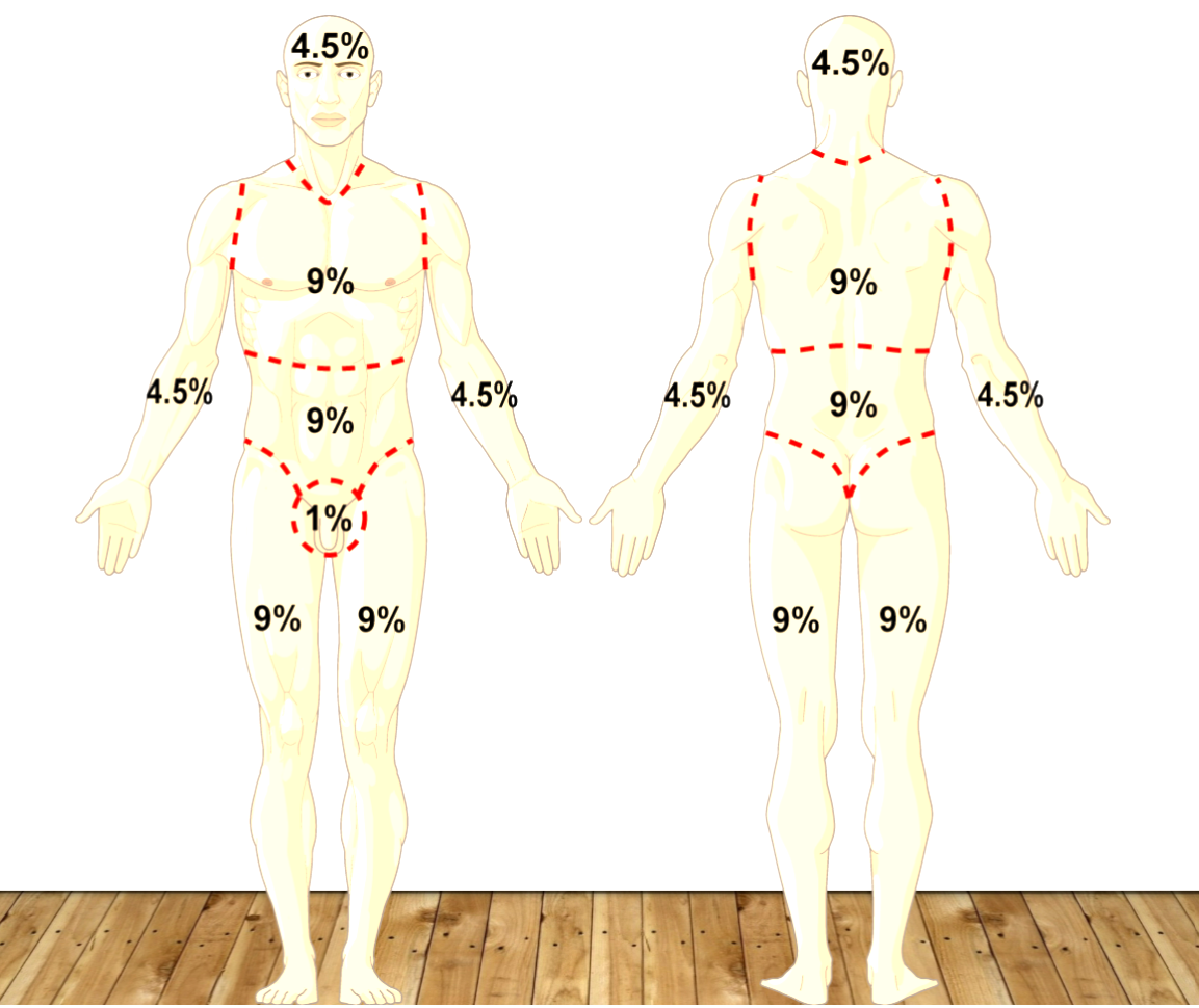
rule of nines
total head 9% (4.5% front and 4.5% back)
total back 18%
total chest/abdomen 18%
total each arm 9% (4.5% front and 4.5% back)
total each leg 18% (9% front and 9% back)
premium 1%
fluid resuscitation calculations - Parkland formula
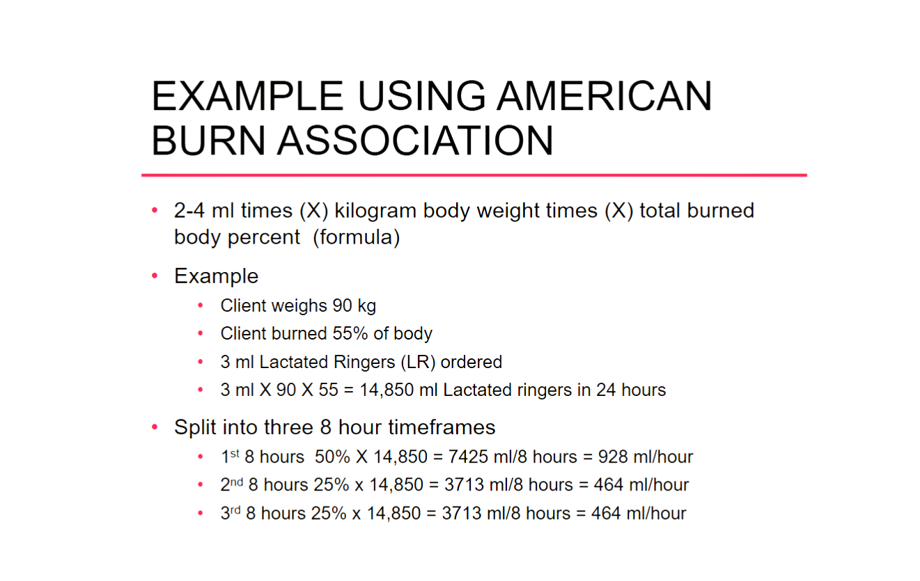
4mL x TBSA (total body surface area burned) x pt wt in kg = mL of LR in 24 hours
divided in the following ways:
50% in the first 8 hours
25% in the second 8 hours
25% in the third 8 hours