Chapter 32 Circulation and Cardiovascular Systems
1/80
There's no tags or description
Looks like no tags are added yet.
Name | Mastery | Learn | Test | Matching | Spaced |
|---|
No study sessions yet.
81 Terms
Some invertebrates lack a circulatory system because they have a thin body wall that makes a circulatory system unnecessary. Hydras have cells in an external layer that are exposed to water and can independently exchange gases and rid itself of wastes. These layers are able to pass nutrients Some invertebrates, like nematodes, use coelomic fluid for transport purposes.
Describe the common features that determine why invertebrates, such as sponges, cnidarians, and flatworms, do not require a circulatory system. (LO)
Blood is always contained within blood vessels, while hemolymph is not. Blood is purely blood, while hemolymph is a mixture of blood and interstitial fluid.
Explain two differences between blood and hemolymph. (LO)
In an open circulatory system, the heart pumps hemolymph via vessels into tissue spaces that are often enlarged into saclike sinuses. The hemolymph eventually drains back to the heart. In some open systems, hemolymph is pumped into a dorsal aorta, which empties into the hemocoel. Then the hemolymph is drained back to the heart through openings called ostia. In open circulatory systems, blood is pumped from the heart into a system of blood vessels. Gas and nutrient for waste exchange occurs in the capillaries. The blood then moves back to the heart for repumping.
Compare and contrast the open circulatory system of an arthropod with the closed system of an annelid. (LO)
circulatory system
in animals, an organ system that moves substances to and from cells, usually via a heart, blood, and blood vessels.
blood
fluid circulated by the heart through a closed system of vessels; type of connective tissue.
hemolymph
circulatory fluid that is a mixture of blood and interstitial fluid; seen in animals that have an open circulatory system, such as mollusks and arthropods.
open circulatory system
arrangement of internal transport in which blood bathes the organs directly, and there is no distinction between blood and interstitial fluid.
closed circulatory system
a type of circulatory system where blood is confined to vessels and is kept separate from the interstitial fluid.
Arteries carry blood away from the heart and are thick. Capillaries are the sight of gas and waste exchange. They are microscopic and very narrow. Veins carry blood to the heart and are much thinner than arteries.
Distinguish the structure and functions of arteries, veins, and capillaries. (LO)
In animals with a one-circuit pathway the heart has one atrium and one ventricle. Blood is pumped to the gills, where it is oxygenated. After passing through the gills, blood returns to the dorsal aorta, which distributes blood throughout the body. In animals with a two-circuit pathway have blood pumped from the right atrium to the right ventricle to the lungs, where it is oxygenated. This is called the pulmonary circuit. Then blood enters the left atria, the left ventricle, and is pumped out into the entire body. This is called the systemic loop.
Compare the path of blood in animals with a one-circuit circulatory pathway vs. a two-circuit pathway. (LO)
Fish have one atrium and one ventricle. Amphibians and most replies have two atria and one ventricle. Crocodiles, birds, and mammals have two atria and two ventricles.
Identify the number of atria and ventricles in each type of vertebrate animal: fish, amphibians, most reptiles, crocodilians, birds, and mammals. (LO)
cardiovascular system
vertebrates have a closed circulatory system called a ___________________
heart
The muscular ___________ keeps blood circulating through the blood vessels.
atria
The _____ are the chambers of the heart that receive blood.
ventricles
The ____________ pump blood into arteries.
artery
blood vessel that transports blood away from the heart
capillaries
microscopic blood vessel; gases and other substances are exchanged across the walls of a capillary between blood and tissue fluid.
vein
blood vessel that arises from venues and transports blood toward the heart.
arteriole
vessel that takes blood from an artery to capillaries.
venule
vessel that takes blood from capillaries to a vein.
systemic circuit
circulatory pathway of blood flow between the tissues and the heart
pulmonary circuit
circulatory pathway between the lungs and the heart
heart
muscular organ whose contraction causes blood to circulate in the body of an animal
myocardium
major portion of the heart consisting mostly of cardiac muscles; its muscle fibers are branched and tightly joined together
pericardium
sack in which the heart resides. It secretes a lubricating fluid
endocardium
inner lining of the heart
septum
partition or wall that divides two areas; the septum in the heart divides the right half from the left half.
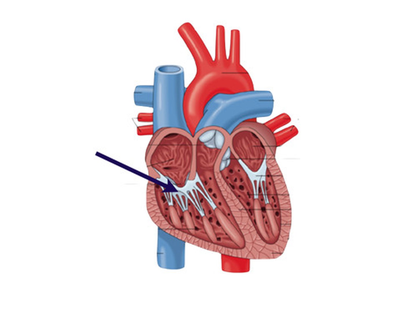
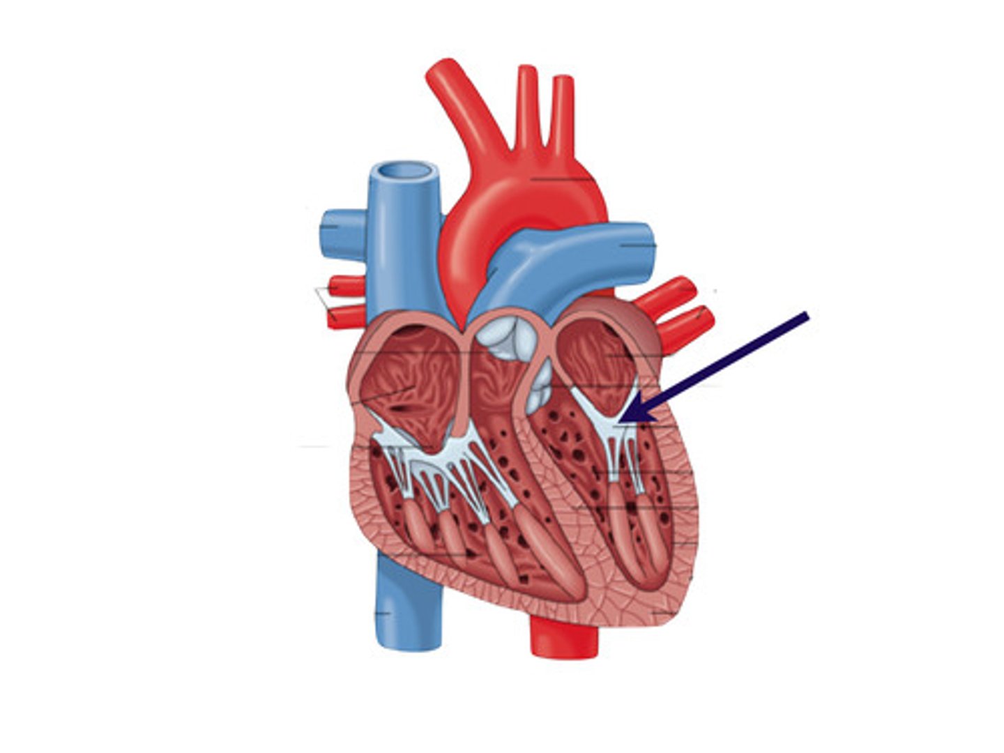
atrium
chamber; particularly an upper chamber of the heart lying above a ventricle
ventricle
cavity in an organ, such as a lower chamber of the heart
chord tendineae
strong fibrous tendons the support the heart valves and prevent them from inverting when the heart contracts
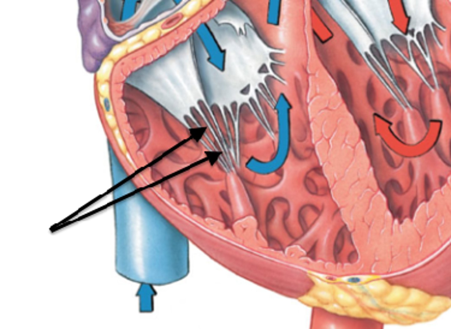
atrioventricular valves
heart valve located between an atrium and a ventricle
tricuspid valve
valve on the right side of the heart the has three cusps/flaps
bicuspid valve
valve on the left side of the heart that has two cusps
semilunar valve
valve resembling a half moon located between the ventricles and their attached vessels.
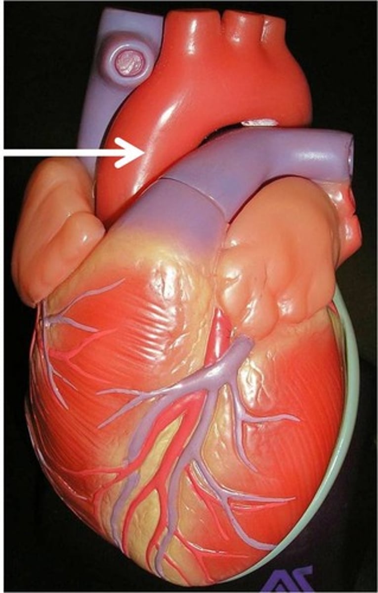
The human heart consists of the left and right atrium and the left and right ventricle. The valves include the bicuspid, tricuspid, aortic semilunar, pulmonary semilunar valves.
List the major components of the human heart, including the four chambers and four valves. (LO)
Blood enters the right atrium through the inferior vena cava and superior vena cava. The right atrium sends the blood through the tricuspid valve to the right ventricle. The right ventricle sends blood through the pulmonary semilunar valve to the lungs, where it is oxygenated. Pulmonary veins take the blood into the left atrium. The blood leaves the left atrium through the bicuspid valve. It enters the right ventricle, where it is sent through the aortic semilunar valve into the aorta and to the rest of the body.
Trace the path of blood through the human heart, lungs, and major vessels leading to the lower leg. (LO)
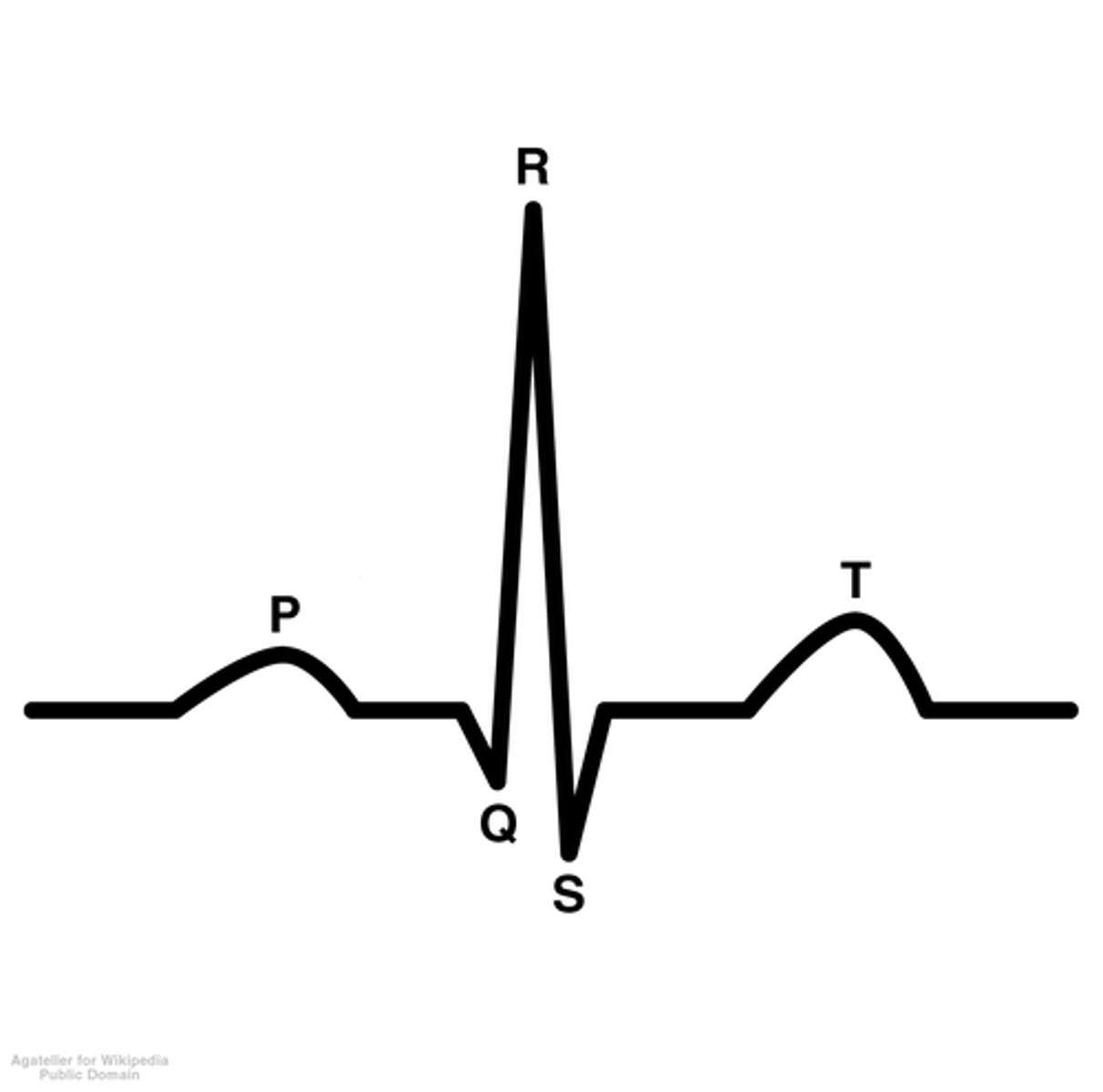
The SA node is the "pacemaker" found in the upper dorsal wall of the right atrium. It initiates the heartbeat by sending out an impulse that causes the atria to contact. The AV love is found in the base of the right atrium hear the septum. When stimulated by impulses from the SA node, it sends out impulses through the septum to cause the ventricles to contract. An ECG is a recording of these electrical charges. The P wave represents excitation occurs just before atrial contraction. The QRS complex signals that the ventricles are about to contract. The electrical changes that occur as the ventricular muscles recover produce the T wave.
Discuss how the SA and AV nodes control the contractions of the heart muscle, and how these electrical changes result in the characteristic patterns seen on an ECG. (LO)
Hypertension is high blood pressure, often caused by a narrowing of arteries. Atheroseclerosis is an accumulation of soft masses of fatty material. Stroke is a condition resulting when an arteriole in the brain bursts or becomes blocked by an embolism. A heart attack is damage to the myocardium due to blocked circulation in the coronary arteries. Angina is a condition characterized by thoracic pain resulting from occluded coronary arteries; may precede a heart attack.
Describe the major categories of cardiovascular disease that occur in the United States (LO)
cardiac cycle
one complete cycle of systole and diastole for all heart chambers.
systole
contraction period of the heart during the cardiac cycle
diastole
relaxation period of a heart chamber during the cardiac cycle
cardiac output
blood volume pumped by each ventricle per minute (Not total output pumped by both ventricles)
Lub
caused by the vibrations of the heart when the atrioventricular valves close
dub
is heated when vibrations occur due to the closing of the semilunar valves.
pulse
vibration felt in arterial walls due to expansion of the aorta following ventricle contraction.
pacemaker
cells of the sinoatrial node of the heart; electrical device designed to mimic the normal electrical patterns of the heart.
electrocardiogram (ECG)
recording of the electrical activity associated with the heartbeat.
aorta
In humans, the major systemic artery that takes blood from the heart to the tissues.
vena cava
large systemic vein that returns blood to the right atrium of the heart in tetrapods; either the superior or inferior vena cava
portal system
pathway of blood flow that begins and ends in capillaries, such as the portal system located between the small intestine and liver.
blood pressure
force of blood pushing against the inside wall of blood vessels.
hypertension
form of cardiovascular disease characterized by blood pressure over 140/95 (over 45) or 130/90 (under 45)
atherosclerosis
form of cardiovascular disease characterized by the accumulation of fatty materials (usually cholesterol) in the arteries.
stroke
condition resulting when an arteriole in the brain bursts or becomes blocked by an embolism; cerebrovascular accident.
heart attack
damage to the myocardium due to blocked circulation in the coronary arteries; myocardial infarction.
angina pectoris
condition characterized by thoracic pain resulting from occluded coronary arteries; may precede a heart attack.
Red blood cells transport oxygen and help transport carbon dioxide. White blood cells fight infection. Platelets aid in clotting.
List the major types of blood cells, and their functions. (LO)
RBCs are manufactured in the red bone marrow of the skull, ribs, vertebrae, and the ends of long bones. The hormone erythropoietin is produced when an enzyme from the kidneys acts on a precursor made by the liver and stimulates production of red blood cells. Before being released from bone marrow, the RBCs lose their nucleus and synthesize hemoglobin. Red blood cells have a life span of about 120 days; then they are destroyed chiefly in the liver and spleen
Identify the major cellular and molecular events that result in a blood cell. (LO)
In the ABO system the presence or absence of type A and type B antigens on red blood cells determines the persons blood type. In the Above system, there are four blood types: A,B,AB, and O. Rh is another important antigen. Rh+ has the Rh factor on red blood cells; Rh- lacks the antigen. Rh-negative individuals do not have antibodies to Rh factor but make them if exposed to Rh+ blood.
Compare and contrast the ABO and Rh blood classification systems. (LO)
Capillary exchange is is control by osmotic pressure and blood pressure. Blood that enters a capillary at the arterial end is rich in oxygen and nutrient, and it is under pressure created by the pumping of the heart. Osmotic pressure tends to cause water to move from tissue fluid into blood, and blood pressure tends to cause water to move in the opposite direction. At the arterial end of a capillary, the osmotic pressure is lower the blood pressure, so water exits water exits the capillary at this end. Mid way along the capillary blood pressure is lower and the two forces cancel each other out. At the venule end of a capillary, the blood pressure is even lower, so water tends to move into the capillary.
Define capillary exchange and describe the two major forces involved. (LO)
plasma and formed elements (cells and platelets)
What are the two components of blood?
plasma
In vertebrates, the liquid portion of blood; contains nutrients, wastes, salts, and proteins.
They keep the blood pH near 7.4 and maintain the blood osmotic pressure, so water has a tendency to enter capillaries.
What do salts and proteins do?
antibody
protein produced in response to the presence of an antigen; each antibody combines with a specific antigen.
formed elements
portion of blood that consist of erythrocytes leukocytes, , and platelets (thrombocytes)
red blood cells
erythrocyte; contains hemoglobin and carries oxygen from the lungs or gills to the tissues in vertebrates.
Hemoglobin
iron-containing respiratory pigment occurring in vertebrates red blood cells and in the blood plasma of some invertebrates.
antigen
foreign substance, usually a protein or polysaccharide, that stimulates the immune system to react, such as to produce antibodies.
agglutination
clumping of red blood cells due to a reaction between antigens on red blood cell plasma membranes and antibodies in the plasma.
White blood cell
leukocyte, of which there are several types, each having a specific function in protecting the body for invasion by foreign substances and organisms.
granular leukocytes
form of white blood cell that contains spherical vesicles (granules) in its cytoplasm.
neutrophil
granular leukocyte that is the most abundant white blood cells; first to respond to infection
basophil
white blood cell wit ha granular cytoplasm; able to be stained with a basic dye
eosinophil
white blood cell containing cytoplasmic granules that stain with acidic dye.
monocyte
type o granular leukocyte that functions as a phagocyte, particularly after it becomes a macrophage, which is also an antigen-presenting cell.
macrophage
in vertebrates, large phagocytic cell derive from a monocyte that ingests microbes and debris.
lymphocyte
specialized white blood cell that functions in specific defense; occurs in two forms- T lymphocytes and B lymphocytes.
platelet
component of blood that is necessary to blood clotting.
clotting
also called coagulation, the response of the body to an injury in the vessels of the circulatory system; involves platelets and clotting proteins.
tissue fluid
fluid that surrounds the body's cells; consists of dissolved substances that leave the blood capillaries by filtration and diffusion.
lymph
fluid, derived from tissue fluid, that is carried in lymphatic vessels.