lateral leg and sole of foot
1/54
There's no tags or description
Looks like no tags are added yet.
Name | Mastery | Learn | Test | Matching | Spaced | Call with Kai |
|---|
No analytics yet
Send a link to your students to track their progress
55 Terms
whats the main role of the muscles in the lateral leg
evasion and plantar flexexion
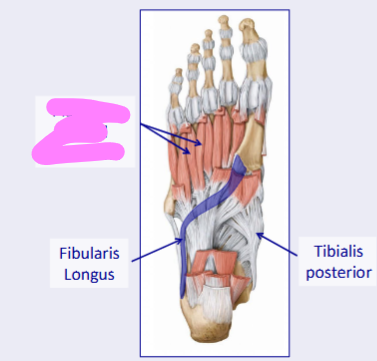
wha are the muscles in the lateral leg
fibularis brevis and fibularis longus

what mscle s shown in purple
fibularis longus
whats the orogin and insertion of the fibularis lngus
the head of the fibla, insertion onto the base of 1st metatarsal
whats the innervatn of the fiularis longus
superficial fibular nerve
hats the roles of the fibularis longus
eversion and plantar flexion
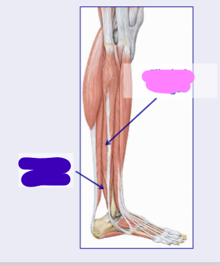
whats the muscle shown in blue
fibularis brevis
whats the orogin and insertion of the fibularis brevis
orogin on the inferior fibia and insertion on base of the 5th metacarpal
whats the innervation of fibularis brevis
superficial fibular nere
whats the role of fibularis brevis
foot eversion and plantar flexion
vwhat nerve is shown in blue
the common fibular nerve
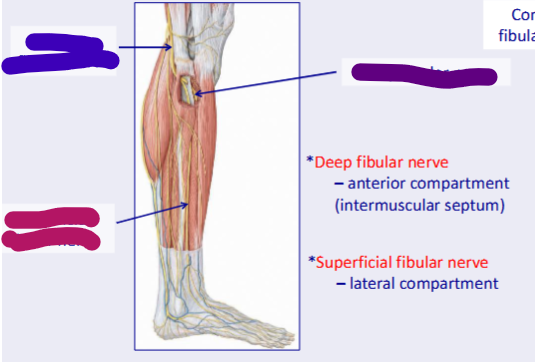
what nerve is shown in purple
the deep fibular nere

what nerve s shown in pink
the superficial fibular nerve
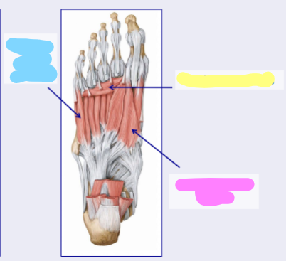
vthe first layer of the foot is shown here, what is the most superficial layer shown in pink
the plantar apperneurosis
how many layers/ groups of muscles are there in the foot
4
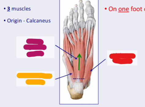
what muscle of the first layer is shown in pink
flexor digitorum brevis
whats the orgin and insertion of the flexor digitorm brevis
orogin on the calcanes, insertion on middle phalange of 4 digits
whats the innervation of the flexor digitorum brevis
median plantar nerve
whats the role of flexor digitorum brevis
flexes the lateral 4 digitis

whats the muscle of the 1st layer shown in red
the abductor hallusis
whats the orogin and inserton of abductor hallsus
originates on the calcaneus, inserts on the proximal palynx of 1st digit
whats the innervatin of the abductor hallusis
medial plantar nerve
whats the role of abductor hallusis
abduction of the 1st digit

what muscle f the 1st layer is shown in orange
the abductor digiti minimi
whats the orogin and insertion of abductor digiti minimi
orogin on the calcaneus, insertion on the proximal phalnx of 5th digit
whats the innervation of the abductor digiti minimi
lateral plantar nerve
whats the role of the abductor digiti minimi
abduction and flexion of the 5th digit

what muscles of the 2nd layer are shown in green
the lumbricals
whats the insertion of the lumbricals
orogins onto the flexor digitorum longus insertion onto the extensor hoods to lateral 4 digits

what component of the 2ns layer of the fot is hilghted in blue
the flexor digitorum longus
what component of the 2nd layer of the foot is highlighted in purple
the flexor hallusis longus

what muscle of the 2nd layer of the foot is shown in light blue
the quadratus plante

whats the orogin and insertion of the quadratus plante
orogin on the clacaneus, insertion on the flexor digitorum longus

what component of the 2nd layer of the foot is shown in dark blue
the medial plantae nerve
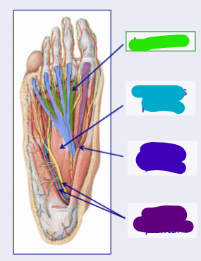
what component of the 2dn layer of the foot is shown in purple
the lateral plantar nerve

what muscle of the 4th layer of thr foot is shown in blue
the flexor digiti minimi
whats the roogin and insertion of the flexor digiti minimi
orogin on the 5th metacarpal, inserton on the lateral side f the base of proximal phalymx
whats the role of the flexor digiti minimi
flexes the proximal phalynx at metatarsalphalangeal joint
whats the innervation of the flecor figiti minimi
laeral plantar nerve
what muscle is shown in yellow
the abductor hallusis
whats the orogin and insertion of adductor hallucis
orogin is head dependant, insertion on metatarsophlangeal jointe of uter toes
what are the 2 heads of the adductor hallusis
the oblique head and transverse head
what the role of adductor hallucis
adduction of the 1st digit

what muscle is shown in pink
the flexor allucs brevis
what msucle of the 4th layer of the foot is shon in pink
the plantar and dorsal interossi

what bone in the foot is shown in red
the talus
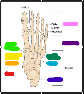
vwhat bone is shown in ornage
the navicular

what bne is shown in yellow
the intermediate cuniform

what bone is shown in light green
the medial cuniform

what bone is shown in dark blue
the calcaneus

what bone is shown in light blue
the cuboid bone

what bone is shown in dark green
the lateral cuniform

what group of bones is shown in dark purple
the metatarsles

what group of bones is shown vin light pink
the phalanges - proximal middle and distal