Radiology Lecture 2: X-Ray Filtration & Collimation Terms
1/69
There's no tags or description
Looks like no tags are added yet.
Name | Mastery | Learn | Test | Matching | Spaced | Call with Kai |
|---|
No analytics yet
Send a link to your students to track their progress
70 Terms
Removes low-energy x-rays
Filtration
Collimation
Limits the size of the x-ray beam
- Reduces the radiation dose
- Reduces scattered radiation
Inherent filtration
X-rays produced at target go in all directions; the process where most are absorbed by the tube or tube head
Other x-rays exit the tube head through the diaphragm at the window to become the _________
Primary beam
Primary beam photons have __________
Wide range of energy (some very strong, some weak)
Added filtration is used to _______________ from the primary beam before they hit the patient
Absorb the low energy photons
What is filtration measured in?
mm Al
What is the typical range of inherent filtration?
Usually 0.5 to 2.0 mm Al
What is added filtration?
Aluminum plate at opening of tubehead
What must added filtration be with kVp lower than 70?
Must be 1.5 mm Al
What must added filtration be with kVp at 70 or above?
Must be 2.5 mm Al
Added filtration is the _____________ at __________ of tubehead
Aluminum plate; base
By removing low energy photons, there is a _________ proportion of high energy photons
Greater
Filtration ___________ the average energy of the x-ray photons in the primary beam, but ________ number of photons
Increases; decreases
Energy (quality) of primary beam photons increased with:
Increased kVp
Increased added filtration
Number (quantity) of primary beam photons increased with:
-Increased mA
-Increased exposure time
-Increased kVp
-Decreased added filtration
Collimator
Opening in a plate on outer surface of the added filtration
What does the collimator determine?
Size and shape
What is collimation a combination of?
Collimator and the position indicating device at the end
What does the position indicating device (PID) do?
Limits size of beam and reduces area exposed
- Reducing absorbed dose
- Reducing scattered photons
What is the best PID shape?
Rectangular; reduces radiation dose by 65%
What is the best PID length?
Long; 16" reduces radiation dose by 35%
What PID is used in UDM radio clinic?
12" SFD rectangular
What is the inverse square law?
Intensity of radiation varies inversely with the square of the distance between source and absorbing material
T/F: Intensity is increased with long distance
False
- Intensity is increased with short distance
- Decreased with long distance
Length of PID affects ___________
Intensity of radiation
Changes in PID length require ______________
Changes in exposure time
Shorter PIDs will produce ____________ on the sensor than long PIDs, with the difference proportional to the ________ of the difference in length.
- Image will be ________
More intense radiation; square
- Darker
Longer PIDs will produce ____________ on the sensor than short PIDs, with the difference proportional to the ________ of the difference in length.
- Image will be ________
Less intense radiation; square
- Lighter
To compensate for inverse square law,
- Short PID requires ________ exposure time
- Long PID requires ________ exposure time
Short; long
- Note: Difference in time varies with the square of the difference in distance
New exp time / Old exp time = (New PID length)² / (Old PID length)²
Example of changing exposure time to compensate for inverse square law:
- Original PID is 8", original exposure time is 0.2 sec
- New PID is 16", what exposure time should be used?
- X sec/0.2 sec = (16")^2/(8")^2= 4
- X sec= 0.2 sec * 4 = 0.8 sec
- Exposure time with 16" must be 0.8 sec to maintain the same darkness of the image as 12 impulses with an 8"
What makes the image darker?
Overexposure
- Too high kVp, mA, and/or exposure time
- Too short PID
What makes the image lighter?
Underexposure
- Too low kVp, mA, and/or exposure time
- Too long PID
What are three possible interactions?
1. Transmission
2. Absorption
3. Scatter
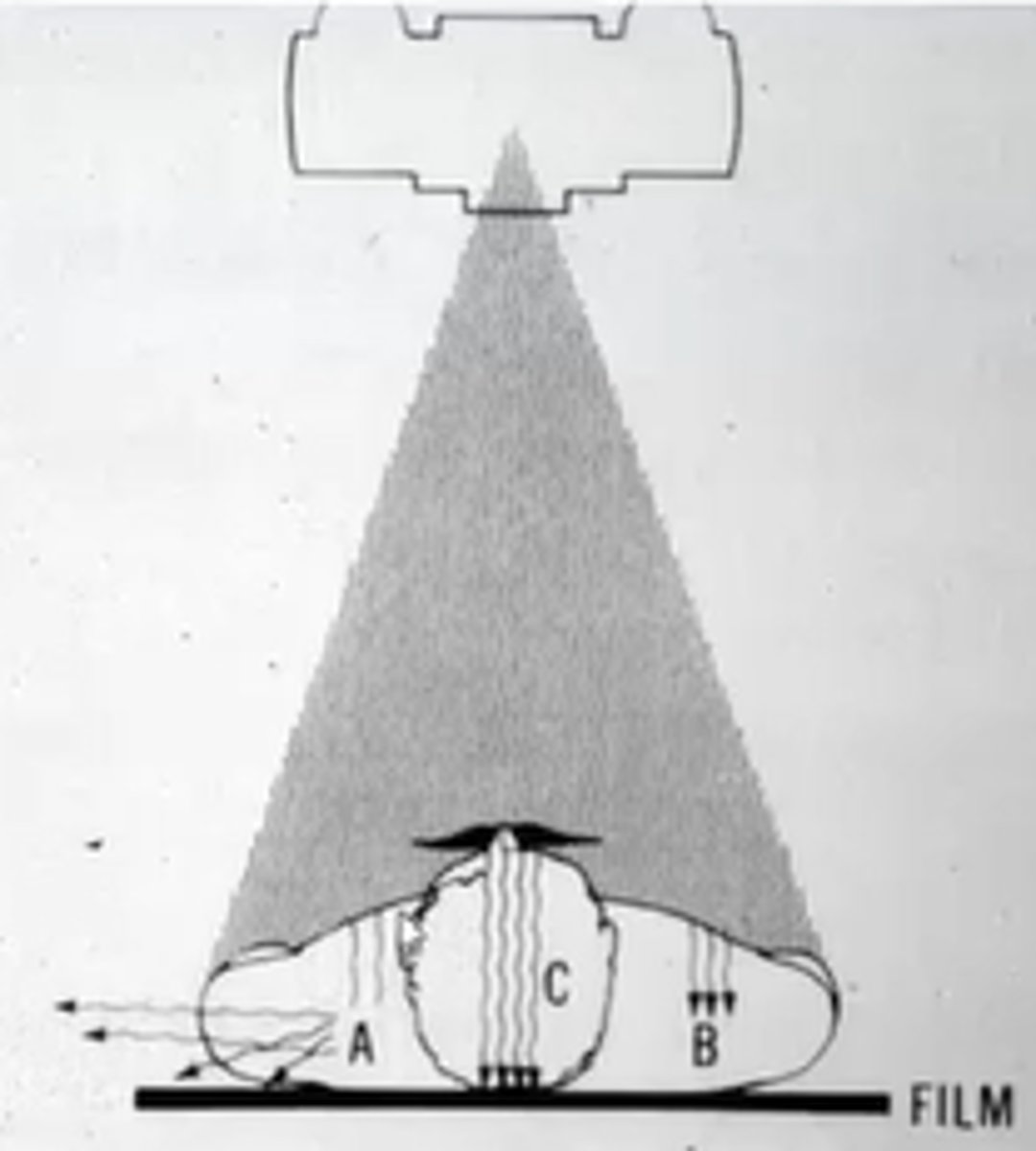
Label A, B, and C
A= scatter
B= absorption
C= transmission
What color images does transmission, absorption, and scatter make?
Transmission= dark
Absorption= light areas
Scatter= bounce off in diff. directions
What is the incidence of transmission, absorption, and scatter?
Transmission = 9%
Absorption = 27%
Scatter = 64%
What are the two types of scatter?
coherent scatter and compton scatter
What is Coherent (Rayleigh, elastic) scatter?
- Emits photon with same energy
- 7%
What are Compton scatter?
- Emits photon with lower energy
- 57%
What is the most common interaction of x-rays with matter?
Compton scattering
Fog
Undesirable darkening of the image
- Caused by scatter
What is transmission more common in? How does it appear?
Soft tissues are radiolucent
What is absorption more common in? How does it appear?
Hard matter are radiopaque
Absorption occurs through the _________
Photoelectric effect
Where is scatter more likely to occur with?
High energy x-rays
During a dental radiographic exposure, which is the most common interaction of x-ray photons and the patient?
A. Transmission
B. Absorption
C. Coherent scatter
D. Compton scatter
D. Compton scatter
Dark images have ________ radiographic density
High
Light images have ________ radiographic density
Low
What is radiographic density?
The degree of darkness on the radiograph
Radiographic density is increased with....
- High energy x-rays
- Soft tissues
- Thin tissue sections
- High vKp, mA, and exposure time
- Short PID
= Darker image
Greater x-ray exposure leads to ________
Increased radiographic density
Thinner parts allow __________, which causes images to be ________
- More transmission
- Dark (increased radiographic density
Thicker parts of a stepwedge ________________; less _______________ causes ____________
- Absorb more radiation
- Transmission of x-rays to film/sensor
- Light appearance
What is radiographic contrast?
Difference in density (darkness) between different parts of the image
What are the two components of radiographic contrast?
- Subject contrast
- Viewing conditions
What does subject contrast result from?
Differential absorption of x-rays through different tissues in the patient's body
= Different degrees of darkness

How does a high kVp affect subject contrast?
- High kVp→ gradual decrease in transmission→ gradual decrease in darkness→ less difference= low subject contrast
How does a low kVp affect subject contrast?
- Low kVp→ drastic transmission→ greater difference in darkness= high subject contrast
High kVp x-rays produce many shades of gray between black and clear (low contrast). This is called a ______________
Long gray scale or long-scale contrast
Low kVp x-rays produce few shades of gray (high contrast). This is called _________
Short gray scale or short-scale contrast
What is contrast controlled by?
Degree of exposure (how dark is the image?)
- Underexposure = too light
- Overexposure = too dark
Latitude
Range of exposures in which contrast and density are acceptable
Post-acquisition enhancement
Change brightness and contrast in digital images to correct for small errors in exposure time
T/F: Post-acquisition enhancement will fix a radiograph with severe errors in exposure time
False; it will NOT
What is fog?
Degree of unwanted darkness on radiograph
What causes fog?
Scattered radiation
How is scattered radiation/fog reduced?
By using long, rectangular PIDs
What does viewing x-rays in a darker room do?
Allows detection of subtle differences in darkness
What are the rules for viewing radiographs?
Don't look at them on a tablet PC in the bright clinic if you are trying to detect caries, periapical disease, or periodontal defects
- You will probably miss something
Radiographs should be examined in a darkened, quiet room