Pathophysiology of dyspepsia, GERD and PUD
 Call Kai
 Learn
Learn Practice Test
 Spaced Repetition
 Match
Match Flashcards
 Knowt Play
1/36

There's no tags or description
Looks like no tags are added yet.
Name | Mastery | Learn | Test | Matching | Spaced | Call with Kai | Chat |
|---|
No analytics yet
Send a link to your students to track their progress
37 Terms
Dyspepsia
is a symptom not a diagnosis
collection of upper GI symptoms lasting at least one month (not every day in the month but ongoing over the whole month)
where epigastric pain is the dominant feature
other symptoms:
NV
burping or bloating (upper abdomen)
early satiety
uncomfortable post-prandial
heartburn
Functional dyspepsia
is a diagnosis of elimination
rules out everything else through other investigations
is not PUD, GERD, esophageal or gastric cancer
Red flags for dyspepsia
Vomiting
frequent and >7 days
Bleeding
hematemesis (vomiting bright red blood)
coffee ground emesis
dark tarry stools
Anemia/Anorexia/Abdominal mass
unexplained dizziness or fatigue, pale
can have subclinical bleeding from ulcer or malignancy
Dysphagia
choking when eating or drinking
difficulty swallowing
odynophagia - pain with swallowing
patients >60 years
greater risk of malignancies
**severity of dyspepsia does not infer severity of condition
Which patients are able to self-treat for dyspepsia?
patients without red flags (VBAD
could still have GERD, PUD or malignancy but risk is low enough
called uninvestigated dyspepsia
Esophagogastric junction
where esophagus meets stomach, not a clear division, will see a change in the tissues that occur
comprised of:
lower esophageal sphincter (LES)
squamocolumnar junction (z-line)
Lower esophageal sphincter (LES)
made of 2 muscle layers
internal: thickened smooth muscle of distal esophagus (~4cm)
external: crural part of diaphragm (~2cm)
supports the sphincter by applying pressure
Squamocolumnar junctions (z-line)
transition from one type of epithelium to mucosa
esophageal mucosa: stratified squamous cells
gastric mucosa: columnar cells covered by protective layer of mucus
Normal function of LES
prevents backflow of stomach contents
some degree of gastroesophageal reflux is normal after meals
saliva neutralizes acidic content
secondary peristaltic waves help reflux return
contracted (closed) at rest and relaxes (opens) with swallowing
GERD
GER becomes pathological when reflux is sustained, leading to symptoms or complications
generally due to increased frequency of transient LES relaxation
less significant mechanisms:
decreased LES tone at rest
delayed gastric emptying
NOT due to increased gastric acid secretion
Non-modifiable GERD risk factors
age
esophageal tissue less resistant to damage
diminished salivation
pregnancy
increased intraabdominal pressure - pushes against diaphragm
progesterone decreases LES tone
family history
likely predisposition to decreased LES tone
scleroderma
muscle of LES replaced with connective tissue
Zollinger-Ellison syndrome
tumour (gastrinoma) leads to excessive acid production - overwhelming protective factors
hiatal hernia
Hiatal hernia
lower part of esophagus and upper part of stomach push through the diaphragm and into chest cavity
loss of external LES support
larger angle between esophagus and curve of stomach (angle of His)
happens from increased intraabdominal pressure
more common in:
>50 years
obesity
smoking
can undergo surgery to fix
Modifiable GERD risk factors
obesity
increased intraabdominal pressure
increased risk for hiatal hernia
smoking
lowers LES pressure
hyposalivation
medications
lowers LES pressure
anticholinergic, benzos, CCB, nitrates, opioids, tricyclic antidepressants
alcohol/caffeine
lowers LES pressure
eating chocolate/peppermint
lowers LES pressure
eating fatty/fried foods
delays gastric emptying
eating large meals
increased pressure on LES
increased volume for reflux
sleeping (recumbent position)
swallowing occurs less often
gravity unable to assist clearance of reflux
GERD symptoms
classic symptoms
heartburn - throat to upper stomach along midline
direct irritation of esophageal mucosa
regurgitation - gastric contents into back of throat
may be sour taste or burning
may experience hypersalvation (“water brash”)
other symptoms
dyspepsia
dysphagia
usually reflects esophageal erosion or stricture (complications)
extraesophageal symptoms
chronic cough or throat clearing
tooth erosion
asthma (aspiration into bronchial tree)
Complications of GERD
require additional diagnostics for definitive diagnosis
erosive esophagitis (EE)
esophageal stricture
Barrett’s esophagus
Erosive esophagitis (EE)
exposure of stomach acid on the epithelium causes inflammation (defense mechanism)
persistent inflammation causes erosion and decreases peristalsis
30% of GERD progress to EE
could have EE and be asymptomatic
Esophageal stricture
damaged mucosa and musculature from EE is replaced with scar tissue
not as flexible as before
leads to narrowing of esophagus lumen
will experience symptoms of choking or pain/difficulty to swallow
Barrett’s esophagus
esophageal epithelium is replaced with columnar cell, better able to withstand GER
primary risk factor for esophageal adenocarcinoma
monitoring very important once developed
15% GERD progress to Barrett’s esophagus and 6% Barrett’s progress to cancer
Risk factors developing:
male
> 50 year
Caucasian
obesity
smoking
symptoms of GERD >5 years
Potential exacerbations of GERD
spicy or acidic food
local irritation
medications that can induce or worsen esophagitis due to direct mucosal injury
bisphosphonates
Aspirin and NSAIDs
potassium and iron salts
tetracycline, doxycycline and clindamycin
Diagnosis of GERD
classic symptoms that are moderate-to-severe (impacts daily activity) OR occur >2x/week even if mild
……OR…….
diagnostic evidence of complications, regardless of symptoms
in the absence of classic symptoms, other causes need to be ruled out
Role of additional investigations
when appropriate:
if alarm features present (VBAD)
GERD is refractory to therapy
when not needed:
most patients with classic symptoms - if undergo and find no damage called non-erosive reflux disease (NERD)
those with risk factors of Barrett’s esophagus for screening purposes
Types of investigations used for GERD
esophageal manometry
measures the pressure in the esophagus during swallowing to measure how well functioning
24-hr pH impedance
how much acid in esophagus, so if increased frequency or degree of stomach acid reflex
x-ray with barium contrast
swallow barium and watch how moves with x-ray to identify if have stricture - lumen will get smaller while barium is travelling
endoscopy with biopsy
biopsy is only way to diagnose Barrett’s
most common
GER in infants
extremely common but is not a disease
peaks at 4 months and resolves usually at 1 year
increased susceptibility due to:
very little solid intake (easier to reflux)
anatomy predisposes (LES not fully matured and angle of His naturally blunted)
often lying down, or semi-recumbent
GERD in infants
if suspected needs to be referred:
refusing to eat
irritable
not keeping anything down
multiple mimickers - as long as child seems healthy and gaining weight is okay
Gastric cell types
surface mucous cells
secrete mucous and bicarbonate
parietal cells
secrete H+ and Cl-
produce intrinsic factor
neck mucous cells
secrete mucous and bicarbonate
chief cells
secrete pepsinogen and lipase to breakdown the contents of food
enteroendocrine cells
3 types of cells that secrete regulatory hormones
Enteroendocrine cells
enterochromaffin-like cells
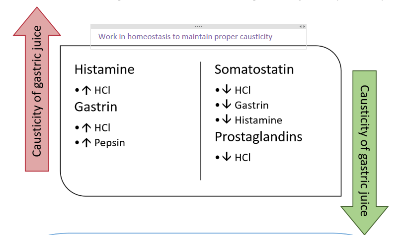
produce histamine (stimulating parietal cells)
G-cells
produce gastrin (stimulates parietal, chief cells, and peristalsis)
D-cells
stimulated by acid presence
produce somatostatin (inhibits G-cells, parietal cells, and enterochromaffin cells)
the “breaks”
negative feedback loop for HCl
How much gastric juice is produced by gastric mucosa?
~1.5L per day
How is the homeostatic causticity maintained?
along with the mucosal defenses
mucous and bicarbonate production
stimulated by prostaglandins
mucosal blood flow
delivers oxygen and bicarbonate
facilitates healing of mucosal damage
Peptic ulcer disease (PUD)
develops in the stomach or in the duodenum when causticity of gastric juices overwhelm mucosal defenses
causticity damages the GI mucosa goes deep enough through epithelium to the muscularis mucosa underneath
exposed muscle later (can be few mm or cm deep)
if deep enough can cause perforation
can lead to slow (anemia) or severe (haematosis) bleeding, perforation or obstruction
Causes of PUD
Primary
NSAID use
4x increased risk
particularly gastric ulcers
Helicobacter pylori infection
6-10x increase risk
historically large percentage of ulcers - but now know how to treat
Secondary
Zollinger-Ellison Syndrome
other medications especially used alongside NSAIDs
directly toxic to the barrier of the stomach
on own not generally a risk
acetaminophen > 2-3g/day, bisphosphonates, glucocorticoids, clopidogrel, sirolimus, spironolactone, SSRIs, oral potassium
Cause of NSAID-induced ulcers
systemic effects - inhibition COX
COX2 pro-inflammatory prostaglandins (inhibiting decreased pain and inflammation)
COX1 gastroprotective prostaglandins (inhibiting disrupts stomach’s balance)
dominant
local effects - direct epithelial irritation
NSAIDs = carboxylic acids and unionized in stomach
once absorbed across gastric mucosa ionize and cause local toxicity
coating tablet not preventative because this is minor pathway
Risk of PUD with NSAID use
dose
choice of agent
highest risk: ketorolac
lower risk: ibuprofen, diclofenac, naproxen
lowest risk: COX2 selective agents (but not zero)
duration of therapy
want shortest because less time for ulcer to develop
can get back to homeostasis
Use of combo therapy
other risk factors:
advanced age
history of PUD
smoking/alcohol
family history
concurrent med use
concomitant infection with H. pylori
Helicobacter pylori infection
gram-negative
helical, rod-shaped
motile
adapted to survive gastric (acidic) environment
acquired commonly in childhood because of fecal-oral route
leads to colonization of gastric mucosa
can have for entire life and does not always cause ulcer
Cause of H. pylori-induced ulcer
using flagella, moves through mucosa layer and land on epithelial cells
attach to epithelial cells and start producing urease
urease combines urea and H2O to make CO2 and ammonia
ammonia will neutralize the gastric acid and makes it more suitable for H. pylori to live and proliferate
during proliferation it produces enzymes that disrupt the integrity of gastric mucosa and toxins that damage local epithelium
will move along mucosa, and will mucosal damage leads to cellular damage
creates a gap in protection causing caustic stomach contents to reach mucosa and create ulcer
H. pylori can also lead to?
chronic gastritis
gastric adenocarcinoma
gastric mucosa-associated lymphoid tissue (MALT) lymphoma
NSAID-negative and H. pylori-negative PUD (“stress ulcer”)
can be caused from critical illness because body is under great stress
can be causes from MAJOR emotional stress
PUD symptoms
most are asymptomatic until complications develop
anemia symptoms
hematemesis or coffee-group emesis
black or tarry stools
severe acute abdominal pain
if there are symptoms - generally non-specific and often fluctuate (cycle of spontaneous healing and relapse)
dyspepsia
nausea
bloating
early satiety
± GERD symptoms
some patient may find food worsens and get weight loss
Diagnosis of PUD
definitive requires visualization
non-invasive H. pylori testing recommended for patients < 60 years