Spinal Cord Anatomy, Tracts, Fasciculi, Myelination
1/83
There's no tags or description
Looks like no tags are added yet.
Name | Mastery | Learn | Test | Matching | Spaced | Call with Kai |
|---|
No analytics yet
Send a link to your students to track their progress
84 Terms
__% of new spinal cord injuries cases are male
78
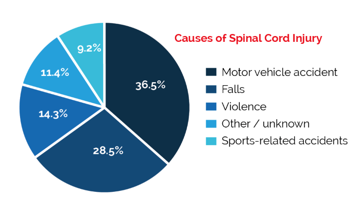
what are some causes of spinal cord injury?
motor vehicle accident
falls
violence
sports-related accidents
Brain and Spinal Cord covered by ______ and is collectively known as the…?
meninges
central nervous system
what is the network of spinal and cranial nerves that are linked to the brain and the spinal cord (somatic and autonomic divisions) called?
peripheral nervous system (PNS)
what are the 3 layers of Meninges that surround tissues of the CNS, but not the PNS?
dura mater (out)
arachnoid
pia mater (in)
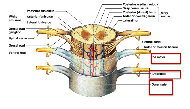
Blood vessels and cerebrospinal fluid are located where?
subarachnoid space (between the arachnoid and pia)
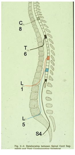
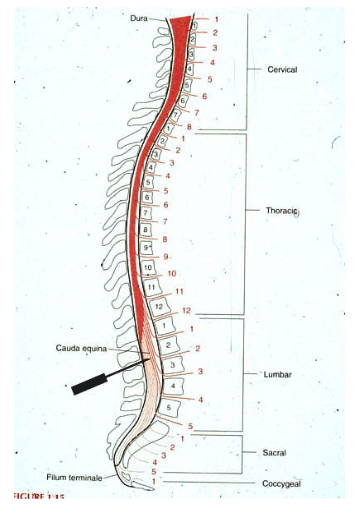
Dissection of central nervous system, including spinal cord with its # pairs of spinal nerves
31
how many of each spinal nerve pairs are there?
cervical
thoracic
lumbar
sacral
coccygeal
8 Cervical
12 Thoracic
5 Lumbar
5 Sacral
1-3 coccygeal
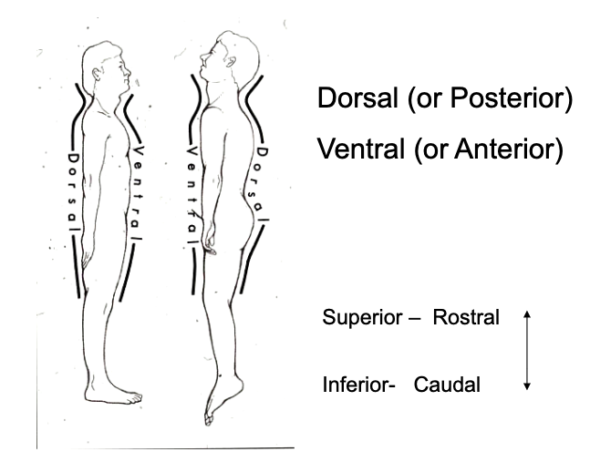
what are alternative terms for:
anterior
posterior
superior
inferior
anterior → ventral
posterior → dorsal
superior → rostral
inferior → caudal
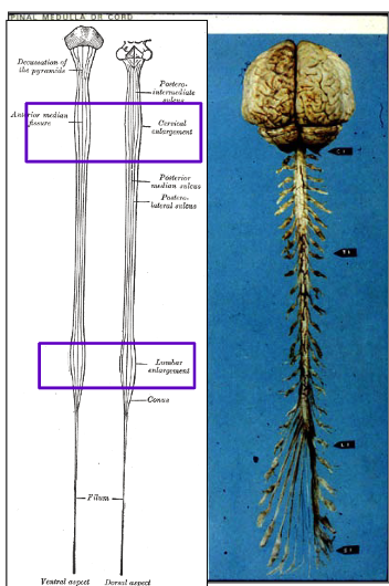
The Spinal Cord is increased in diameter in which regions? why?
cervical and lumbar regions
to accommodate the neurons required for the upper and lower extremities.
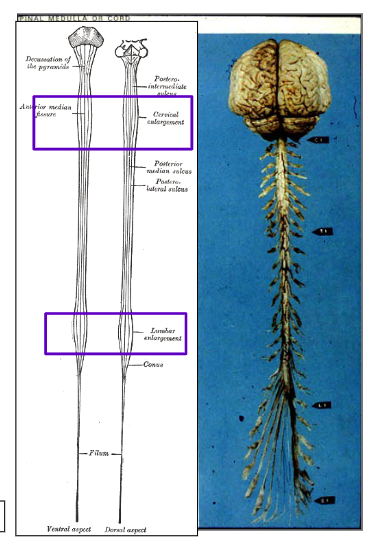
where are the cervical and lumbar enlargements of the spinal cord located?
cervical enlargement → C5-T1
lumbar enlargement → L2-S3
how can we differentiate the anterior (ventral) surface of the spinal cord from the posterior (dorsal) surface?
anterior (ventral) surface has a single spinal artery that supplies the anterior 2/3 of the cord
anterior spinal a.
posterior (dorsal) surface has a pair of spinal arteries
dorsal spinal a.
what is the bloody supply to the spinal cord?
anterior and posterior spinal arteries
what is the termination of the spinal cord called?
conus medullaris
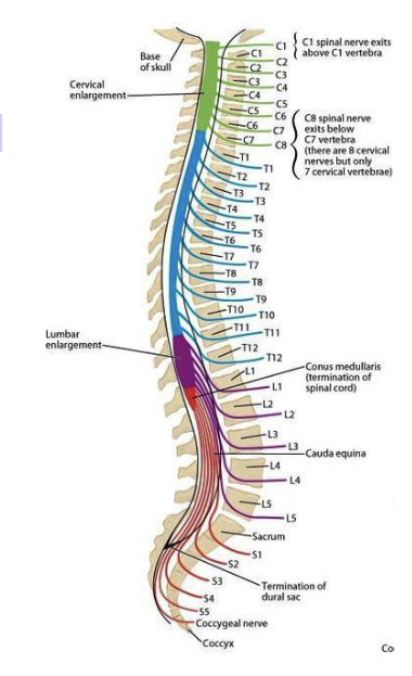
what is the conus medullaris? which vertebrae does it end at?
tapered inferior end of the SC,
ends between vertebrae L1/L2
due to differences in growth of spinal cord and spinal column

what is the cauda equina?
Origin of spinal nerves extending inferiorly from conus medullaris.

The length of nerve roots becomes progressively shorter/longer from cervical to sacral levels of the spinal cord stemming from differences in the growth of the spinal cord and the spinal column.
longer
Spinal taps can be safely performed if the needle is inserted where?
below the 2nd lumbar vertebrae
because adult spinal cord ends between lumbar vertebrae 1 and 2
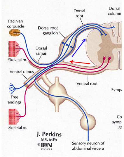
the part of the cord that gives rise to a pair of spinal nerves is called…?
spinal cord segment
describe the structure of a spinal cord segment
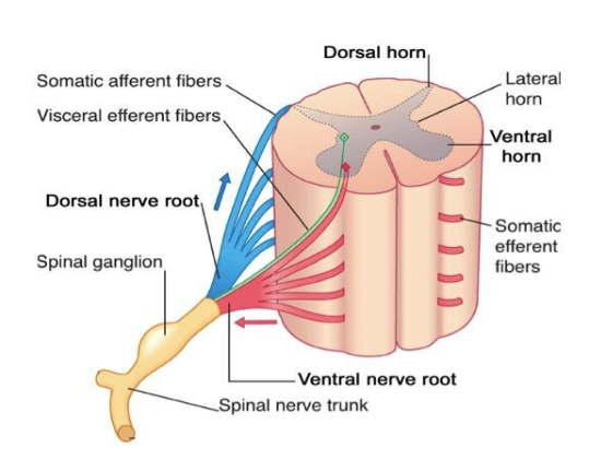
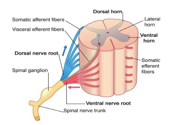
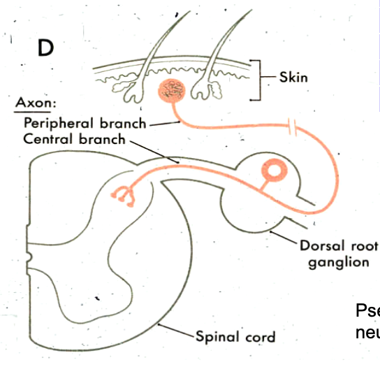
where are Primary Sensory Neurons are located ?
in the Dorsal Root Ganglia
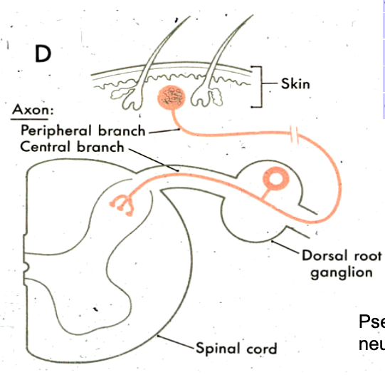
what type of neuron are the primary sensory neurons located in dorsal root ganglia? (multipolar, pseudounipolar, bipolar, etc.)
pseudounipolar
where are motor neurons located in the spinal cord?
ventral horn of spinal cord
sensory neurons
where are cell bodies located?
afferent vs efferent
type of neurons (___polar)
cell bodies in dorsal root ganglion
afferent
unipolar
roots project into dorsal horn of spinal cord gray matter or into the white matter of the dorsal funiculus
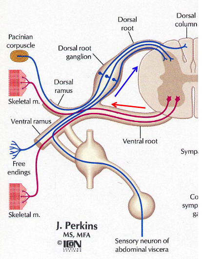
motor neurons
where are cell bodies located?
afferent vs efferent
type of neurons (___polar)
cell bodies in ventral horn of spinal cord gray matter
efferent to skeletal muscles
multipolar
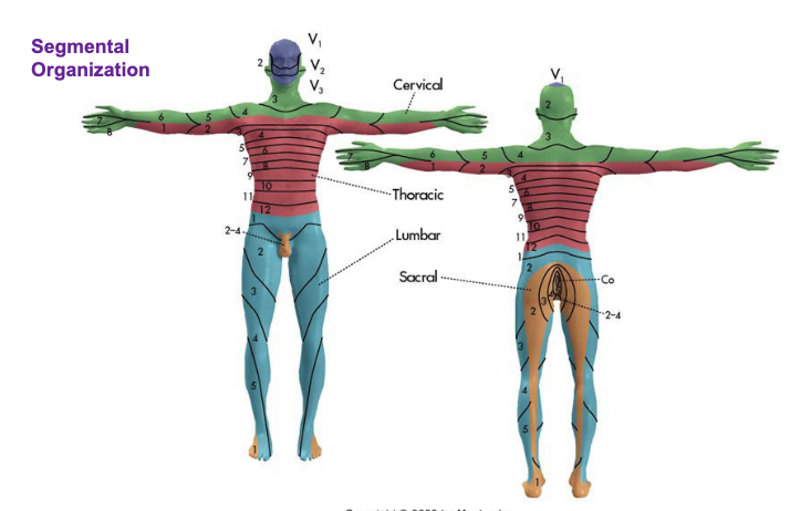
what is a dermatome map?
Cutaneous territories innervated by spinal nerves (dermatomes)
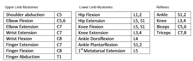
The muscles innervated by a single nerve root constitute a
myotome
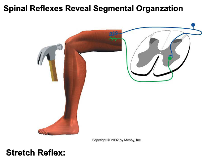
what is the stretch reflex?
Striking the patellar tendon activates muscle spindle primary endings, which then monosynaptically excite alpha motor neurons that innervate the stretched muscle
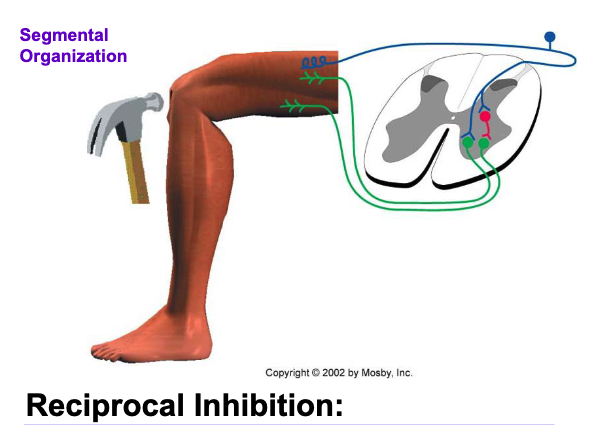
what is reciprocal inhibition?
Striking the patellar tendon initiates a stretch reflex (excitation of the quadriceps) and it also causes inhibition of the motor neurons to the antagonist hamstring muscles
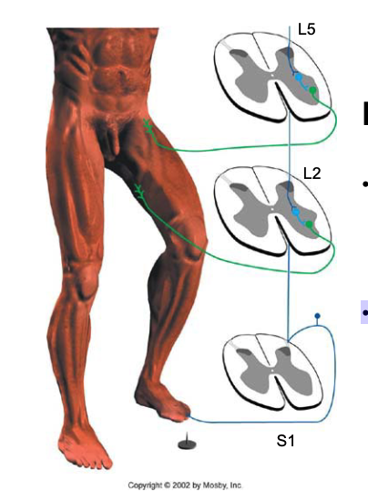
what is the flexor reflex?
This reflex involves several segments, and all connections are polysynaptic.
A nociceptive fiber from the foot enters the spinal cord at S1 and activates motor neurons to iliopsoas and hamstring muscles
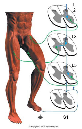
what is the crossed extension reflex?
Stepping on a tack initiates a flexor reflex.
It also causes excitation, of the contralateral antagonist muscles.
Contraction of the contralateral quadriceps helps the leg with the nonpunctured foot to support the body
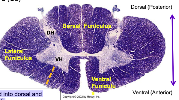
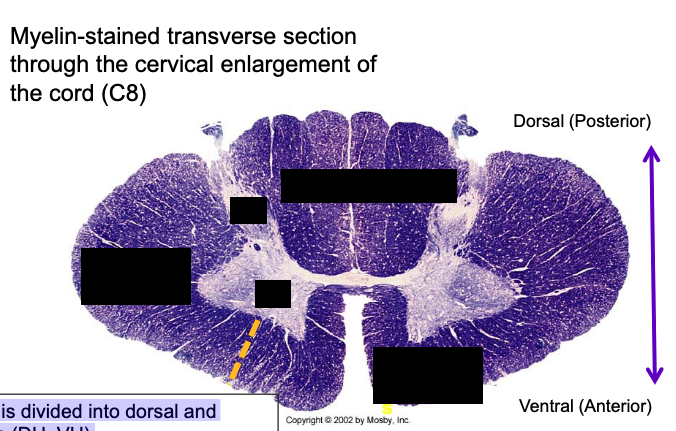
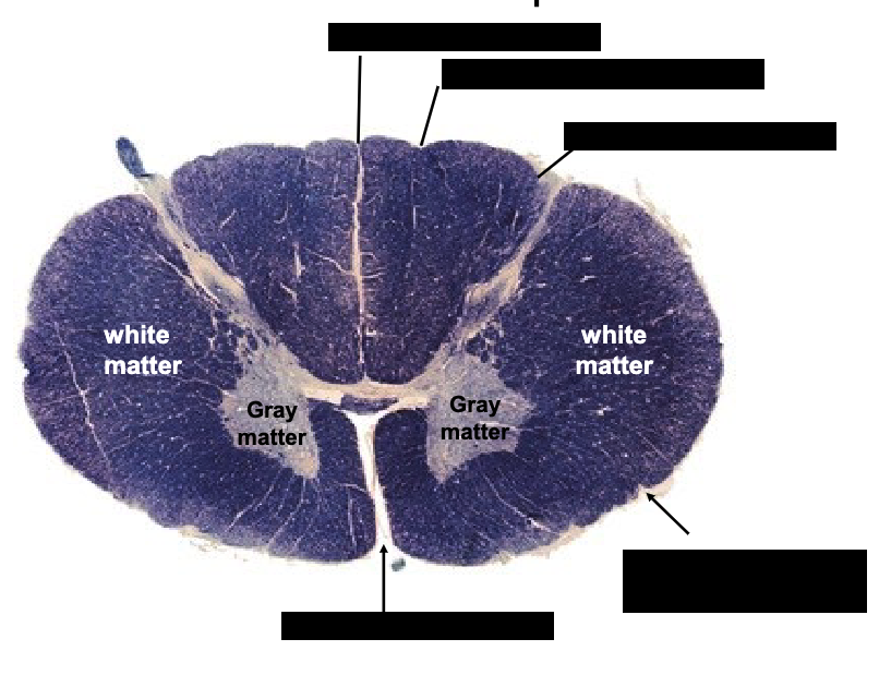
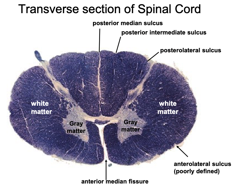
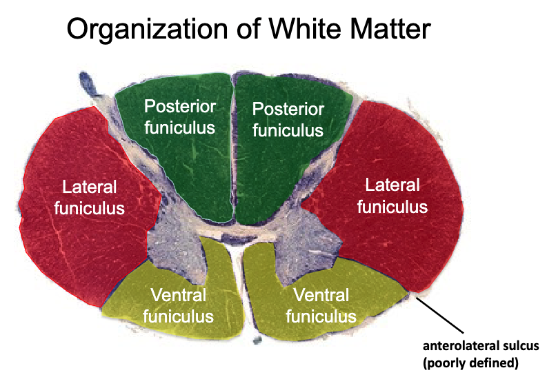
gray matter is divided into…?
white matter is divided into…?
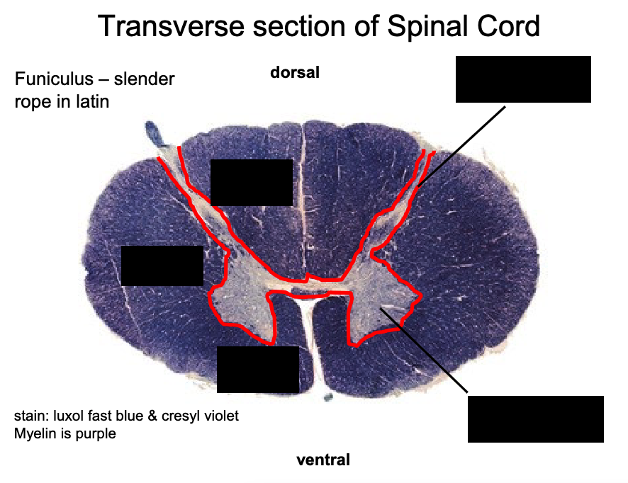
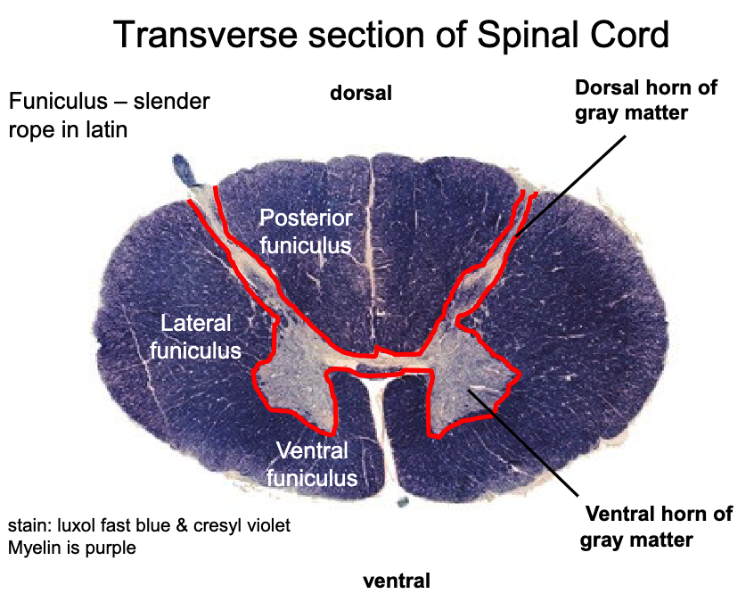
Gray matter is divided into dorsal and ventral horns (DH, VH)
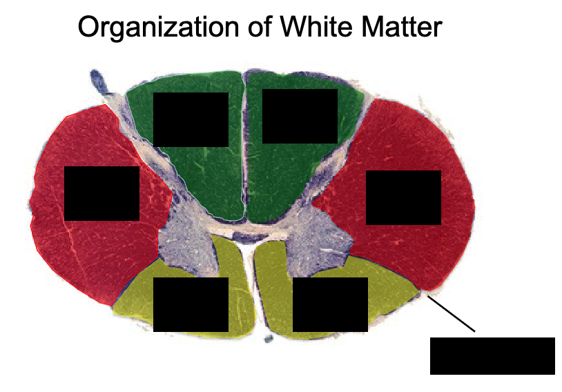
White matter is divided into funiculi (dorsal {posterior}, ventral {anterior}, lateral)
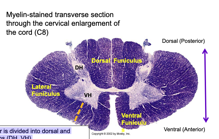
what is a funiculus?
bundle of nerve fibers (how the white matter is divided)

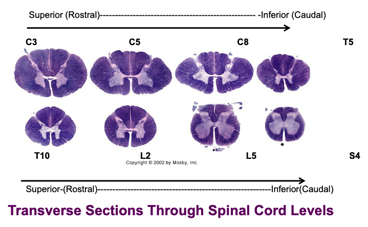
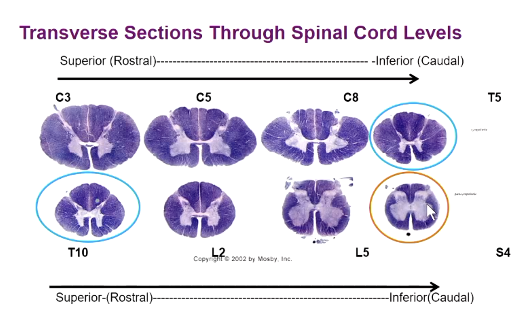
what are some similarities and differences throughout the spinal cord?
all have ventral/dorsal horns and faniculi
cervical regions = largest parts of cord (most white matter)
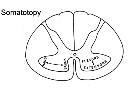
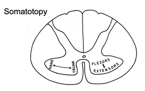
The motor neuron bodies in the ventral horn are positioned according to…?
the muscles they innervate
For more distal-lateral muscles neurons are lateral
Neurons for flexors are more dorsal
extensors are ventral
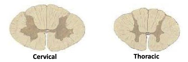
why do the transverse sections of cervical and thoracic spinal cords look so different?
cervical sections involve brachial plexus (lots of muscles in arm so more lateral location of motor neuron bodies in ventral horn)
thoracic sections involve the torso which has less muscles so less gray matter)
where are the autonomic neurons located in the spinal cord?
preganglionic sympathetic → T1-L2
preganglionic parasympathetic → S2-S4
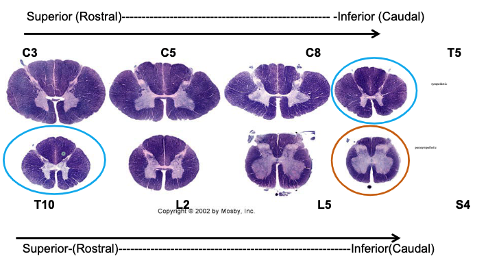
Preganglionc sympathetic neurons for the autonomic NS are visible in T5 and T10. Which area are they located in?
bulge in central part between dorsal and ventral horns
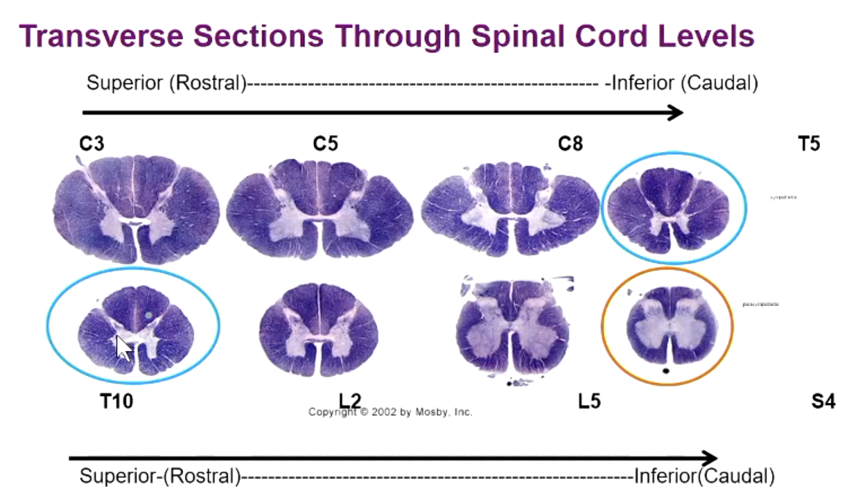
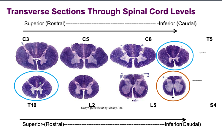
Preganglionic parasympathetic neurons for the pelvic viscera found in S2-S4. Which area are they located in?
no bulge but also central part between dorsal and ventral (similar location to sympathetic neurons)
what levels is the substantia gelatinosa nucleus located at?
all levels
what levels is the body of posterior horn nucleus located at?
all levels
what levels is the ventral horn nucleus located at?
all levels
what levels is the Clarke’s nucleus located at?
T1-L2
what levels is the intermediolateral column nucleus located at?
T1-L3
what levels is the parasympathetic nucleus located at?
S2-S4
what levels is the phrenic nucleus located at?
C3-C5
what is the function of the intermediolateral column?
Preganglionic sympathetic
what is the function of the parasympathetic nucleus?
Preganglionic parasympathetic
what is the function of the phrenic nucleus?
motor neurons-diaphragm
what is the function of the substantia gelatinosa nucleus?
modulate transmission of pain and temperature information
what is the function of the body of posterior horn nucleus?
sensory processing
what is the function of the ventral horn nucleus?
motor neurons
what is the function of the Clarke’s nucleus?
posterior spinocerebellar cells
what are the 5 basic functional groups of myelinated axons within the spinal cord?
long ascending fibers
long descending fibers
efferent (motor) fibers
afferent (sensory) fibers
propriospinal fibers
what are long ascending fibers?
sensory “bottom-up” projections from DRG destined to reach the cortex, thalamus, and brainstem
what are long descending fibers?
motor fibers “top-down” from the cortex, brainstem destined to reach motor neurons in the ventral horn that project to peripheral muscles
what are efferent (motor) fibers?
exiting projections from motor or autonomic neurons from the ventral horn
what are afferent (sensory) fibers?
incoming projections from dorsal root ganglion (DRG) to reach the dorsal horn
what are propriospinal fibers?
3 propriospinal tracts (dorsal, lateral, ventral) that ascend, descend, cross, and don’t cross that stay intrinsic to the spinal cord and interconnect different levels –Reflexes described by Dr Leavis
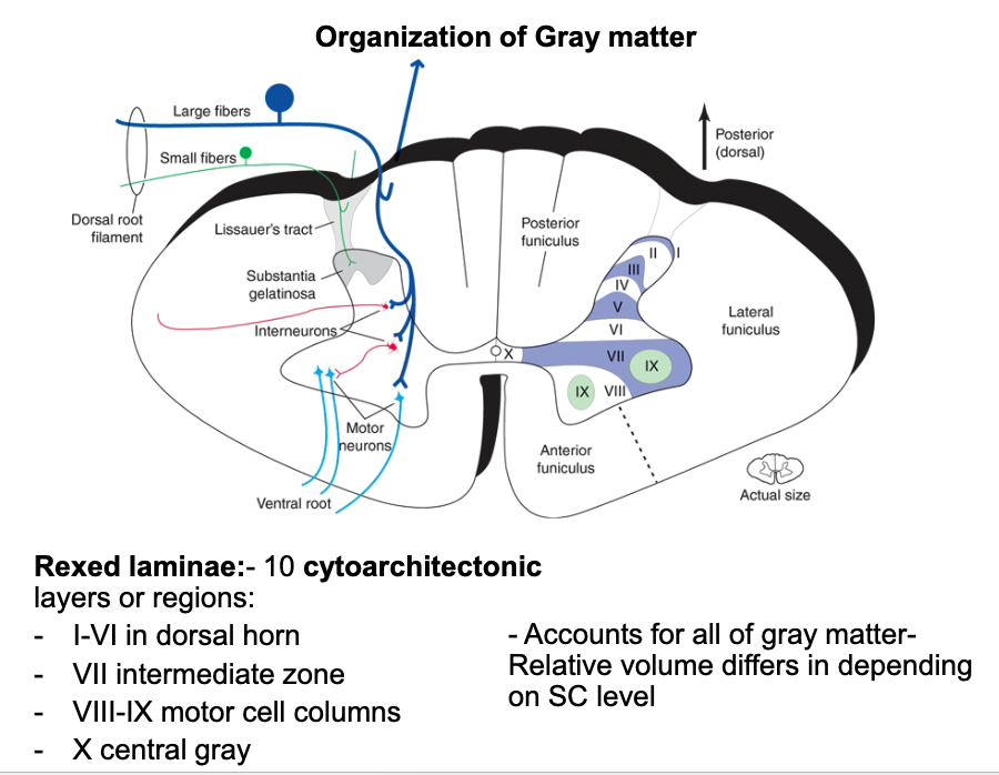
how is the gray matter organized?
rexed laminae (10 cytoarchitectonic layers/regions)
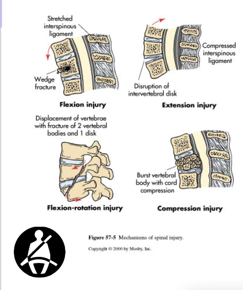
what are the mechanisms of spinal injury: trauma?
Hyperextension: Excessive posterior movement of head or neck. Extension injuries.
Hyperflexion: Excessive anterior movement of head onto chest. Flexion Injuries.
Compression: weight of the head or pelvis into a stationary neck or torso. Diving into shallow water.
Rotation: Excessive rotation of head/neck or torso
what is paresis?
injury caused by lesion on spinal cord but some muscle strength is preserved
what is plegia/paralysis?
complete loss of muscle function
what is a quadriplegic (tetraplegic)?
Injury of the cervical spinal cord
Some Patients can move their arms using the segments above the injury (e.g., in a C7 injury, the patient can still flex their forearms, using the C5 segment)
what is paraplegia?
Injury of the thoracic or lumbo-sacral cord, or cauda equina
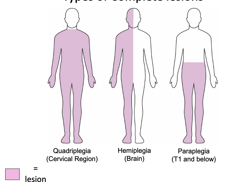
what is hemiplegia?
Paralysis of one half of the body
Usually in brain injuries (e.g., stroke)
quadriplegia is caused by injury in which region? what does this affect?
cervical region
all 4 extremities affected
paraplegia is caused by injury in which region? what does this affect?
injury in thoracic, lumbar, or sacral segments
lower extremities affected
what are the types of complete lesions?
quadriplegia (cervical region)
hemiplegia (brain)
paraplegia (T1 and below)
what are types of incomplete lesions?
Anterior Cord Syndrome
Posterior Cord Syndrome
Central Cord Syndrome
Brown – Sequard Syndrome
incomplete lesions are caused by…?
compression or loss of blood flow or compression of cord

what is anterior cord syndrome?
motor paralysis below the level of the lesion as well as the loss of pain and temperature at and below the level of the lesion
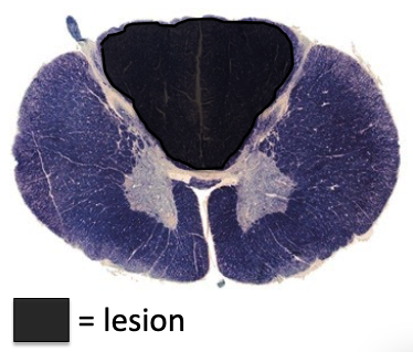
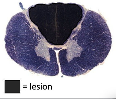
black = lesion
what is posterior cord syndrome?
(rare)
loss of proprioceptive sensation, fine touch, pressure, and vibration below the lesion
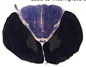
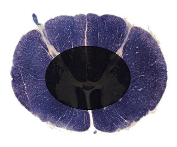
a lesion in the black area would cause what type of lesion?
Anterior Cord Syndrome
motor paralysis below the level of the lesion as well as the loss of pain and temperature at and below the level of the lesion
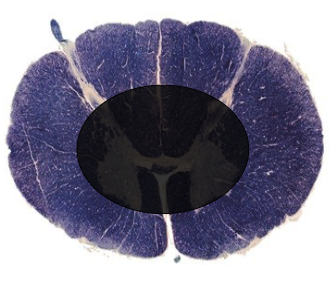
a lesion in the black area would cause what type of lesion?
Posterior Cord Syndrome (rare)
loss of proprioceptive sensation, fine touch, pressure, and vibration below the lesion
what is central cord syndrome?
loss of motion and sensation in arms and hands, damage occurs due to spondylosis due to osteoarthritis
what is the most common type of incomplete lesion?
central cord syndrome
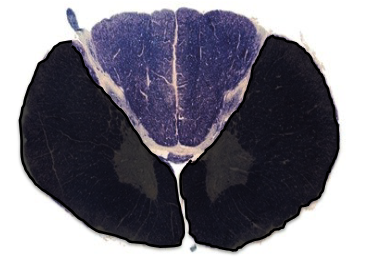
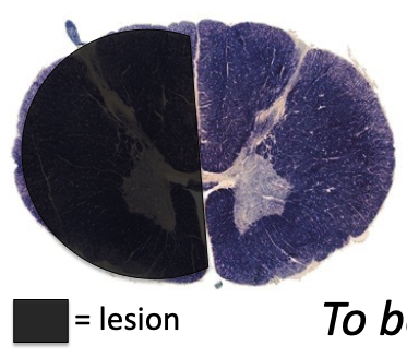
a lesion in the black area would cause what type of lesion?
central cord syndrome
what is brown-sequard syndrome?
hemisection of the spinal cord resulting in paralysis and loss of proprioception on the ipsilateral side as the injury or lesion, and loss of pain and temperature sensation on the contralateral side as the lesion
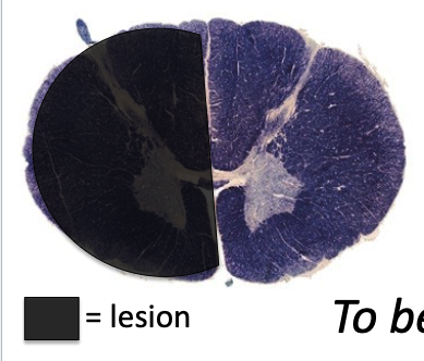
a lesion in the black area would cause what type of lesion?
Brown – Sequard Syndrome
hemisection of the spinal cord resulting in paralysis and loss of proprioception on the ipsilateral side as the injury or lesion, and loss of pain and temperature sensation on the contralateral side as the lesion