Biochemistry Exam 1
 Call Kai
 Learn
Learn Practice Test
 Spaced Repetition
 Match
Match Flashcards
 Knowt Play
1/155

There's no tags or description
Looks like no tags are added yet.
Name | Mastery | Learn | Test | Matching | Spaced | Call with Kai | Chat |
|---|
No analytics yet
Send a link to your students to track their progress
156 Terms
Biochemistry
The fundamental understanding of the molecular basis for the functioning and malfunctioning of living tissue, including the study of the building blocks of life
Buffer
A chemical pair, typically a weak acid and its conjugate base, that helps maintain a specific environment required for healthy cell function
pH
pH is a measure of hydronium ion (H3O+) concentration
Strong Acid Dissociation
Acids that dissociate completely (~100%) when added to water, such as Hydrochloric Acid (HCl)
Weak Acid Dissociation
Acids that dissociate only partially (
What type of logarithm is used in the pH scale?
Logarithm base 10 (log10)
What does a change of 1 pH unit represent in terms of [H3O+]?
A 10-fold change
What does a change of 2 pH units represent in terms of [H3O+]?
A 100-fold change
What does a change of 3 pH units represent in terms of [H3O+]?
A 1000-fold change
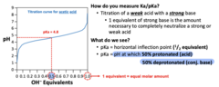
pKa Definition (Titration)
The horizontal inflection point on a titration curve (0.5 equivalents of base) where a group is 50% protonated and 50% deprotonated
pH=pKa when 50% disassociation occurs
Henderson-Hasselbalch Equation
The formula pH = pKa + log10([Conjugate Base]/[Acid]), used to calculate biological pH or relative acid-base concentrations
Ka
acid disassociation constant (how strong an acid is)
H30+ x Conj Base/ Conj Acid= Products/ Reactants
what is Ka of a strong acid
large number (in millions)
What is the Ka of a weak acid?
small number
pKa of a strong acid
-6.3 SMALL
pKa of a weak acid
4.8 LARGE
how do you measure Ka/pKa
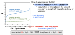
titration of a weak acid with a strong base (1 equivalent of a strong base is the amount necessary to completely neutralize a strong or weak acid)
what is the ratio needed to titrate a weak/strong acid with a strong base
1:1
pH
weak acid protonates
pKa>pH
weak acid deprotonates
Effective Buffer Range
A buffer system is most effective at resisting pH changes within ±1 pH unit of its pKa
what are the factors that make a buffer good
pH range
having ALOT of the buffer
amino acid structure
amino group (NH2), carboxyl group (COOH), R group
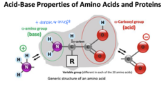
why is there a range of pKa's for amino/carboxyl groups for different amino acids
environment/interaction with the R-groups makes a difference
Isoelectric Point (pI)
The specific pH at which a molecule, such as an amino acid, carries no net electrical charge; it is marked by a vertical inflection point on a titration curve (in the middle)
why is pI significant
this is where a buffer becomes ineffective
titration of glycine
there is no dissociable R-group, just 1 acid and 1 base
-causes there to be two ideal pKa's for glycine because of the dissociation of either titratable species
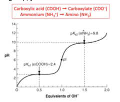
what do we see for ionizable species with glycine
# equivalents of OH- = # of ionizable species (pKa)
causes a strict order based off the pKa that COOH goes first then NH3+
glutamic acid titration
has 3 equivalents of strong OH- which are required to titrate the 3 ionizable groups of glutamic acid so ends up having 3 effective buffering ranges
Histidine Buffering
One of five amino acids with buffering side chains (pKa ~6.0); it allows hemoglobin to function as a buffer during gas transport
is histidine a good buffer when not bound
no its pKa is too low but when bound to hemoglobin its a good buffer
Blood pH Homeostasis
In healthy individuals, blood pH is strictly maintained within a narrow range of 7.35 to 7.45
Primary Blood Buffers
Biological pH in the blood is regulated by carbonic acid/bicarbonate (pKa 6.1), proteins like hemoglobin (pKa ~6.8), and phosphoric acid/phosphate (pKa 7.2)
Saliva pH Range
Healthy saliva typically ranges from pH 6.2 to 7.5; it is an "open system" influenced by factors like diet, medication, and disease
why is saliva important in the mouth
contains multiple buffers which helps neutralize acid
is a natural lubricant to help reduce abrasions
contains minerals
is a natural rinsing agent for plaque
has antimicrobial properties to help limit bacterial colonization
xertostomia
lack of saliva that causes dry mouth
can adversely affect oral health
what causes xerostomia
consequence of some diseases (sjogrens or diabetes 1 or 2)
side effect of certain medications
Critical Saliva pH
The threshold of 5.5; if saliva pH drops below this, tooth enamel begins to demineralize, increasing cavity risk
what minerals does saliva contain to help with early decay
calcium/phosphate
is phosphate higher in saliva or blood
saliva
Stimulated Saliva Buffering
gustatorily stimulated, significantly increases the concentration of bicarbonate in saliva, making it a more effective buffer than resting saliva
GERD
gastroesophageal reflux disease, treated with diet/lifestyle changes, medications for acid reduction, and sugar free gum to help stimulate saliva reproduction
acidosis
alkalosis
> pH 7.45, net gain of base in body fluids
how does bicarbonate and carbonic acid regulate blood pH
bicarbonate:carbonic acid
20:1 ratio
how can a reduction in bicarbonate influence blood pH
it lowers the pH causing borderline acidosis
how can an increase in bicarbonate influence blood pH
it raises the pH causing borderline alkalosis
what system regulates CO2 in blood
respiration
what type of pH regulation does respiration do in the blood
quick regulation and becomes a minor contribution overall to blood pH
what system regulates acid and bicarbonate (HCO3-) in the blood
kidneys
what type of regulation does kidney do in the blood
slower pH regulation and becomes a major contribution to pH of blood
respiration response to acidosis
increased respiration rate, which causes blood co2 to decrease and blood h2co3 to lower and increased pH to create homeostasis
respiration response to alkalosis
a decreased respiration rate causes a blood co2 to increase and blood h2co3 to increase and lowers pH to crease homeostasis
what is the kidneys response to acidosis
it lowers blood acid and increases hco3 and increases pH
what is the kidneys response to alkalosis
it increases blood acid and decreases hco3 and lowers pH
Respiratory Acidosis
Excessive acid in body fluids caused by a buildup of CO2 in the blood due to hypoventilation
what are common causes of respiratory acidosis
Neuromuscular disorders, chest muscle weakness, chest deformities/injuries, chronic lung disease, and sedative drug abuse
Metabolic Acidosis
Excessive acid buildup or bicarbonate loss (which is acid gain)
metabolic acidosis causes
diabetic acidosis (if there is no sugar in the blood the body uses fats which creates ketones which are acids)
hyperchloremic acidosis (loss of bicarbonate and increased chloride)
lactic acidosis (lactic acid build up)
kidney disease
aspirin poisoning
Respiratory Alkalosis
Excessive base in body fluids caused by insufficient CO2 in the blood
what causes respiratory alkalosis
anxiety (hyperventilation)
lack of O2 from high altitude
Metabolic Alkalosis
Excessive base caused by acid loss or bicarbonate gain
what causes metabolic alkalosis
dehydration which causes changes in body's water distribution
repeated vomiting
excessive sweating
abuse of diuretics
diarrhea
kidney disease (can cause either)
Peptide Bond Formation
A covalent amide linkage formed through a condensation reaction (releasing H2O) between the alpha-carboxyl group of one amino acid and the alpha-amino group of another
Peptide Bond Character
It possesses partial double bond character, making the bond rigid and restricted in rotation, which keeps the atoms in a single plane
what causes peptide bond structure to be rigid
the alpha carbon and the plane that the peptide bonds they end up depend on R-groups
Trans vs. Cis Configuration
Most amino acids prefer the trans configuration to avoid steric clash (~1,000:1 ratio)
which amino acid is the exception to the the trans configuration favoring
proline because the side chain comes back and is covalently linked to amide
Phi (φ) and Psi (ψ) Angles
Phi is the rotation around the Ca-N bond and Psi is the rotation around the Ca-C bond; these angles help dictate secondary structure
what dictates protein function
protein structure which in turn is dictated by amino acid structure
Primary Protein Structure
The linear sequence of amino acids in a polypeptide chain, determined by the DNA sequence
Secondary Protein Structure
Local structural elements like alpha-helices, beta-strands/sheets, and beta-turns, stabilized by hydrogen bonds between main chain atoms
Alpha-helix Characteristics
A right-handed corkscrew structure (~4 residues/turn)
stabilized by H-bonds between amide protons (n+4) and carbonyl oxygens (n) MAIN CHAIN ATOMS
present in globular and fibrous proteins
which amino acids do alpha-helices not normally contain
glycine and proline
why is glycine not normally found in secondary structures
it has a small R group (H)
causes it to be really flexible
common in loops and required for beta turns
Beta-strand
Beta-strands are extended structures (~2.3x longer than alpha-helices)
open phi and psi angles
usually associated with other strands to form beta sheet
stabilized by main chain atoms
beta sheets
created of multiple beta strands
can be parallel or anti parallel
amide protons of one strand hydrogen bond to the carbonyl oxygens of the second strand
stabilized by main chain atoms
which is more stable: parallel or anti-parallel beta sheets
parallel
Beta-turn
A 4-amino acid motif (often X-Pro-Gly-X) that allows a polypeptide to reverse direction; Proline and Glycine are essential for these tight corners
which form of proline creates beta turns
cis formation
which positions in a beta turn is usually hydrogen bonded
position 1 and 4
Proline as "Helix Breaker"
Because its imino side chain is covalently linked to the amide nitrogen, it lacks an amide hydrogen for H-bonding and disrupts regular secondary structures
which two amino acids are required for beta turns
glycine (allows the turn to still connect to the rest) and proline (causes the turn)
what stabilizes primary structure
covalent linkage between adjacent amino acids
tertiary Protein Structure
The 3D arrangement of secondary elements in a single polypeptide, caused by hydrophobic effect that has a thermodynamic explanation
thermodynamic explanation of tertiary structures
local folding begins in secondary structure usually spontaneously (-delta G), some may require help from chaperones, delta S of water helps to drive tertiary structures because of the hydrophobic effect, environment really matters
so delta S of water is favorable and delta H of protein is favorable
free energy equation
delta g (free energy) = delta h (enthalpy) - TdeltaS (entropy)
what do tertiary proteins usually have a role in
extra-cellular systems
what stabilizes tertiary/quartenary protein structure
stabilized by R-group interactions including H-bonds, van der waals, electrostatic forces, and disulfide bonds
Quaternary Protein Structure
The specific 3D association of multiple discrete polypeptide subunits (e.g., hemoglobin), the polypeptides may be the same or different, causes allostery within these proteins
allostery
regulation of the activity of a protein by the binding of an effector molecule to a site other than the active site
Thermodynamics of Folding
Driven by a decrease in free energy (ΔG); while protein entropy decreases, the hydrophobic effect increases water entropy (ΔS), making the process spontaneous and rapid
Hydrophobic Effect
The tendency of nonpolar side chains to cluster in the protein interior to minimize contact with water, serving as the primary driver of folding
what can errors in protein folding cause
disease like alzheimers, CF, mad cow disease, inherited emphysema, and certain cancers
what can influence final structure of protein folding
posttranslational modification and chaperones (atp), disulfide isomerase, and cis-trans isomerase, and temperature
Protein Modularity
The organization of proteins into domains, allowing structurally similar catalytic sites to work on different substrates via distinct specificity sites
why does protein modularity matter
helps drive evolution
Serine Protease Catalytic Triad
A functional module in enzymes like trypsin consisting of POLAR aa.- Aspartate (D102), Histidine (H57), and Serine (S195- which usually doesn't normally ionize) involved in a charge relay system
what does trypsin cut after
lys and arg
what does chymotrypsin cut after
phe, tyr, trp (large hydrophobic)