Pulmonary path II: Obstructive Lung Diseases
 Call Kai
 Learn
Learn Practice Test
 Spaced Repetition
 Match
Match Flashcards
 Knowt Play
1/28

There's no tags or description
Looks like no tags are added yet.
Name | Mastery | Learn | Test | Matching | Spaced | Call with Kai | Chat |
|---|
No analytics yet
Send a link to your students to track their progress
29 Terms
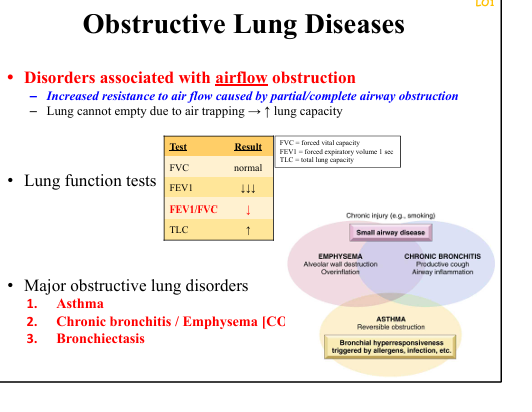
Major obstructive lung diseases
Asthma, Chronic bronchitis, Emphysema, Bronchiectasis.
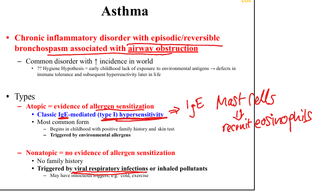
Asthma definition
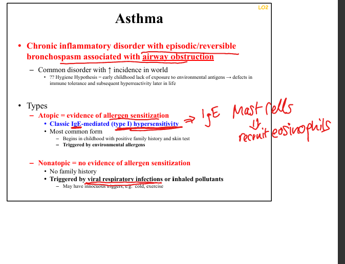
Chronic inflammatory disorder with episodic, reversible bronchospasm causing airway obstruction.
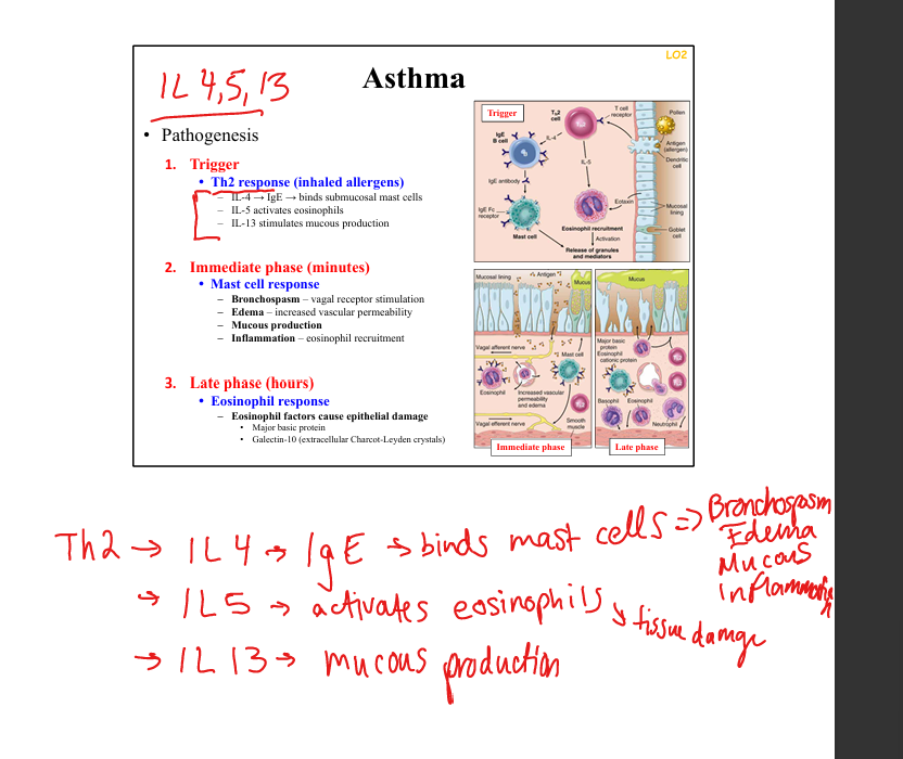
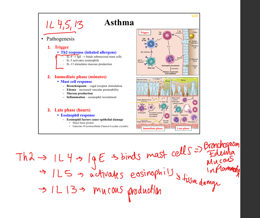
Asthma immune pathogenesis
TH2-mediated(inhaled allergens): IL‑4 → IgE, IL5 activates eosinophils, IL13 stimulates mucous production
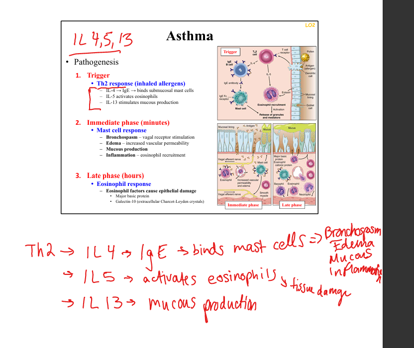
Immediate phase of asthma
Mast cell degranulation → bronchospasm, edema, mucus production.
Late phase of asthma
Eosinophil-mediated epithelial damage (major basic protein, Charcot‑Leyden crystals).
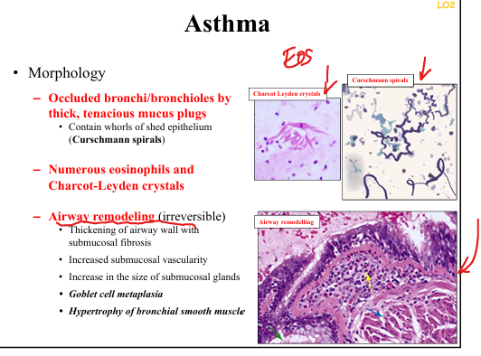
Asthma airway remodeling
Goblet cell metaplasia, smooth muscle hypertrophy/hyperplasia, submucosal gland enlargement, basement membrane thickening.
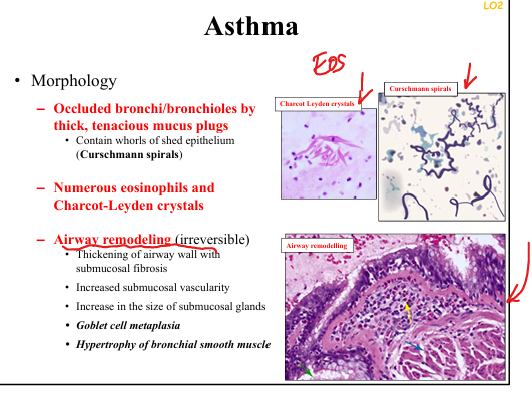
Classic sputum findings in asthma
Curschmann spirals(epithelium that has been shed) + Charcot‑Leyden crystals(destroyed eosinophils).
Triggers for atopic asthma
Allergens, dust, pollen, pets
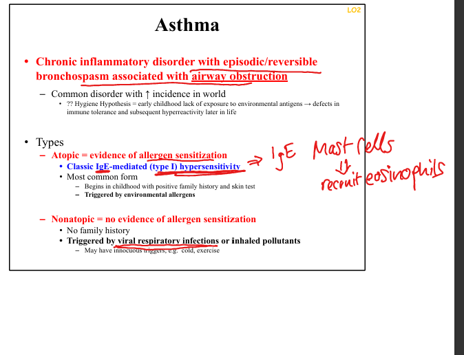
Triggers for non-atopic asthma
Viral infections, pollutants, cold air, exercise
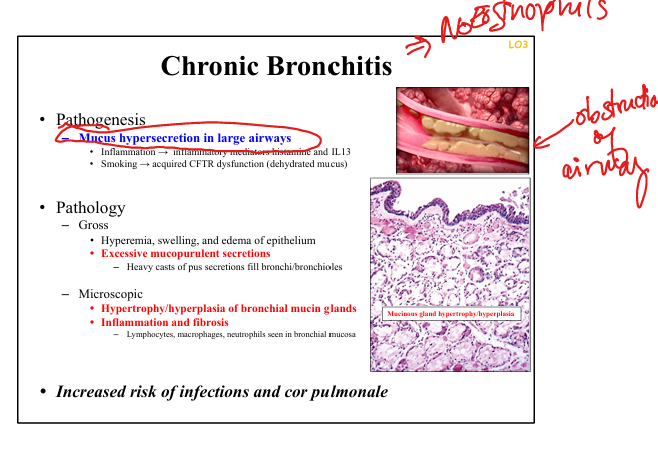
Chronic bronchitis definition
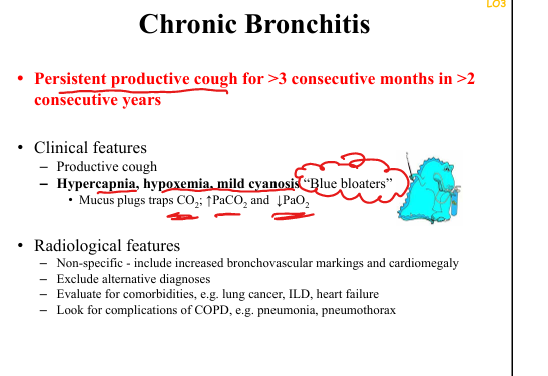
Productive cough ≥3 months per year for ≥2 consecutive years.
Chronic bronchitis pathogenesis
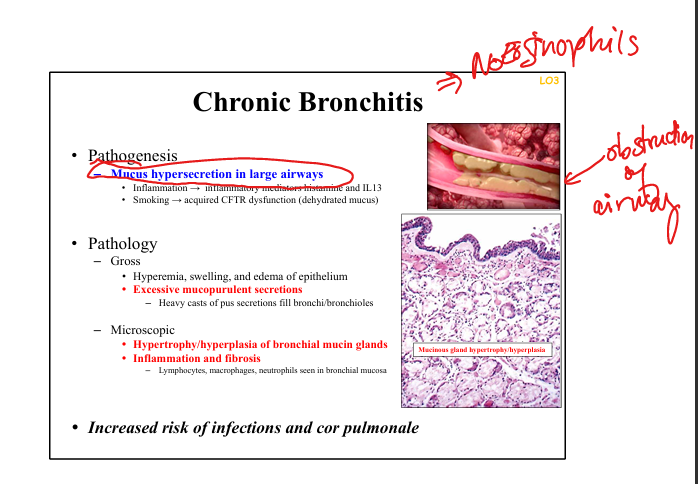
Smoking → mucous gland hyperplasia, goblet cell hyperplasia, CFTR dysfunction → dehydrated mucus.
Chronic bronchitis clinical features
Productive cough, wheezing, hypercapnia, hypoxemia, mild cyanosis (“blue bloater”).
Chronic bronchitis gross pathology
Hyperemia, edema, mucopurulent secretions filling bronchi.
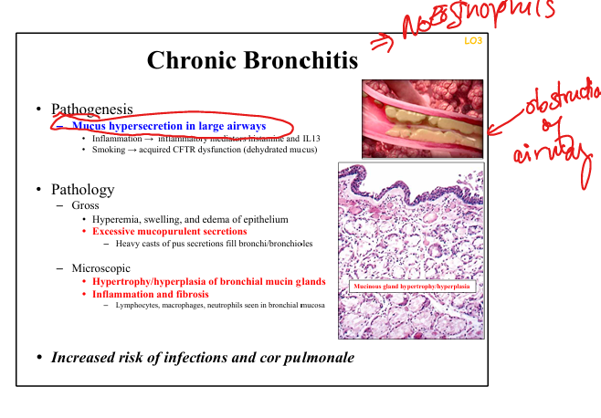
Chronic bronchitis microscopic pathology
Mucinous gland hypertrophy/hyperplasia, chronic inflammation, fibrosis.
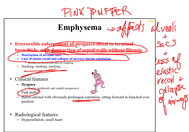
Emphysema definition
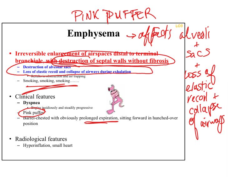
Permanent enlargement of airspaces distal to terminal bronchiole with destruction of septal walls without fibrosis.
Emphysema pathogenesis
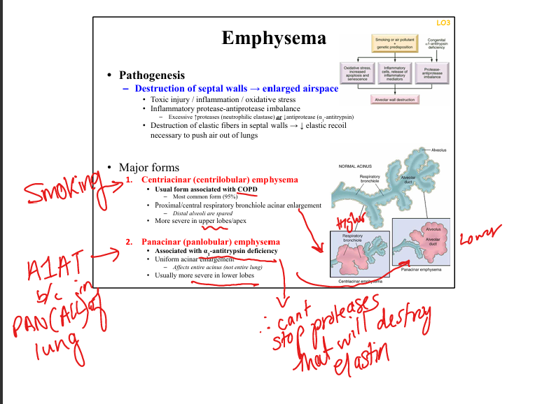
Protease–antiprotease imbalance (↑neutrophil elastase, ↓A1AT), oxidative stress, inflammation»leads to destruction of septal walls and enlarged air space
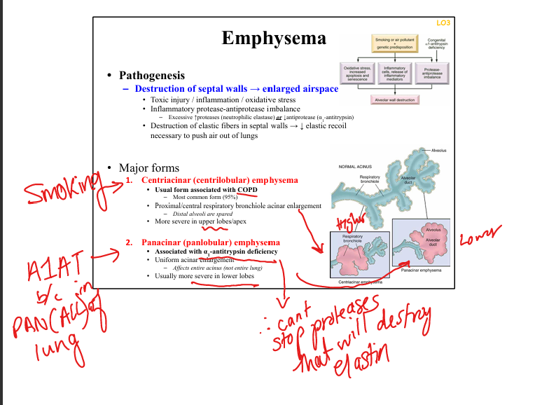
Centriacinar emphysema
Smoking-related, upper lobes
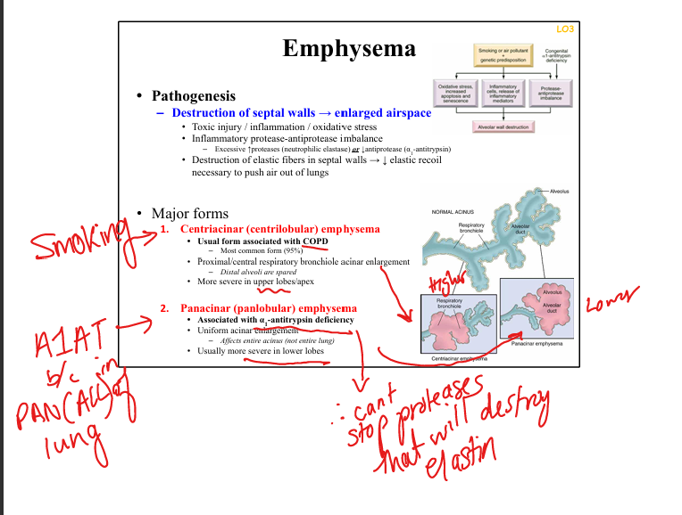
Panacinar emphysema
A1AT deficiency, lower lobes
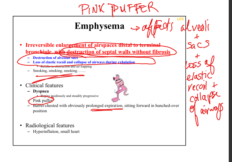
Emphysema clinical features
Dyspnea, prolonged expiration, barrel chest, “pink puffer.”
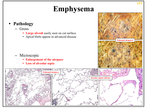
Emphysema gross pathology
Enlarged airspaces, apical blebs, bullae.
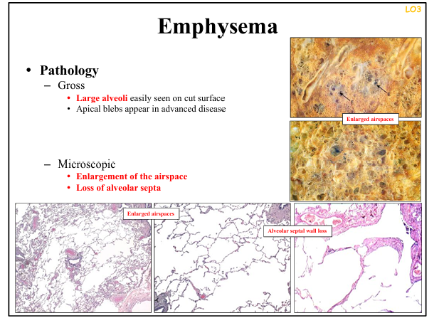
Emphysema microscopic pathology
Loss of alveolar septa, enlarged airspaces, minimal fibrosis.
Why emphysema causes air trapping
Loss of elastic recoil → airway collapse during exhalation.
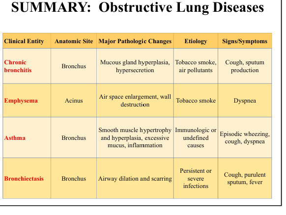
COPD= (two diseases
Chronic bronchitis + emphysema causing airflow limitation
Blue bloater vs pink puffer
Blue bloater = chronic bronchitis (hypoxemia, hypercapnia). Pink puffer = emphysema (dyspnea, hyperventilation).
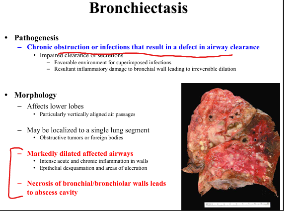
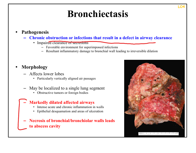
Bronchiectasis definition
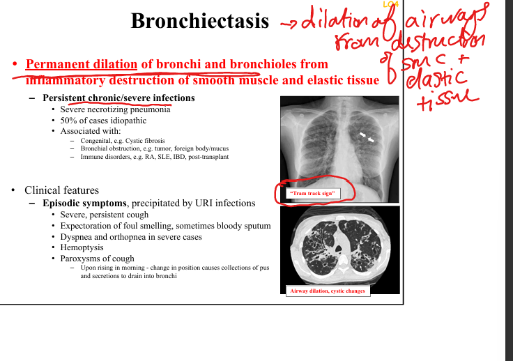
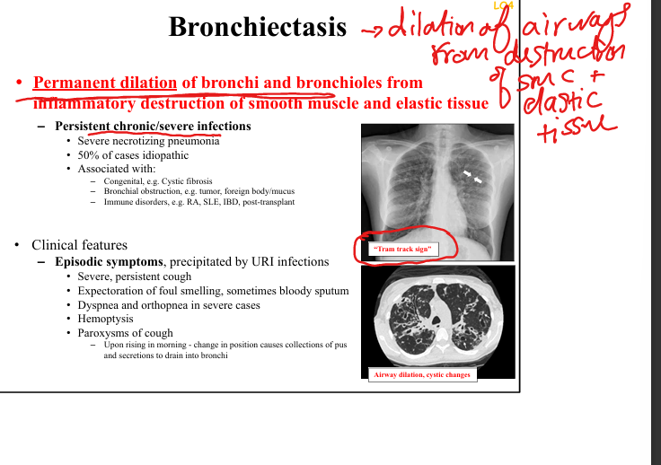
Permanent dilation of bronchi/bronchioles due to inflammatory destruction of smooth muscle + elastic tissue.
Bronchiectasis pathogenesis
Chronic obstruction + infection → impaired clearance → inflammation → airway wall destruction.
Bronchiectasis clinical features
Chronic cough, foul-smelling sputum, hemoptysis, recurrent infections, worse in morning.
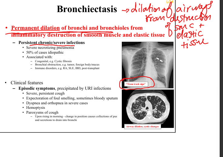
Bronchiectasis radiology
CXR: tram-track sign, peribronchial cuffing. CT: signet-ring sign, lack of tapering, airway visibility to periphery.
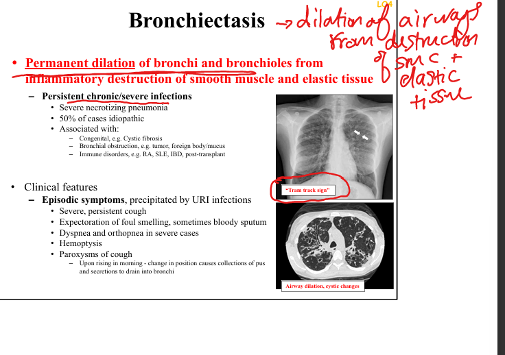
Bronchiectasis gross pathology
Dilated airways filled with pus