Seasons Wk5 LG pt.1
 Call Kai
 Learn
Learn Practice Test
 Spaced Repetition
 Match
Match Flashcards
 Knowt Play
1/161

Earn XP
Description and Tags
Comparing patterns of motility in the esophagus and stomach
Name | Mastery | Learn | Test | Matching | Spaced | Call with Kai | Chat |
|---|
No analytics yet
Send a link to your students to track their progress
162 Terms
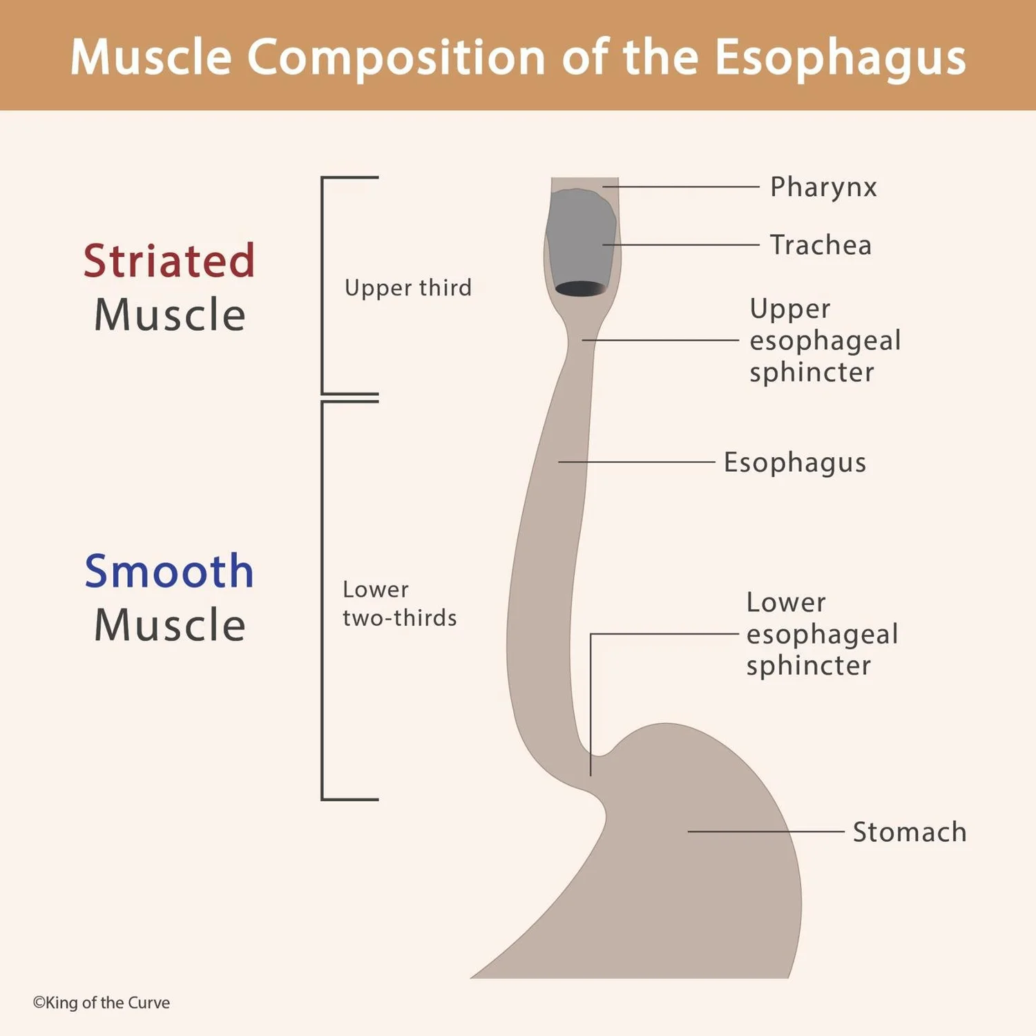
What is the major neural-control transition from the upper esophagus to the stomach?
Answer: Control transitions from voluntary somatic motor control in the upper esophagus to involuntary autonomic and enteric control in the lower esophagus and stomach.
Extra Information:
The upper esophagus contains striated muscle.
The lower esophagus and stomach contain smooth muscle.
What is the primary motility function of the esophagus?
Answer: To rapidly propel food toward the stomach through sequential contractions.
Extra Information:
- The esophagus functions primarily as a conduit.
- Its major propulsive mechanism is peristalsis.
What are the major motility functions of the stomach?
Answer: Storage, mixing and grinding, and controlled gastric emptying.
Extra Information:
- The stomach acts as both a reservoir and a grinder.
- Its movements require coordinated local and autonomic control.
What type of muscle makes up the upper one-third of the esophagus?
Answer: Striated muscle.
Extra Information:
- This region is under direct somatic motor control.
What type of neural control predominates in the upper one-third of the esophagus?
Answer: Somatic motor control.
Extra Information:
- This differs from the autonomic and enteric control of the lower esophagus.
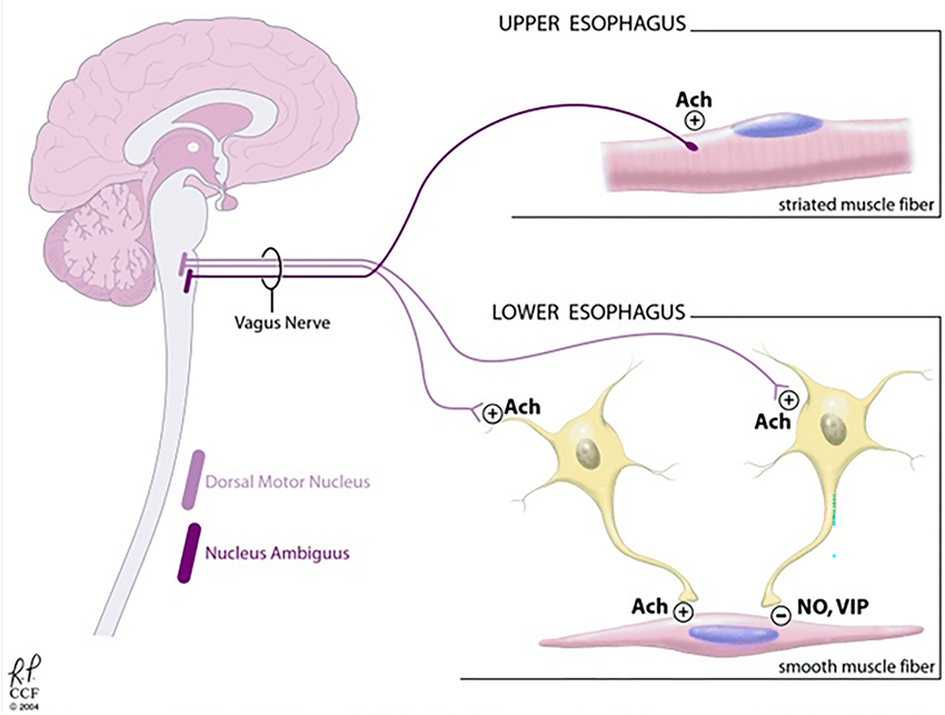
Which cranial nerve provides somatic motor control to the upper esophagus?
Answer: The vagus nerve, cranial nerve X.
Extra Information:
- These motor fibers originate from the nucleus ambiguus.
Where do the somatic motor fibers controlling the upper esophagus originate?
Answer: The nucleus ambiguus of the medulla.
Extra Information:
Their axons travel through the vagus nerve.
They directly stimulate striated esophageal muscle.
What does somatic vagal innervation do in the upper esophagus?
Answer: It directly stimulates striated muscle contraction and regulates upper esophageal sphincter relaxation.
Extra Information:
- This allows coordinated swallowing and entry of the bolus into the esophagus.
What type of muscle makes up the lower two-thirds of the esophagus?
Answer: Smooth muscle.
Extra Information:
Control of this region is autonomic rather than direct somatic motor control.
What systems regulate motility in the lower two-thirds of the esophagus?
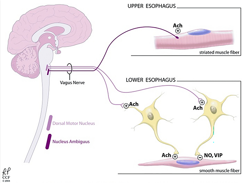
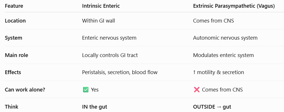
Answer: The intrinsic enteric nervous system and extrinsic parasympathetic vagal pathways.
Extra Information:
These systems coordinate contraction and relaxation around the food bolus.
Where do vagal preganglionic fibers controlling lower esophageal smooth muscle synapse?
Answer: On postganglionic neurons within the myenteric plexus.
Extra Information:
The myenteric plexus is part of the enteric nervous system.
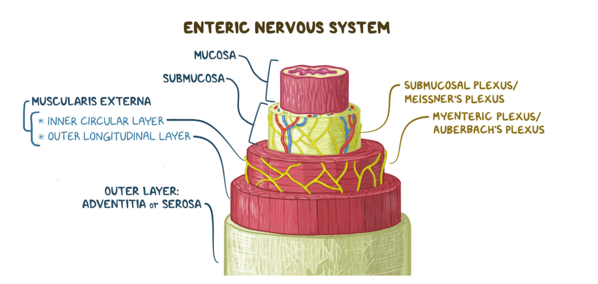
Which neurotransmitter promotes contraction behind an esophageal food bolus?
Answer: Acetylcholine.
Extra Information:
Substance P also contributes to excitatory contraction during peristalsis.
Think about what acetylcholine normally does to GI smooth muscle:
ACh → muscarinic receptors → ↑ intracellular Ca²⁺ → smooth muscle contraction
Which neurotransmitters promote relaxation in front of an esophageal food bolus?
Answer: Vasoactive intestinal peptide and nitric oxide.
Extra Information:
- These inhibitory mediators relax smooth muscle.
- They also participate in lower esophageal sphincter relaxation.
What is the effect of acetylcholine and substance P on gastrointestinal smooth muscle?
Answer: They promote depolarization and contraction.
Extra Information:
- They function as excitatory transmitters in gastrointestinal motility pathways.
What is the effect of vasoactive intestinal peptide and nitric oxide on gastrointestinal smooth muscle?
Answer: They promote smooth muscle relaxation.
Extra Information:
- These inhibitory transmitters are important for receptive relaxation and sphincter relaxation.
What type of muscle makes up the stomach?
Answer: Smooth muscle.
Extra Information:
Gastric motility is therefore controlled through autonomic and enteric mechanisms rather than somatic motor pathways.
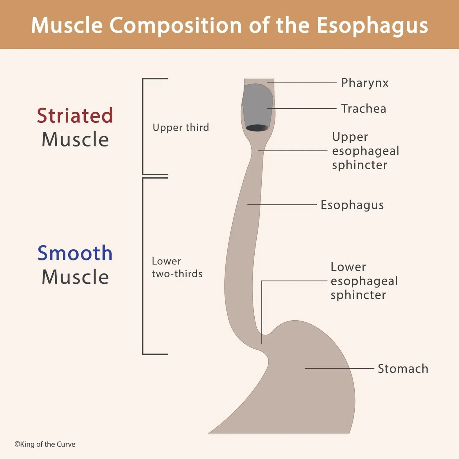
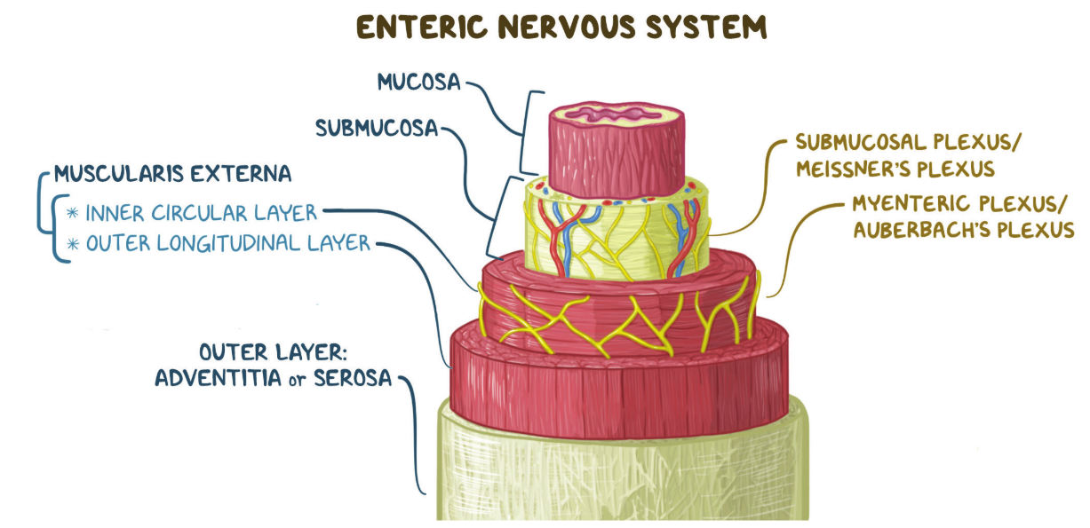
What are the two major plexuses of the enteric nervous system emphasized in the stomach?
Answer: The myenteric, or Auerbach's, plexus and the submucosal, or Meissner's, plexus.
Extra Information:
They perform different major functions within the gastrointestinal wall.
🧠 “Secretions Make Muscles Active”
S = Submucosal
M = Meissner
M = Myenteric
A = Auerbach
Then pair them:
Submucosal–Meissner 💧 = secretion
Myenteric–Auerbach 💪 = motility
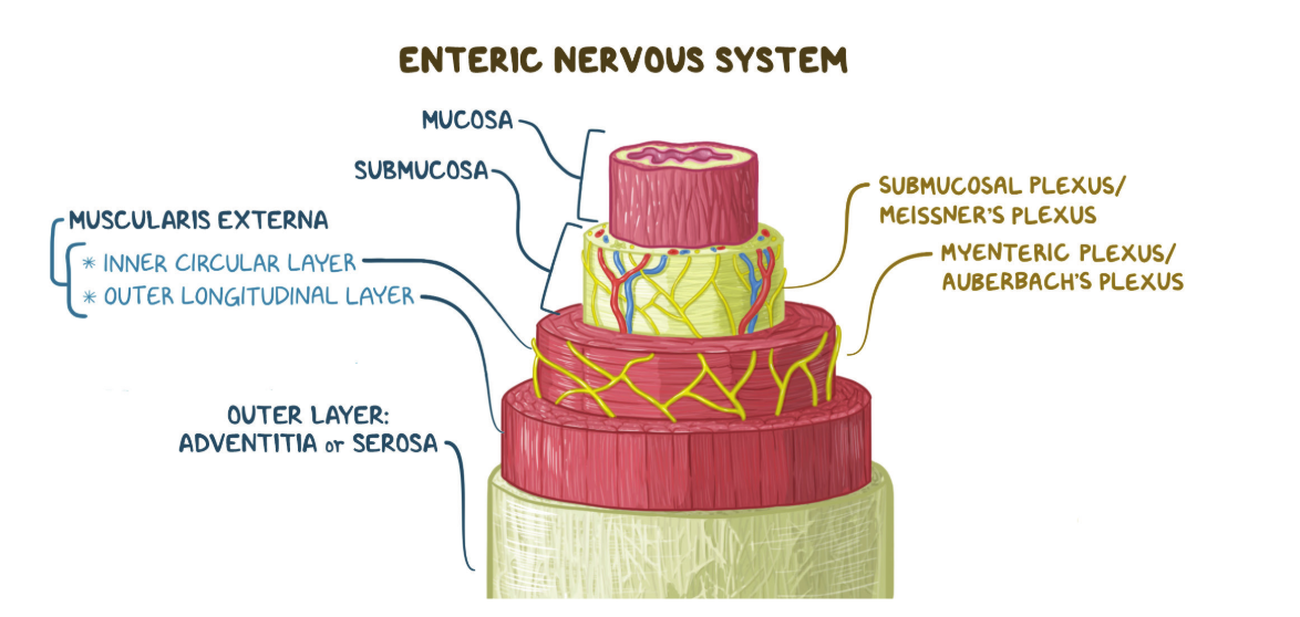
Where is the myenteric, or Auerbach's, plexus located?
Answer: Between the outer longitudinal and middle circular smooth muscle layers.
Extra Information:
Its location allows it to coordinate gastrointestinal smooth muscle activity.
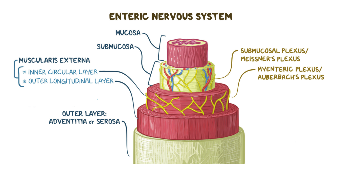
What is the major function of the myenteric plexus?
Answer: To regulate the force and coordination of gastrointestinal motility contractions.
Extra Information:
- It is the enteric plexus most directly associated with motility.
Where is the submucosal, or Meissner's, plexus located?
Answer: In the submucosa.
Extra Information:
- Its major functions differ from those of the myenteric plexus.
What are the major functions of the submucosal plexus?
Answer: Regulation of mucosal secretion and local blood flow.
Extra Information:
- The myenteric plexus is more directly involved in motility.
Which nerve provides the major extrinsic parasympathetic innervation to the stomach?
Answer: The vagus nerve.
Extra Information:
- Vagal pathways interact extensively with the enteric nervous system.
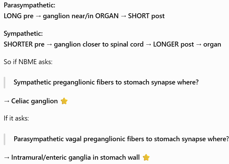
Where do parasympathetic vagal preganglionic fibers to the stomach synapse?
Answer: Within the gastric myenteric and submucosal plexuses.
Extra Information:
- These ganglia are located within the gastrointestinal wall.
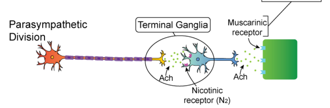
What neurotransmitter is released by vagal preganglionic fibers in the gastric enteric plexuses?
Answer: Acetylcholine.
Extra Information:
- Acetylcholine acts on nicotinic receptors on postganglionic neurons.
Which receptor is activated by acetylcholine released from gastric parasympathetic preganglionic fibers?
Answer: Nicotinic acetylcholine receptors.
Extra Information:
These receptors are located on postganglionic enteric neurons.
ACh released at autonomic ganglion → nicotinic receptor → activates next neuron.
What neurotransmitters are released by excitatory postganglionic neurons controlling gastric motility?
Answer: Acetylcholine and substance P.
Extra Information:
- These transmitters promote gastric smooth muscle contraction.
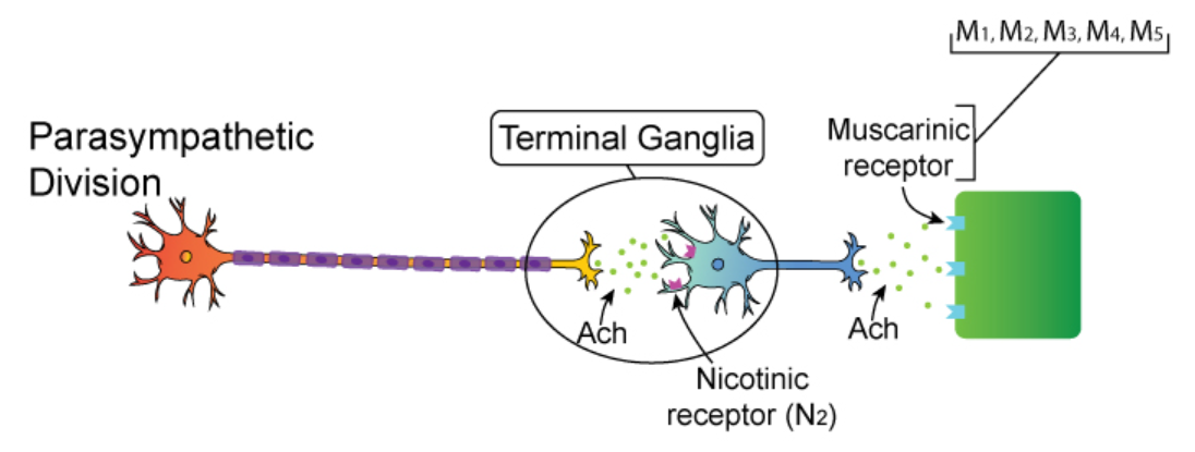
Which receptor on gastric smooth muscle is activated by postganglionic acetylcholine according to the provided material?
Answer: M3 muscarinic receptors.
Extra Information:
Activation promotes smooth muscle depolarization and contraction.
How do excitatory postganglionic fibers affect gastric motility?
Answer: They depolarize smooth muscle and increase the strength and frequency of gastric contractions.
Extra Information:
- Acetylcholine and substance P mediate these excitatory effects.
What transmitters are released by inhibitory postganglionic neurons in the stomach?
Answer: Vasoactive intestinal peptide and nitric oxide.
Extra Information:
- These transmitters relax gastric smooth muscle.
What is a vagovagal reflex?
Answer: A gastrointestinal reflex in which both the sensory afferent and motor efferent limbs travel through the vagus nerve.
Extra Information:
Vagovagal reflexes help coordinate gastric responses to swallowing and distension.
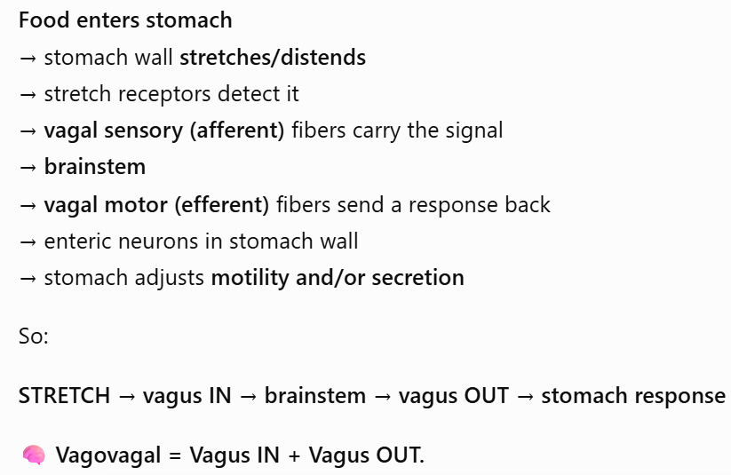
What stimulus can initiate a vagovagal reflex during eating?
Answer: Stretch of the esophagus or stomach detected by mechanoreceptors.
Extra Information:
- The sensory signal travels centrally through vagal afferents.
What gastric response is produced by a vagovagal reflex during swallowing?
Answer: Receptive relaxation of the orad stomach.
Extra Information:
This allows the stomach to accommodate incoming food.
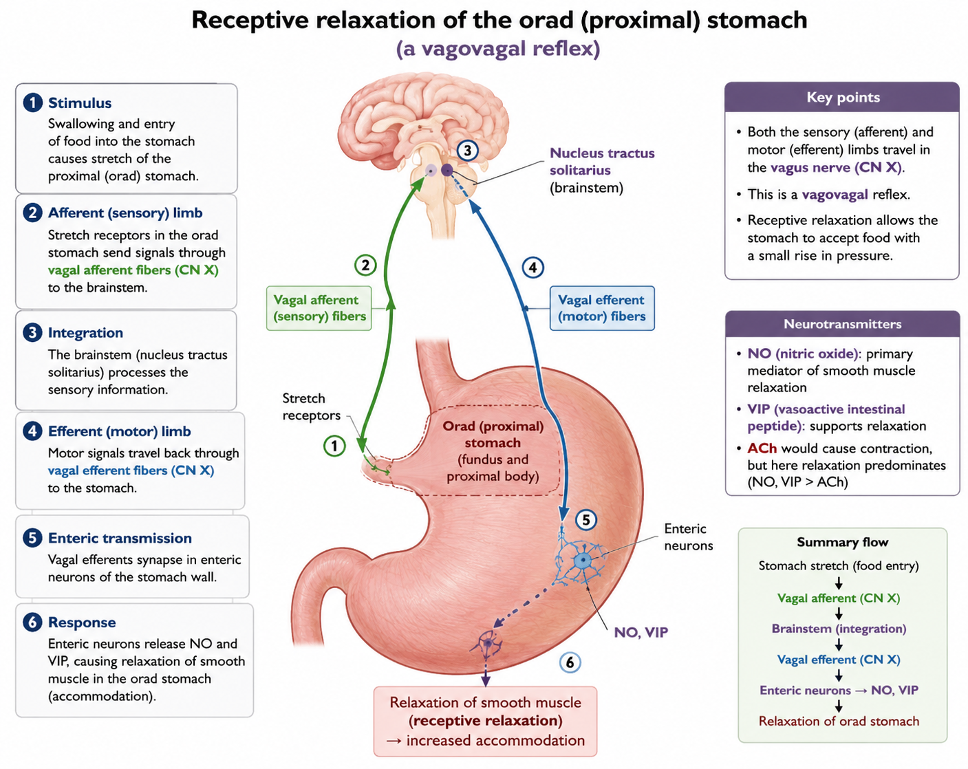
Which transmitters mediate vagovagal receptive relaxation of the stomach?
Answer: Vasoactive intestinal peptide and nitric oxide.
Extra Information:
- Both relax gastric smooth muscle.
What is the purpose of receptive relaxation?
Answer: To allow the proximal stomach to accommodate incoming food without a large increase in intragastric pressure.
Extra Information:
- It primarily involves the fundus and proximal body.
Where do sympathetic preganglionic neurons supplying the stomach originate?
Answer: In the thoracolumbar spinal cord.
Extra Information:
Their fibers ultimately communicate with sympathetic ganglia supplying the gastrointestinal tract.
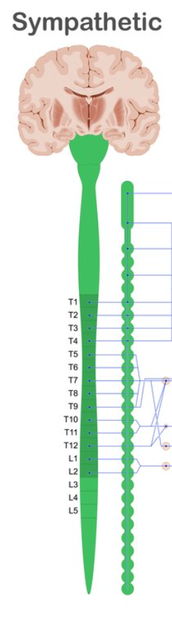
Where do sympathetic preganglionic fibers supplying the stomach synapse according to the provided material?
Answer: In the celiac ganglion.
Extra Information:
Preganglionic sympathetic fibers release acetylcholine at the ganglion.
What neurotransmitter is released by sympathetic preganglionic fibers in the celiac ganglion?
Answer: Acetylcholine.
Extra Information:
- Postganglionic sympathetic fibers use a different major neurotransmitter.
What neurotransmitter is released by postganglionic sympathetic fibers supplying the stomach?
Answer: Norepinephrine.
Extra Information:
Norepinephrine generally inhibits gastrointestinal activity.
NE → α₁ receptors on vascular smooth muscle → contraction → vasoconstriction
What is the overall effect of sympathetic stimulation on gastric motility?
Answer: It inhibits gastric motility and digestion.
Extra Information:
- Sympathetic activity can also promote vasoconstriction and sphincter contraction.
How can norepinephrine inhibit gastrointestinal motility through the enteric nervous system?
Answer: It acts on receptors on myenteric plexus ganglia to suppress gastrointestinal motility.
Extra Information:
- Sympathetic input therefore modifies intrinsic enteric activity.
How can sympathetic stimulation directly affect gastrointestinal blood vessels and sphincters?
Answer: It causes vasoconstriction and promotes sphincter contraction.
Extra Information:
- These effects contribute to the overall inhibition of digestion.
Compare the neural control of the upper esophagus, lower esophagus, and stomach.
Answer: The upper esophagus uses somatic vagal control of striated muscle, while the lower esophagus and stomach use autonomic and enteric control of smooth muscle.
Extra Information:
- The lower esophagus relies on the ENS and vagal autonomic fibers.
- The stomach additionally receives sympathetic input through the celiac ganglion.
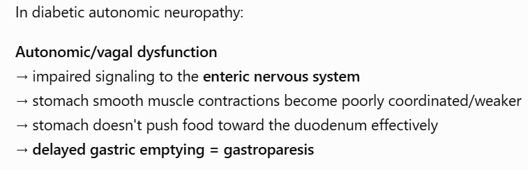
How can diabetic autonomic neuropathy disrupt gastric motility?
Answer: Chronic hyperglycemia can damage vagal autonomic fibers and disrupt extrinsic neural coordination of the stomach.
Extra Information:
- This impairs normal gastric motor function.
What gastric disorder can result from diabetic autonomic neuropathy?
Answer: Gastroparesis.
Extra Information:
Gastroparesis is delayed gastric emptying.
Vagal dysfunction is an important mechanism in the provided material.
What symptoms can result from diabetic gastroparesis?
Answer: Early satiety, abdominal bloating, and vomiting of undigested food hours after eating.
Extra Information:
- These symptoms reflect delayed movement of food out of the stomach.
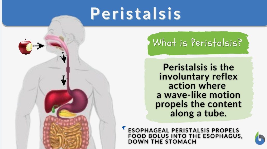
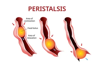
What is esophageal peristalsis?
Answer: A coordinated series of wave-like muscular contractions that propels a food bolus toward the stomach.
Extra Information:
Peristalsis creates coordinated contraction behind and relaxation ahead of the bolus.
What triggers primary esophageal peristalsis?
Answer: The swallowing reflex in the medulla.
Extra Information:
- Primary peristalsis is vagally mediated.
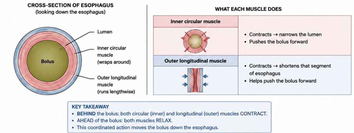
What happens to the inner circular muscle behind an esophageal bolus during primary peristalsis?
Answer: It contracts.
Extra Information:
Contraction narrows the lumen and creates high pressure behind the bolus.
What happens to the outer longitudinal muscle behind an esophageal bolus during primary peristalsis?
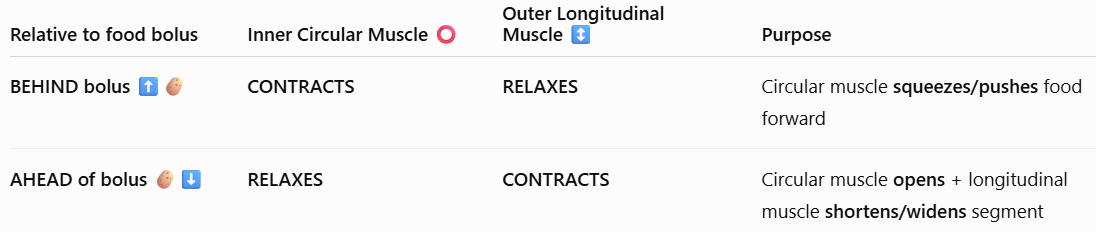
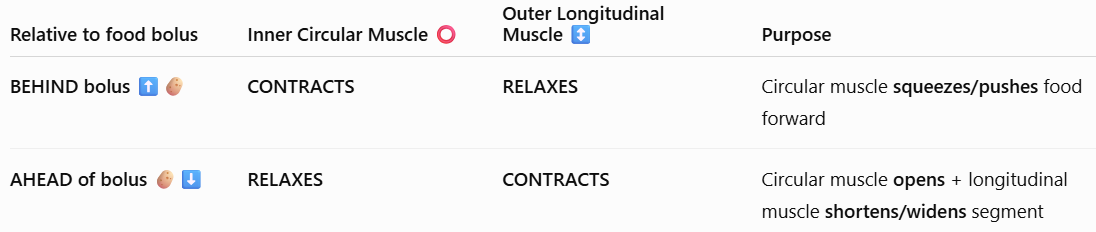
Answer: It relaxes.
Extra Information:
This occurs together with circular muscle contraction behind the bolus.
Which transmitters mediate contraction behind an esophageal bolus?
Answer: Acetylcholine and substance P.
Extra Information:
- These create an excitatory high-pressure region behind the bolus.
What happens to the inner circular muscle in front of an esophageal bolus?
Answer: It relaxes.
Extra Information:
This decreases resistance to forward movement of the bolus.
What happens to the outer longitudinal muscle in front of an esophageal bolus?
Answer: It contracts.
Extra Information:
Longitudinal contraction shortens and widens the receiving segment.
Which transmitters mediate relaxation in front of an esophageal bolus?
Answer: Vasoactive intestinal peptide and nitric oxide.
Extra Information:
- These inhibitory mediators allow the bolus to advance.
How is the lower esophageal sphincter relaxed as a bolus approaches the stomach?
Answer: Vagal pathways stimulate release of vasoactive intestinal peptide and nitric oxide.
Extra Information:
- Lower esophageal sphincter relaxation permits entry of food into the stomach.
Trace primary esophageal peristalsis around a food bolus.
Answer: Swallowing center activation → contraction behind the bolus through acetylcholine and substance P → relaxation ahead through vasoactive intestinal peptide and nitric oxide → lower esophageal sphincter relaxation → entry into the stomach.
Extra Information:
- The coordinated pressure gradient moves the bolus in the caudad direction.
What triggers secondary esophageal peristalsis?
Answer: Local esophageal wall distension caused by residual food that was not cleared by the primary wave.
Extra Information:
- Mechanoreceptors detect the remaining distension.
Which receptors detect retained food and initiate secondary esophageal peristalsis?
Answer: Mechanoreceptors in the esophageal mucosa.
Extra Information:
- They respond to local wall stretch.
Which neural system coordinates secondary esophageal peristalsis?
Answer: The intrinsic myenteric plexus of the enteric nervous system.
Extra Information:
- It is a localized short reflex.
What is the key difference between primary and secondary esophageal peristalsis?
Answer: Primary peristalsis is triggered by swallowing and mediated through vagal control, while secondary peristalsis is triggered by local distension and coordinated by the intrinsic myenteric plexus.
Extra Information:
- Secondary peristalsis clears food left behind after the primary wave.
What is the orad region of the stomach?
Answer: The fundus and proximal body.
Extra Information:
This thin-walled region primarily serves a storage function.
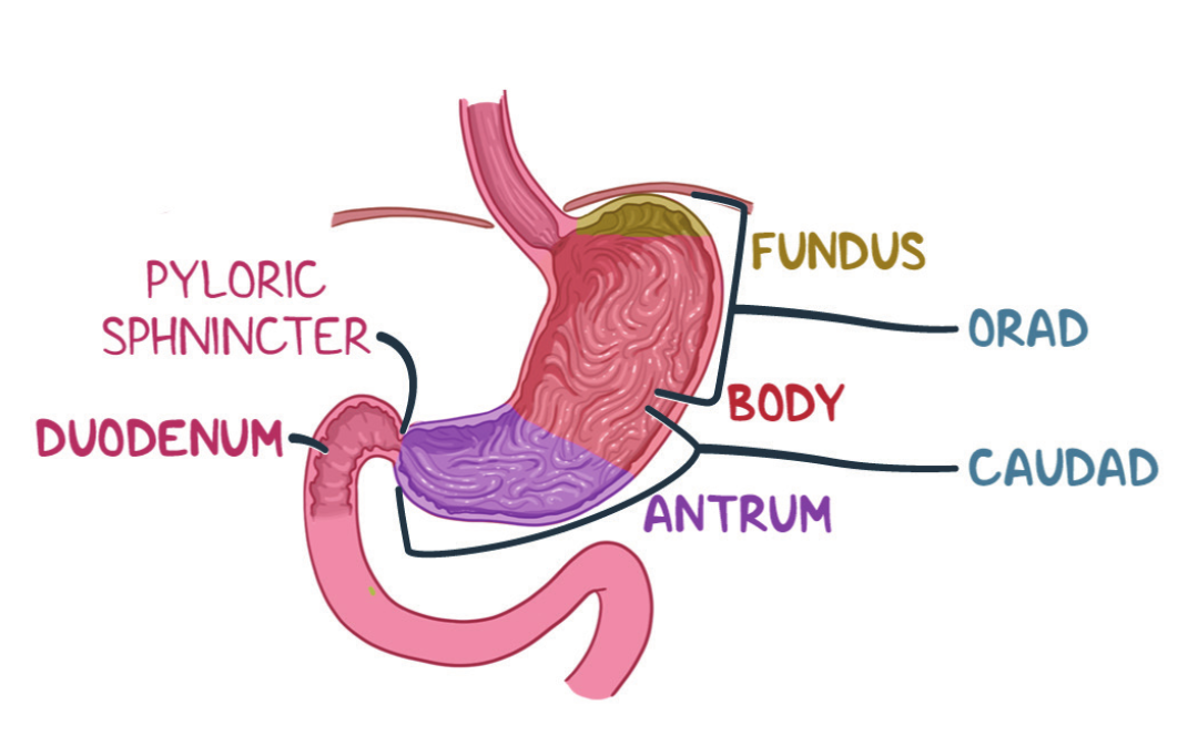
What is the major motility function of the orad stomach?
Answer: Receptive relaxation and storage of incoming food.
Extra Information:
- The region relaxes during swallowing.
Approximately how much food can the orad stomach accommodate through receptive relaxation according to the provided material?
Answer: Up to approximately 1.5 liters.
Extra Information:
- This occurs with only a minimal increase in intragastric pressure.
What is the caudad region of the stomach in the provided material?
Answer: The distal body and antrum.
Extra Information:
This thick-walled region performs mixing and grinding.
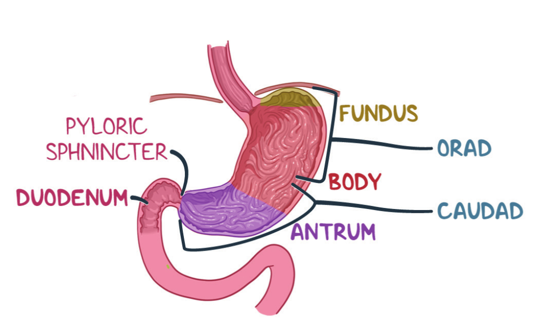
What is the major motility function of the caudad stomach?
Answer: Mixing and grinding gastric contents.
Extra Information:
- Strong contractions help mechanically process food before gastric emptying.
What are the pacemaker cells of the gastrointestinal tract?
Answer: Interstitial cells of Cajal.
Extra Information:
- They generate the basic electrical rhythm of gastrointestinal smooth muscle.
Where are interstitial cells of Cajal located according to the provided material?
Answer: In the myenteric plexus region.
Extra Information:
Their electrical activity spreads to adjacent smooth muscle cells.
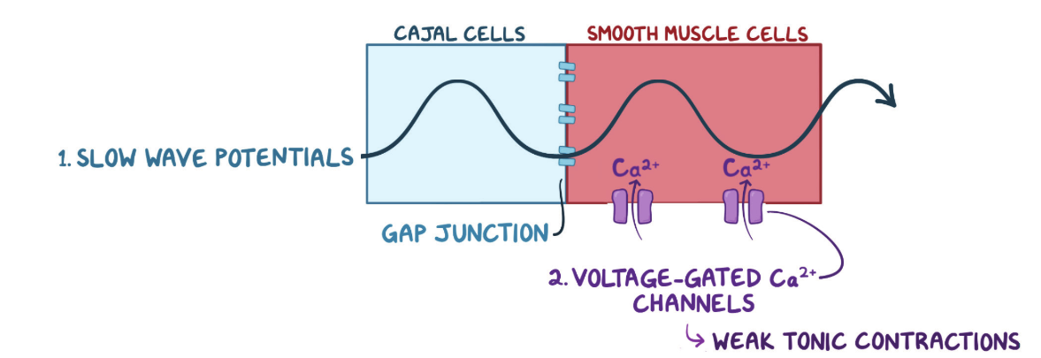
What electrical activity is spontaneously generated by interstitial cells of Cajal?
Answer: Slow waves, also called the basic electrical rhythm.
Extra Information:
- Slow waves consist of cyclic depolarization and repolarization.
What causes depolarization during gastric slow waves according to the provided material?
Answer: Cyclic opening of slow sodium channels followed by voltage-gated calcium channels.
Extra Information:
- This brings positive charge into the cells.
What ion channel activity drives repolarization during gastric slow waves?
Answer: Potassium efflux through potassium channels.
Extra Information:
- Potassium leaving the cell returns the membrane potential toward its resting state.
How do slow-wave potentials spread from interstitial cells of Cajal to adjacent smooth muscle cells?
Answer: Through gap junctions.
Extra Information:
Electrical coupling allows coordinated smooth muscle activity.
What is the gastric basic electrical rhythm frequency?
Answer: Approximately 3–5 slow waves per minute.
Extra Information:
- The stomach has the slowest basic electrical rhythm in the gastrointestinal tract according to the provided material.
What do subthreshold gastric slow waves produce?
Answer: Weak tonic contractions that help maintain stomach tone.
Extra Information:
A slow wave alone does not necessarily generate a strong phasic contraction.
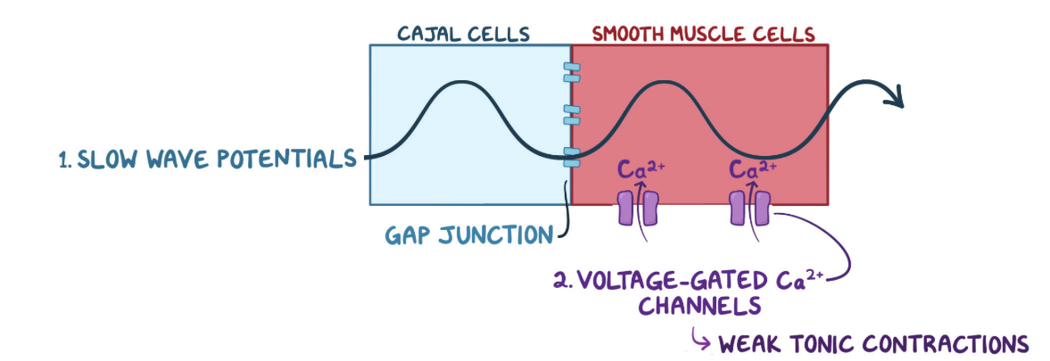
What happens when food or vagal acetylcholine depolarizes gastric smooth muscle past threshold?
Answer: Action potentials spike on top of the slow-wave plateau and trigger strong phasic contractions.
Extra Information:
These stronger contractions contribute to peristaltic mixing and grinding.
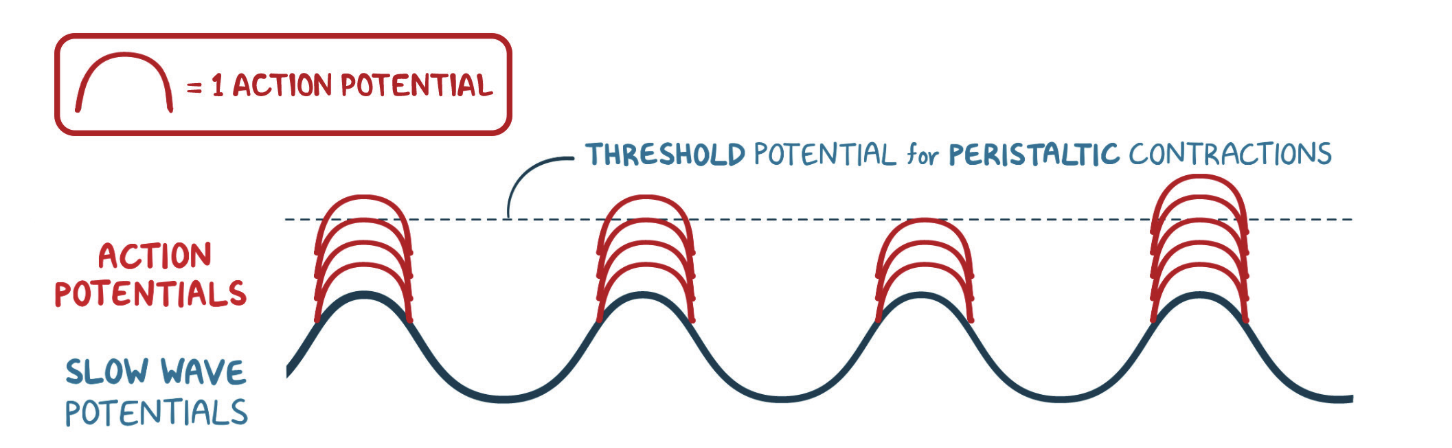
What determines the maximum frequency of gastric contractions?
Answer: The frequency of the slow waves generated by interstitial cells of Cajal.
Extra Information:
- In the stomach, this rhythm occurs approximately 3–5 times per minute.
What determines the strength of gastric contractions?
Answer: The number of action potentials that occur on top of a slow wave.
Extra Information:
- More action potentials produce stronger smooth muscle contractions.
What is the key relationship between gastric slow waves and action potentials?
Answer: Slow waves determine contraction frequency, while superimposed action potentials determine contraction strength.
Extra Information:
- Neural and hormonal stimulation can increase the likelihood and number of action potentials.
What is gastric retropulsion?
Answer: Backward movement of gastric contents caused when a peristaltic contraction approaches a closed pyloric sphincter.
Extra Information:
Retropulsion mechanically shears and mixes gastric contents.
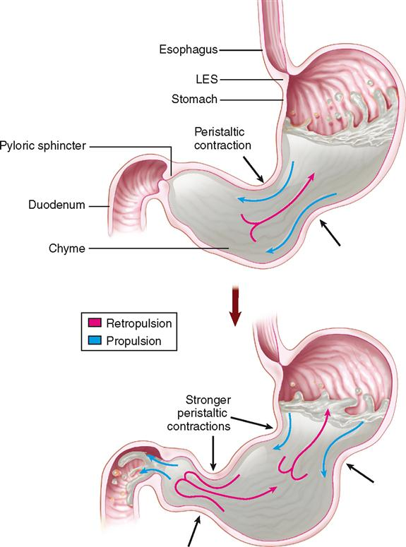
How does closure of the pyloric sphincter contribute to gastric mixing?
Answer: It forces most gastric contents backward into the stomach during an antral contraction.
Extra Information:
- This backward movement is called retropulsion.
What type of gastric contents can pass through the pylorus according to the provided material?
Answer: Liquid chyme containing particles smaller than approximately 1 millimeter.
Extra Information:
- Larger particles are driven backward for additional grinding.
Trace the basic process of gastric mixing and retropulsion.
Answer: Peristaltic wave moves toward pylorus → pyloric sphincter closes → most contents are forced backward → additional mixing and mechanical shearing occur → sufficiently small liquid chyme can enter the duodenum.
Extra Information:
- This process regulates particle size before gastric emptying.
Which parasympathetic neurotransmitter promotes gastric motility?
Answer: Acetylcholine.
Extra Information:
- Parasympathetic stimulation increases contraction strength and promotes gastric emptying.
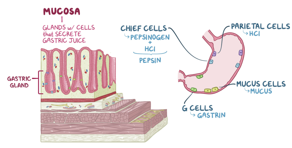
Which hormone released by G cells promotes gastric motility?
Answer: Gastrin.
Extra Information:
Gastrin increases gastric contraction force and promotes emptying.
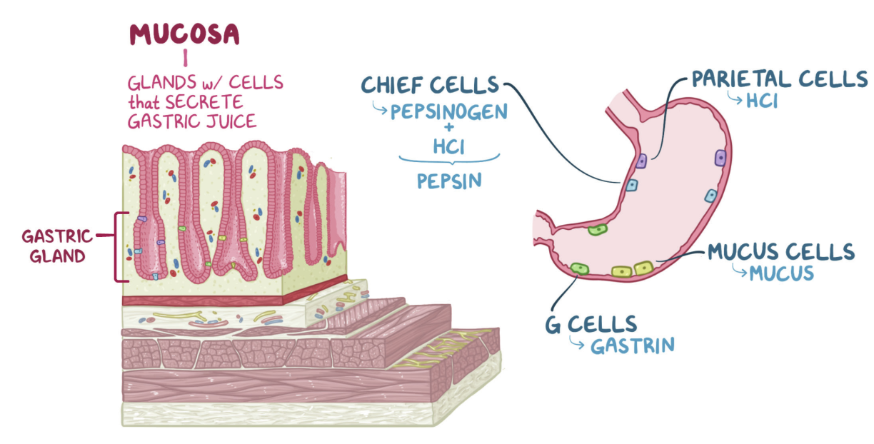
Which cells secrete gastrin?
Answer: G cells.
Extra Information:
Gastrin is one of the endocrine promoters of gastric motility in the provided material.
Which hormone initiates the migrating myoelectric complex during fasting?
Answer: Motilin.
Extra Information:
Motilin is secreted by M cells.
Which cells secrete motilin according to the provided material?
Answer: M cells.
Extra Information:
- Motilin promotes the fasting migrating myoelectric complex.
What is the migrating myoelectric complex?
Answer: A fasting gastrointestinal motility pattern that produces periodic housekeeping contractions.
Extra Information:
- It is stimulated by motilin.
Approximately how often do migrating myoelectric complexes occur during fasting?
Answer: Approximately every 90 minutes.
Extra Information:
- They function as housekeeping contractions between meals.
What are the major neural and endocrine promoters of gastric motility emphasized in the material?
Answer: Parasympathetic acetylcholine, gastrin, and motilin.
Extra Information:
- These signals increase contraction force or promote motility patterns.
What duodenal contents provide inhibitory feedback to slow gastric emptying?
Answer: Acid, fats, and hypertonic chyme.
Extra Information:
- Their presence indicates that the duodenum needs more time to process incoming chyme.
What is the overall purpose of duodenal inhibition of gastric emptying?
Answer: To give the duodenum enough time to neutralize acid and digest nutrients.
Extra Information:
- This prevents the stomach from delivering chyme faster than the duodenum can process it.
Which hormone is released by S cells in response to acid in the duodenum?
Answer: Secretin.
Extra Information:
- Secretin slows gastric emptying.
Which cells release secretin?
Answer: S cells.
Extra Information:
- Duodenal hydrogen ions stimulate its release.
What effect does secretin have on gastric emptying?
Answer: It slows gastric emptying.
Extra Information:
- This provides additional time for duodenal acid neutralization.
Which hormone is released by I cells in response to fatty acids in the duodenum?
Answer: Cholecystokinin.
Extra Information:
- Cholecystokinin slows gastric emptying.
Which cells release cholecystokinin?
Answer: I cells.
Extra Information:
- Fatty acids are an important stimulus for its release.
What effect does cholecystokinin have on gastric emptying?
Answer: It slows gastric emptying.
Extra Information:
- This provides more time for digestion of fats in the small intestine.
What effect does gastric inhibitory peptide have on gastric emptying according to the provided material?
Answer: It inhibits or slows gastric emptying.
Extra Information:
- It participates in inhibitory feedback from the duodenum.
What are the major hormonal inhibitors of gastric emptying emphasized in the material?
Answer: Secretin, cholecystokinin, and gastric inhibitory peptide.
Extra Information:
These hormones provide feedback from the duodenum.
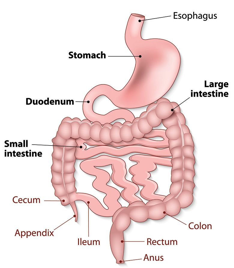
What is the underlying neural defect in achalasia?
Answer: Loss or destruction of inhibitory postganglionic neurons in the myenteric plexus that release vasoactive intestinal peptide and nitric oxide.
Extra Information:
- Loss of these inhibitory neurons prevents normal lower esophageal sphincter relaxation.
Which inhibitory neurotransmitters are deficient in achalasia?
Answer: Vasoactive intestinal peptide and nitric oxide.
Extra Information:
- Their loss prevents appropriate smooth muscle relaxation.