MI-01
1/54
There's no tags or description
Looks like no tags are added yet.
Name | Mastery | Learn | Test | Matching | Spaced |
|---|
No study sessions yet.
55 Terms
who founded microbiology
Leeuwenhoek (1674)
pasteur
koch
leeuwenhoek
founder of microbiology, not a scientist but a lensemaker, first observed small swimming things in water (animalcule)
pasteur
worked on microbial metabolism, immunity, and identification of causative agents of disease.
Discovery or identification of many medically important pathogenic bacteria and viruses took place between 1875 and early 1900’s
koch
proposed rigorous criteria to identify pathogens. Focused on the identification of disease-causing microbes.
what are some typical shapes of bacterial cells
rods
cocci (spheres)
spirochetes
spirochete shape bacteria
highly motile
can evade immune system + penetrate deep into periodontal pockets
What distinguishes a prokaryotic cell from a eukaryotic cell?
Prokaryotes
No membrane bound nucleus (DNA still supercoiled)
No mitochondria or chloroplasts
eukaryotes
Contain a true, membrane bound nucleus
Contain organelles (Mitochondria + Chloroplasts)
eubacteria (“true” bacteria)
Many different groups
Many different shapes
Some are medically important
Multiply by binary fission
No nuclear membrane
Walls contain peptidoglycan, unique to eubacteria
Archaea (primitive bacterial-sized organisms, not bacteria)
Not medically important
Chemically different from Eubacteria, no peptidoglycan
More similar to eukaryotes
Often found in extreme environments (extremophiles)
many believe Archaea are the progenitors to eukaryotic cells
viruses
Composed of DNA or RNA with a protein coat
Must grow on living cells
Not free-living forms of life
Are obligate, intracellular, parasites
viroids
Composed of RNA without a protein coat
Can cause disease in plants (potato spindle tuber disease)
1/80 the size of a virus (200-400 bases long)
prions
Infectious agent is a protein (smallest infectious agent)
Bovine spongiform encephalopathy (Mad Cow Disease)
only problematic if protein misfolds
Creutzfeld-Jacob Disease in humans
About 1/2 to 1/4 the size of viroids
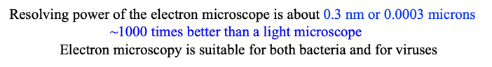
size of microbes
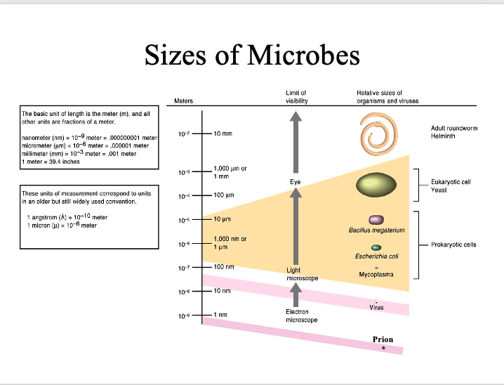
resolution
ability of microscope to separate two objects
light microscope
Resolution of light microscope is ~0.2 mm (microns)
- one-fifth of a bacterial cell width
- Can be used for observing living microorganisms
can see larger structures like nucelus, cell walls, chloroplasts
gram stain can help better visualize
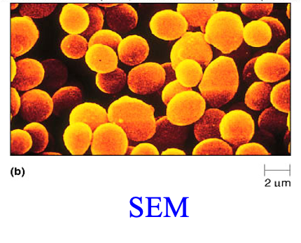
electron microscope
scanning electron microscope (SEM)
allows viewing of three-dimensional view of surfaces
only can see outside surface (cell membrance, cilla, microvilli)
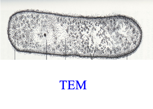
transmission electron microscope (TEM)
looks through the cells or sections of cells
2D
the best approach for looking at cell structure ( all organelles)
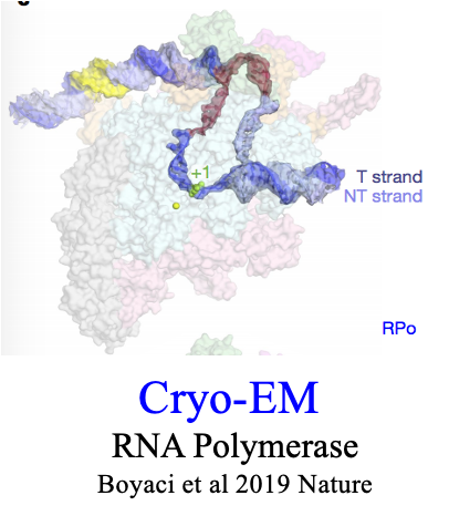
cryogenic electron microscope (cryo-em)
can see molecules and large protein complexes like RNA polymerase
simple stains
Stains all protein and cytoplasm
ex: crystal violet
fix heat to slide by heat before staining
Differential stains
Stains different bacteria differently
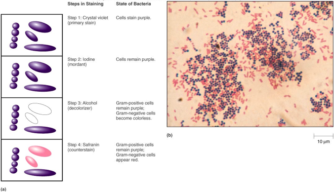
gram stain
most widely used as diagnostic stain
stains differently due to thickness of peptidoglycan layer
**know these steps
IF YOU DONT COUNTERSTAIN GRAN NEG WILL BE COLORLESS
IF YOU DONT DECOLORIZE ALL WILL BE PURPLE
at end colors
gram positive = purple
gran negative = red
arrangements of cells
chains
packets
clusters
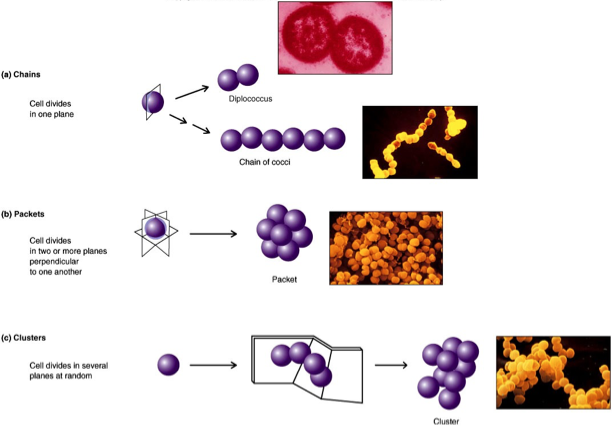
chain
cell divides in 1 plane
packets
cell divides in 2+ planes perpendicular to each other
clusters
cell divides in planes at random
cellular organizations
major structural groupings
cellular organizations: genome
arranged in condensed nucleoid structure
cellular organizations: cytosol/ cytoplasm
Around genome is the cytosol or cytoplasm containing the proteins and ribosomes and other smaller molecules of cell
cellular organizations: cell surface
around all of the above
contains membranes, peptidoglycan and all appendages
Medically important: first part to touch infected host cells
cellular organizations: cellular options

bacterial cell wall
Peptidoglycan is unique to bacteria
Peptidoglycan
-Found in all eubacteria (almost all)
-A network mesh holding cell together
lysozyme
(in tears) dissolves peptidoglycan
antibiotics
inhibit peptidoglycan cross-linking
ex: penicillin
gram negative cell wall
•Gram negative cells have thin peptidoglycan layer
•Thin wall allows removal of "gram-stain material"
•Gram negative cells have two membranes, an outer and an inner membrane
gram positive cell wall
Gram positive cells have thick peptidoglycan
Thick wall does not allow removal of "gram-stain material"
Cell wall is stronger than gram-negative cell wall
Gram positive cells have a single cytoplasmic membrane
sensitive to penicillin G or V ( early gen penicillins)
outer membrane (gram negative)
Outer membrane
Contains lipopolysaccharide (LPS, endotoxin)
composed of:
- lipid A core
Outer membrane prevents access of some antibiotics, like early generation penicillins (PenG or PenV), to their target
porins
proteins that allow material to
enter periplasmic space of cell
periplasm
Material between the two membranes (GRAM NEG ONLYYY)