Light Levels & Logs, Ocular Anatomy
1/5
There's no tags or description
Looks like no tags are added yet.
Name | Mastery | Learn | Test | Matching | Spaced |
|---|
No study sessions yet.
6 Terms
Retinal Anatomy Primer
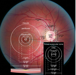
Optic nerve ~ 5 deg (1500 microns)
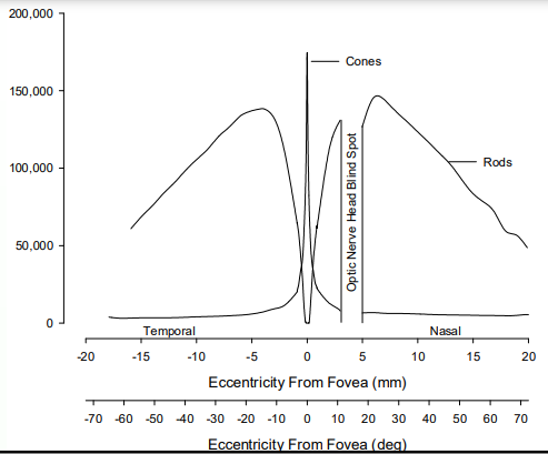
• 15-20 degrees eccentric - distance from foveola
Foveola ~ 1 deg (= rod-free area)

Photoreceptors (#’s & Use)
6 million cones (max cone density 0 deg)
120 million rod (max rod density 20 deg)
optic nerve: nasal

Scotopic vision
Refers to vision at ‘low’ light levels
It is mediated by rod photoreceptors
Rods
• Have a visual acuity of ~ 20/200
Do not play a role in color vision
• Patients with only rods – Rod Monochromats
» Congenital; usually have nystagmus (no foveola, can’t focus)
Photopic vision
Refers to vision at ‘high’ light levels
It is mediated by cone photoreceptors (There are 3 kinds)
Cones
• Have a visual acuity of ~ 20/20
• Play a role in color vision
• Patients with only cones – No congenital cases of true ‘cone monochromats’
– Clinically: Cone monochromats (Patients that have 1 cone type + rods)
– Some eye diseases can present (sort of) like only cone function is present (have rods that don’t work)
» Retinitis pigmentosa (depending on stage)
» Congenital stationary night blindness (CSNB)
Mesopic vision
Refers to vision at ‘medium’ light levels – ‘Twilight’/Moonlight
It is mediated by rod and cone photoreceptors
• Cones are not functioning optimally though
– i.e. Wouldn’t be as good at visual acuity or color vision tasks
• Rods are not necessarily functioning optimally either
– i.e. Wouldn’t be as good at light detection
• But depends on the vision function of interest
– Depending on the vision function of interest, the best system (the one with the lowest threshold) would determine vision function
Visual angles
Objects in space, form a particular image size on our retina
Image size is discussed in terms of angle of subtense
Retinal landmarks can be discussed in angular terms or absolute distances
• Clinically, often assume the optic nerve head is 5 degrees in diameter
• So 1 degree is ~ 300 microns