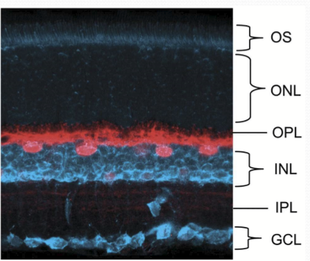
17. Retinal Interneurons
1/34
There's no tags or description
Looks like no tags are added yet.
Name | Mastery | Learn | Test | Matching | Spaced |
|---|
No study sessions yet.
35 Terms
What are bipolar cells?
Cells that photoreceptors and ganglion cells synapse with in the outer plexiform layer. Receptors bind to output signal of glutamate.
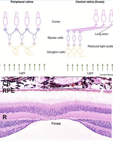
What is the ration of foveal cones to bipolar cells?
1:1
What happens to bipolar cells during the periphery?
Convergence of cones onto multiple bipolar cells occurs.
What do photoreceptors synapse with in the outerplexiform layer?
Bipolar and horizontal cells.
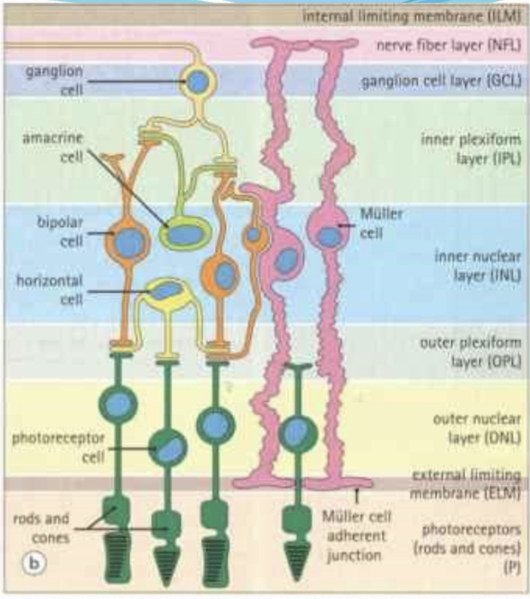
Bipolar cells synapses with what in the inner plexiform layer.
retinal ganglion cells

How many specific bipolar cells are specific for cones?
10 types: on-center or depolarizing bipiolar cells (DBC) OR off-center or hyperporlarizing bipolar cells (HBC)
How many specific bipolar cells are specific for rods?
1 type, DBC.
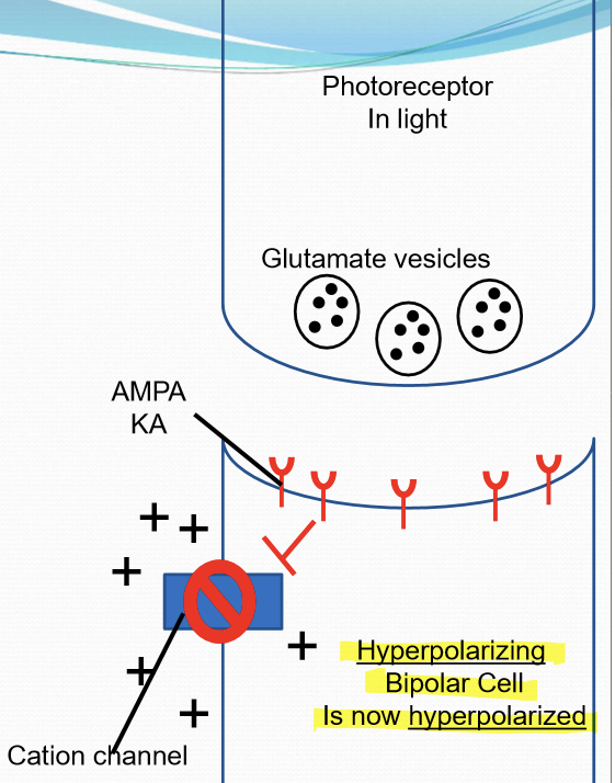
What dy hyperpolarizing bipolar cells do with light?
They hyperpolarize, the reduced glutamate from photoreceptor causes cation channels closure of HBCs. Glutamate from photoreceptors cause cation channel opening of HBCs.
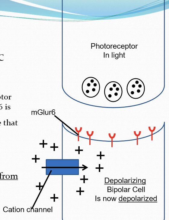
What do depolarizing bipolar cells do with light?
Depolarize. Reduced glutamate from photoreceptor causes cation channel openings of DBCs. Glutamate from photoreceptors cause cation channel closure of DBCs.
What happens at the HBC synapse in light?
Has AMPA and KA glutamate receptors. Ionotropic: when AMPA and KA are stimulated by glutamate, they trigger the opening of cation channels. Cation channel opening will depolarize the cell. UPON light stim, glutamate release is reduced, the HBC cation channel close and the cell becomes hyperpolarized.
What happens at the HBC synapse in light?
Has mGlur6 glutamate receptor. Metabotropic: when mGlur6 is stimulated by glutamate, it activates a G-protein cascade that triggers the closure of cation channels. Cation channels clusre will hyperpolarize the cell. UPON light stimulation, gulatamte release is reduced from PR, the DBC cation channels open, and then the cell becomes depolarized.
What do HBC or DBC do when they are depolarized?
They release glutamate to ganglion cells.
When HBC or DBC are hyperpolarized, what do they do?
They reduce glutamate release to ganglion cells.
What is the firing rate of ganglion cells?
They have a basal rate of firing, with glutamate release increasing ganglion cell firing rate. Reduction of glutamate release decreases ganglion cell firing.
What are horizontal cells responsible for?
Lateral inhibition by:
Feedback mechanism to limit glutamate release from photoreceptors.
Enhances image contrast
Facilitates color discrimination
Assists light adaptation
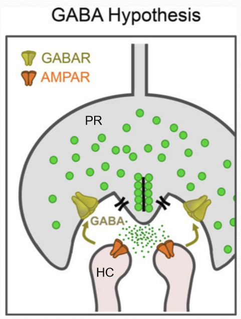
What is the mechanism for lateral inhibition?
Glutamate released by photoreceptors depolarizes HCs
Causes release of GABA to cone receptors
Causes hyperpolarization of photoreceptors
reduces photoreceptor glutamate release
Where are amacrine cells found?
In the plexiform layer.
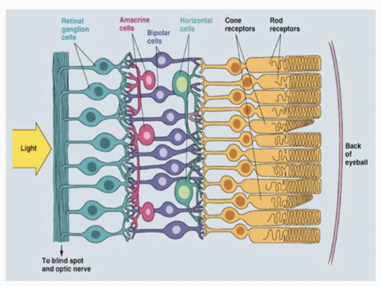
What do amacrine cells synapse with?
Bipolar cells and ganglion cells.
How many types of amacrine cells are in mammals?
~30 types
What receptors are found on amacrine cells?
Most are GABAergic, rest are glycinergic. (inhibitory NTs)
What stimulates amacrine cells?
Gultamate from bipolar cells.
What neurotransmitter do amacrine cells use to alter bipolar cell activity?
GABA
How do amacrine cells influence bipolar cells?
They alter glutamate release from bipolar cells via GABA.
Besides bipolar cells, what other cells can amacrine cells modify?
Other amacrine cells.
How do amacrine cells affect ganglion cell signaling?
They enhance or diminish the signal sent to ganglion cells.
What is the overall function of amacrine cells in visual processing?
They shape the spatial and temporal characteristics of receptive fields of bipolar and ganglion cells.
What are receptive field of a retinal ganglion cell composed of?
The region of photoreceptors and interneurons that are electrically coupled to an individual RGC. Smaller receptive fields allow for greater spatial acuity. Receptive fields in retina overlap too.
What are the two types of bipolar cells involved in signaling to ganglion cells?
Horizontal bipolar cells (HBC) and depolarizing bipolar cells (DBC).
How do HBC and DBC contribute to ganglion cell receptive fields?
They allow receptive fields to send distinct signals based on whether the stimulus is on-center or off-center.
What visual functions are supported by bipolar cell signaling to ganglion cells?
Detection of edges and recognition of contrast.

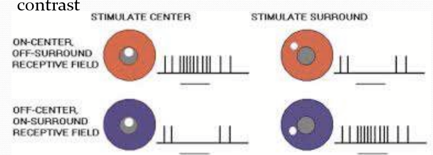
What does CSARF stand for?
Center-Surround Antagonistic Receptive Field.
What is the main function of CSARF in visual processing?
To encode spatial information.
What contributes to the formation of CSARFs?
Distinct types of bipolar cells (HBCs and DBCs)
Inhibition from amacrine and horizontal cells
What are the two types of ganglion cell receptive fields shown in CSARF?
ON-center, OFF-surround
OFF-center, ON-surround
What information is encoded within the RGCs?
A combination of:
RGC firing rate
Type of RGC (midget or parasol RGC)
Location of the photoreceptors that trigger RGC stimulation
Location of RGC axon synapse within the LGN