Structure Week 1: LO's
 Call Kai
 Learn
Learn Practice Test
 Spaced Repetition
 Match
Match Flashcards
 Knowt Play
1/73

There's no tags or description
Looks like no tags are added yet.
Name | Mastery | Learn | Test | Matching | Spaced | Call with Kai | Chat |
|---|
No analytics yet
Send a link to your students to track their progress
74 Terms
LO 1: Describe the bones and muscle layers of the chest wall
Bones
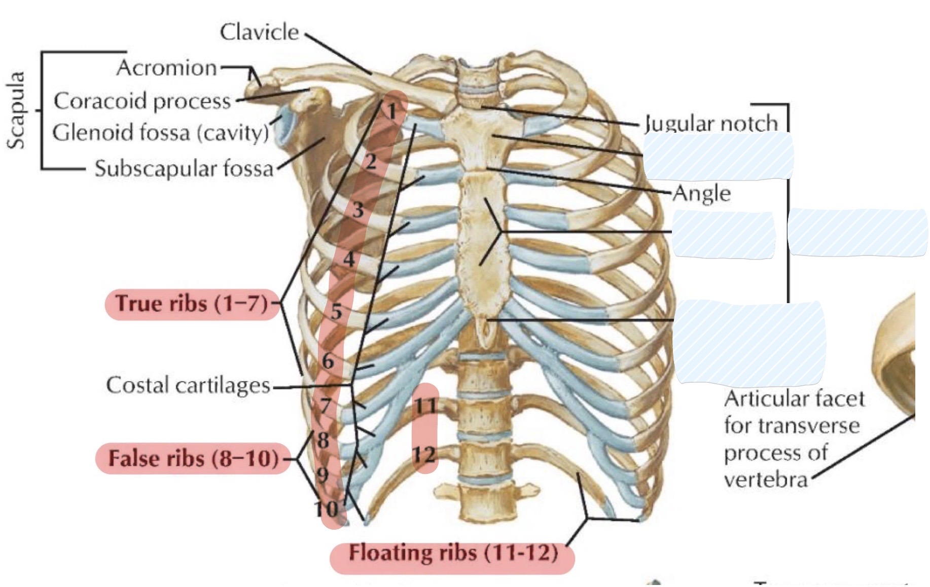
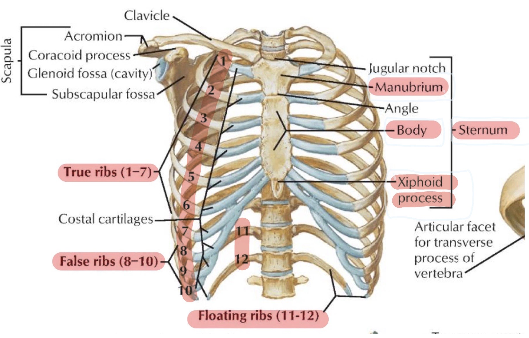
sternum: manubrium, body, xiphoid process (T10)
Ribs (12 pairs)
Thoracic vertebrae (T1-T12)
Muscle Layers
Intercostal muscles: external intercostals, internal intercostals, innermost intercostals
Diaphragm
superficial: pec major & minor
LO 1: what are the 3 components of the sternum
manubrium → The superior portion featuring the jugular (suprasternal) notch and the sternoclavicular joints where it articulates with the clavicles
body → The middle portion of the sternum articulating with ribs 2 through 7
xiphoid process → The inferior-most portion, located at the level of T10, which may ossify and fuse with the body in adulthood
LO 1: what are the 3 components of the manubrium
Jugular (suprasternal) notch → superior portion of sternum
Sternoclavicular joint → where sternum articulates with clavicles
Sternal angle (T4/T5) → critical clinical landmark formed by the junction of the manubrium and the body
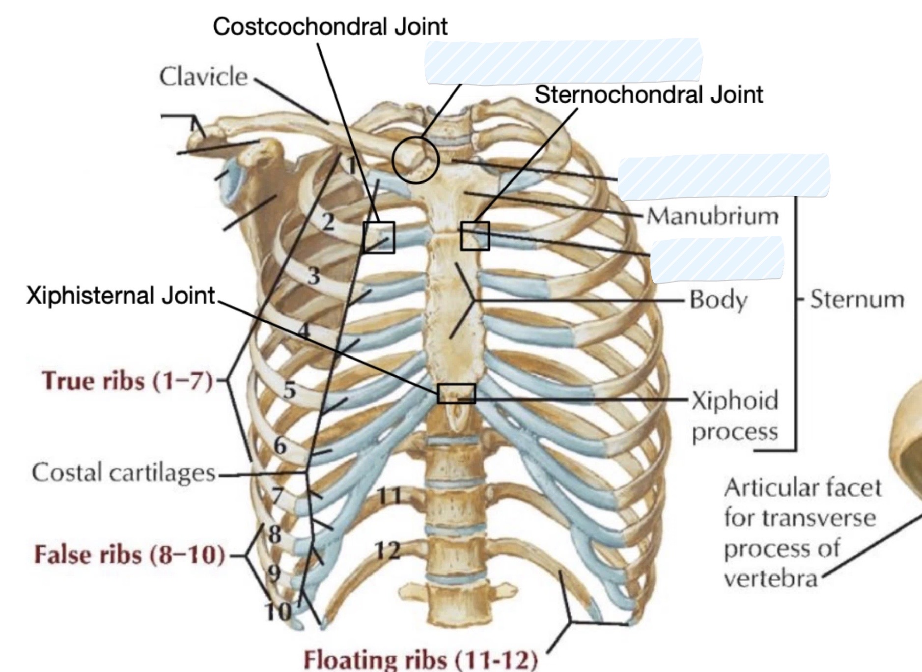
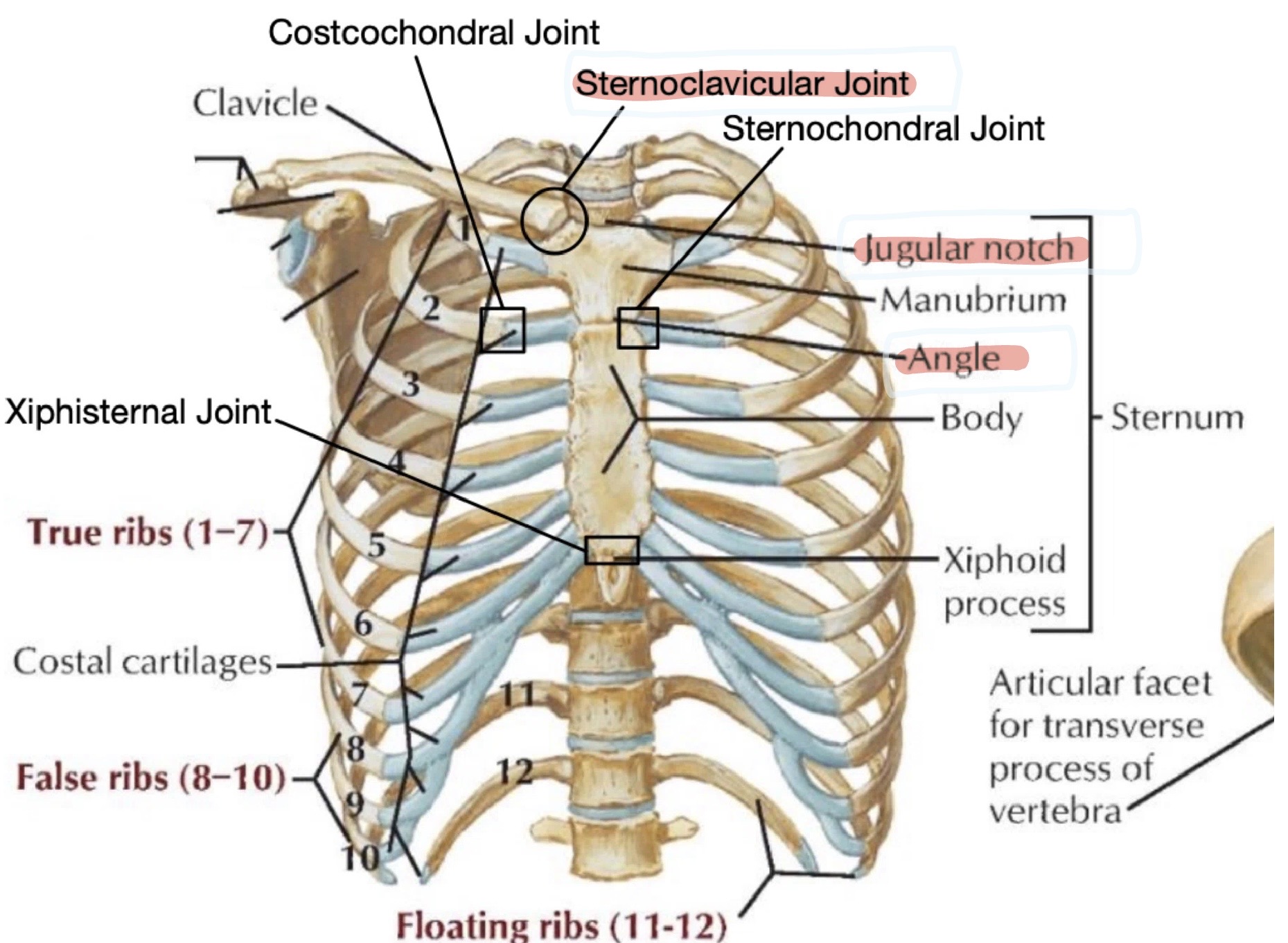
LO 1: what is a subcomponent of the xiphoid process
Xiphisternal joint → The articulation between the xiphoid process and the sternal body
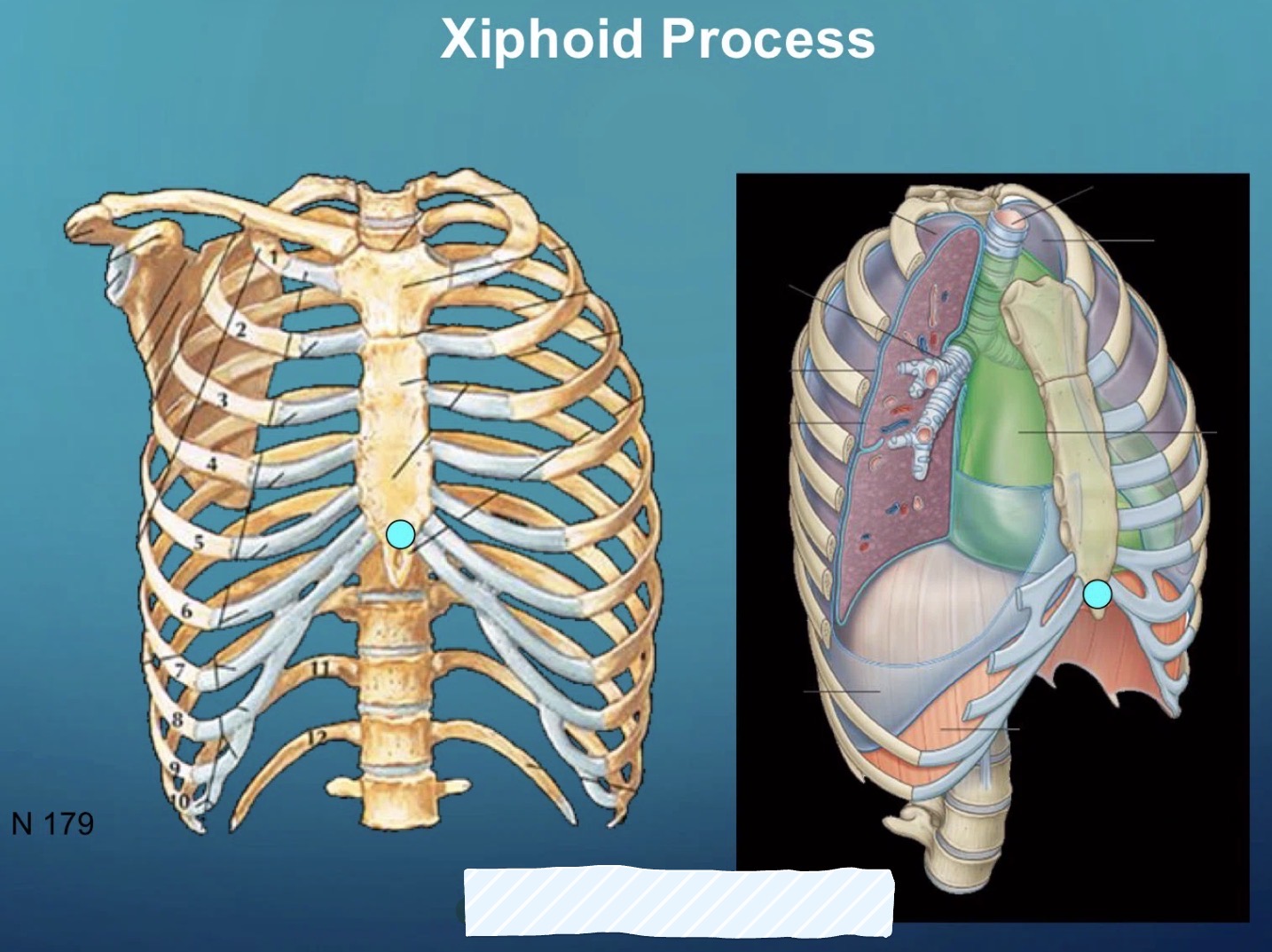
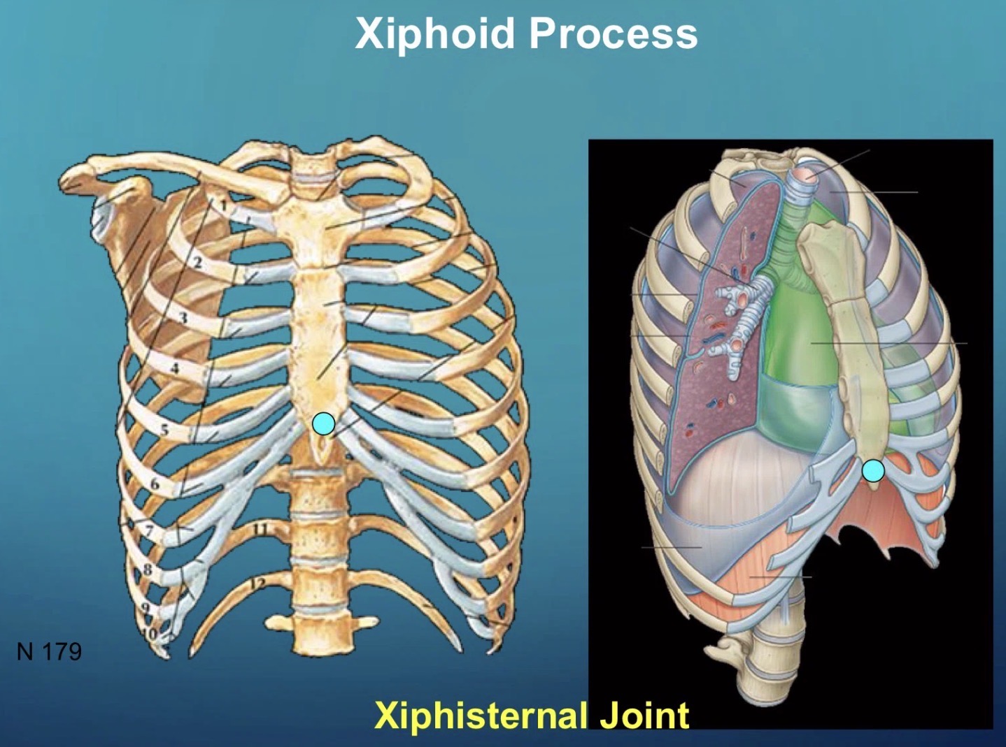
LO 2: what are the 3 classification of ribs & their locations
True ribs (1-7) → Attach directly to the sternum through their own costal cartilages
False ribs (8-10) → Their costal cartilages attach to the cartilage of the rib above, forming the costal margin
Floating ribs (11-12) → Have no anterior attachment and end in the posterior abdominal musculature
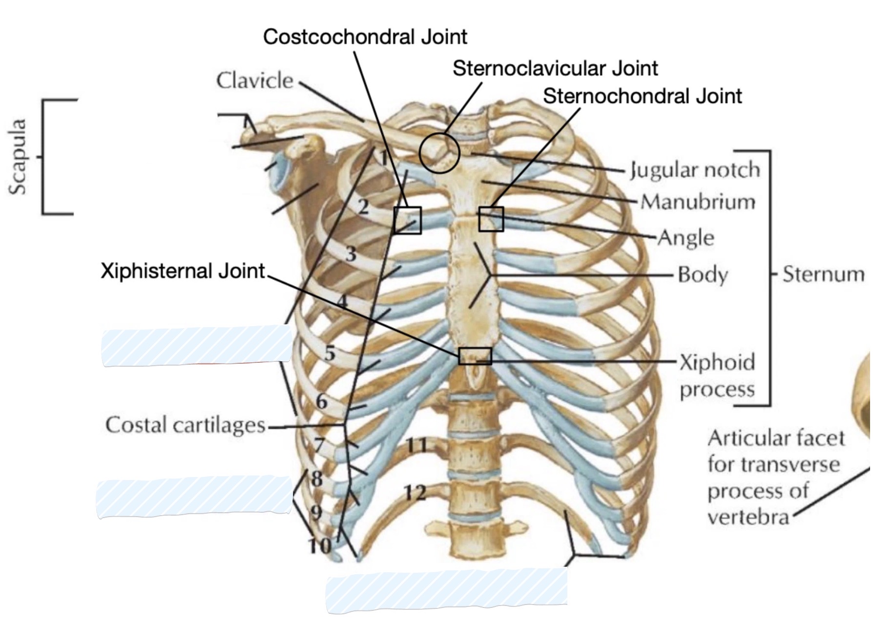
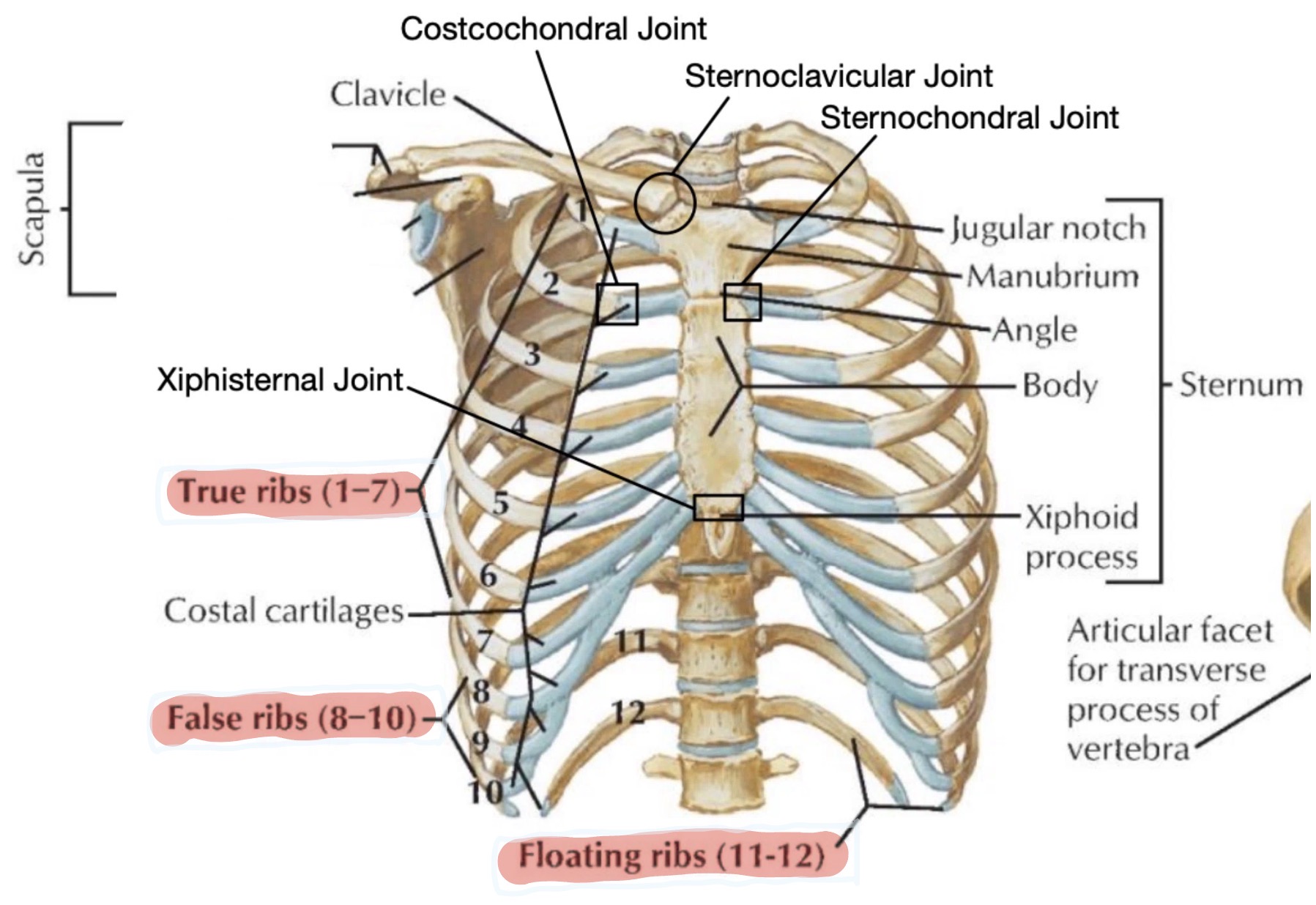
LO 2: What are the features & landmarks of the ribs
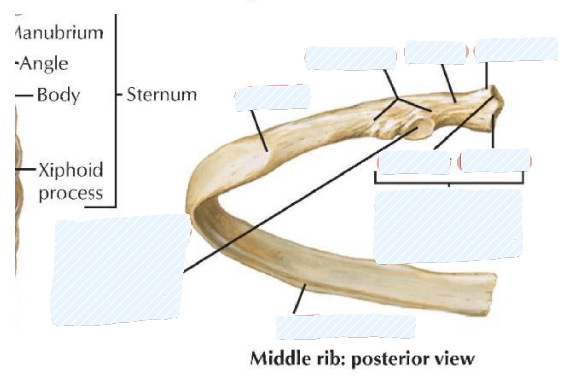
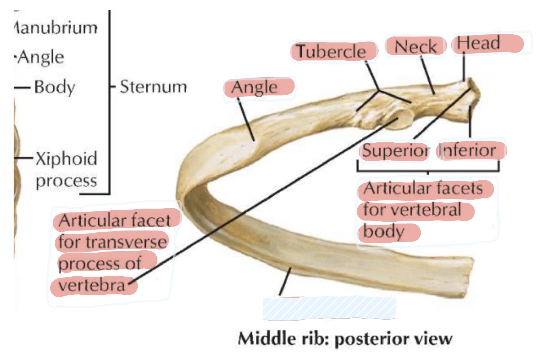
head → Features superior and inferior articular facets for articulation with two adjacent vertebral bodies
neck → Articulate with the transverse process of the vertebra of the same number
tubercules → Articulate with the transverse process of the vertebra of the same number
angle → The point where the rib turns anterolaterally/the site of greatest curvature and most common fracture
costal groove → Located on the internal surface of the inferior border, it protects the intercostal neurovascular bundle
LO 2: what are the 4 atypical ribs
1st Rib: Broadest, shortest, and most curved; has grooves for the subclavian vessels.
2nd Rib: Has a tuberosity for the serratus anterior muscle.
11th and 12th Ribs: Short, rudimentary, and lack a neck or tubercle
LO 2: what does the head of a rib include
Superior/ inferior articular facets
Lo 2: what are the articulations & attachments of the ribs
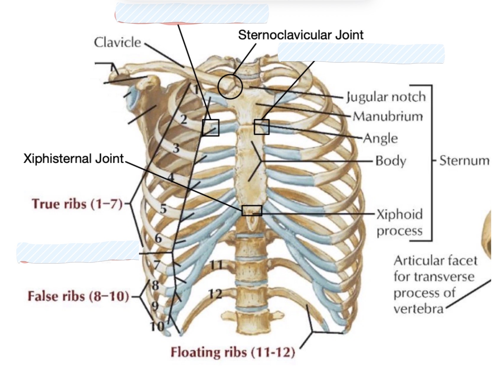
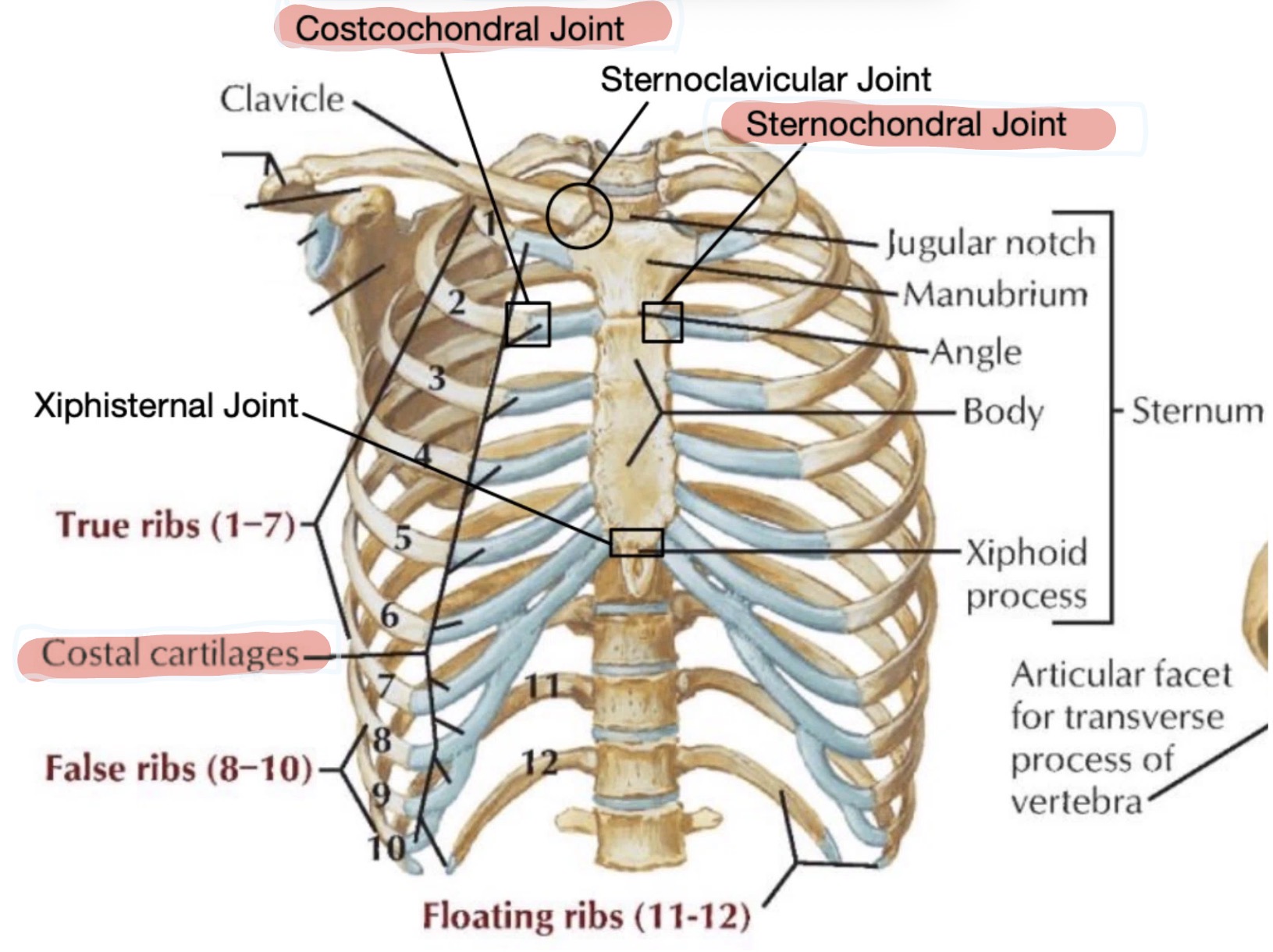
Posterior articulations (costotransverse joint & costovertebral joint)
Anterior articulations (costocondral joint & sternocondral joint)
Costal cartilage
LO 2: Posterior articulations of ttibs
Costotransverse joint
Costovertebral joint
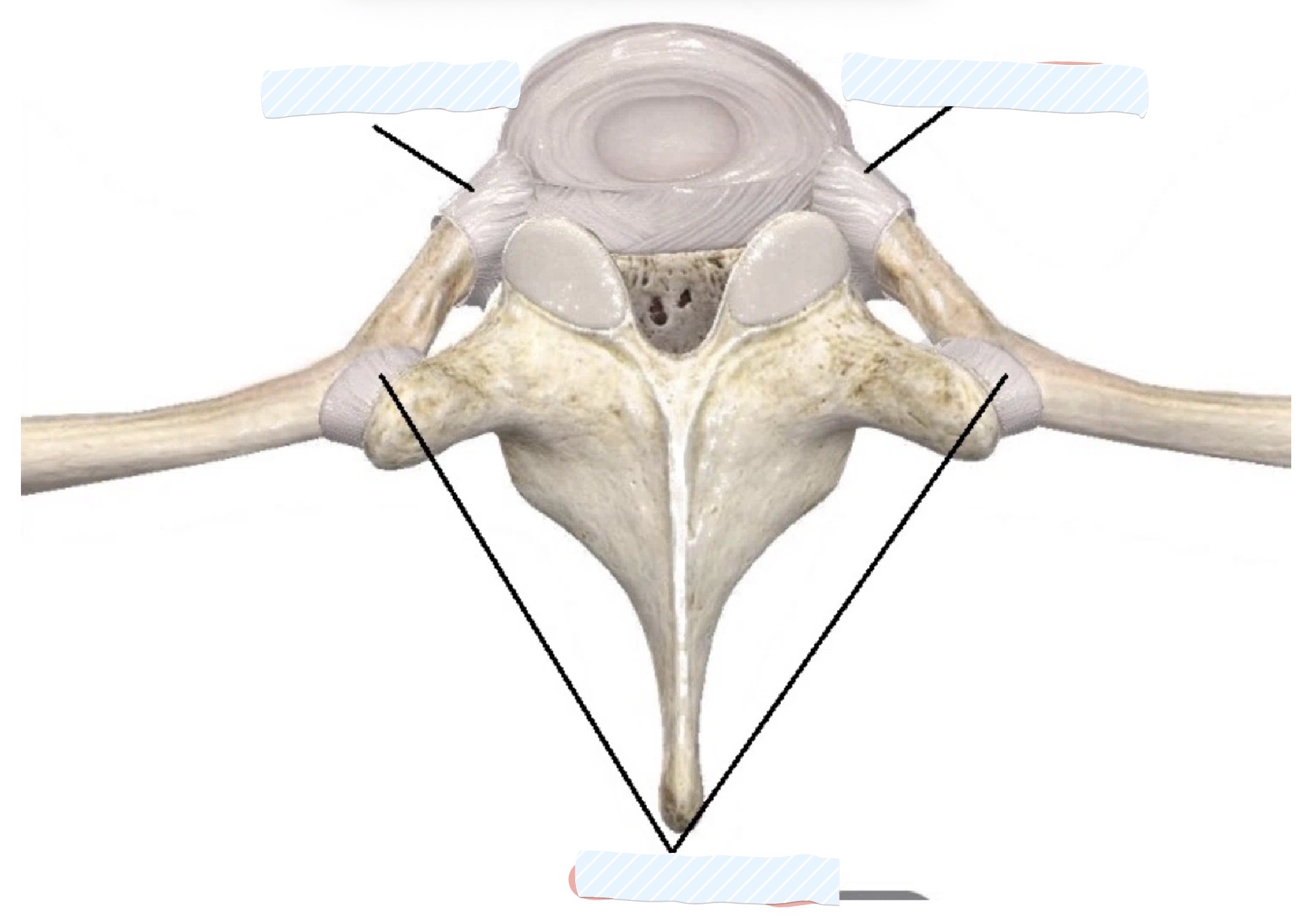
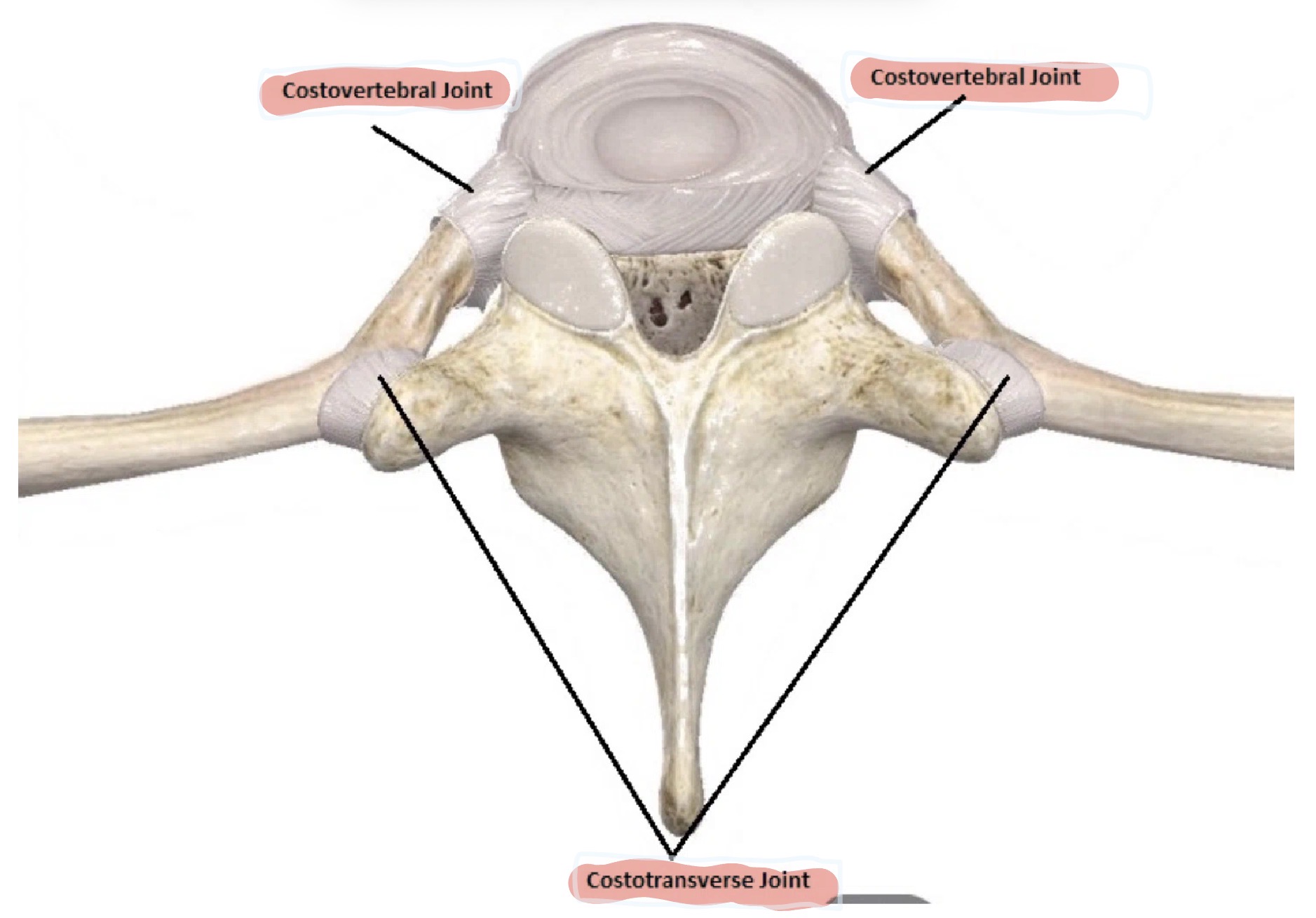
LO 2: Anterior articulations of ribs
Costochondral joint
Sternochondral joint
costal cartilage
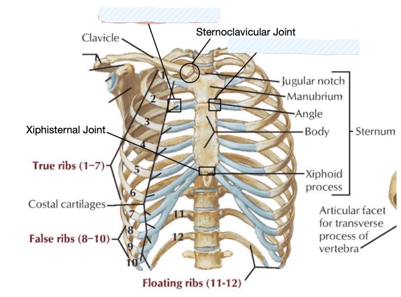
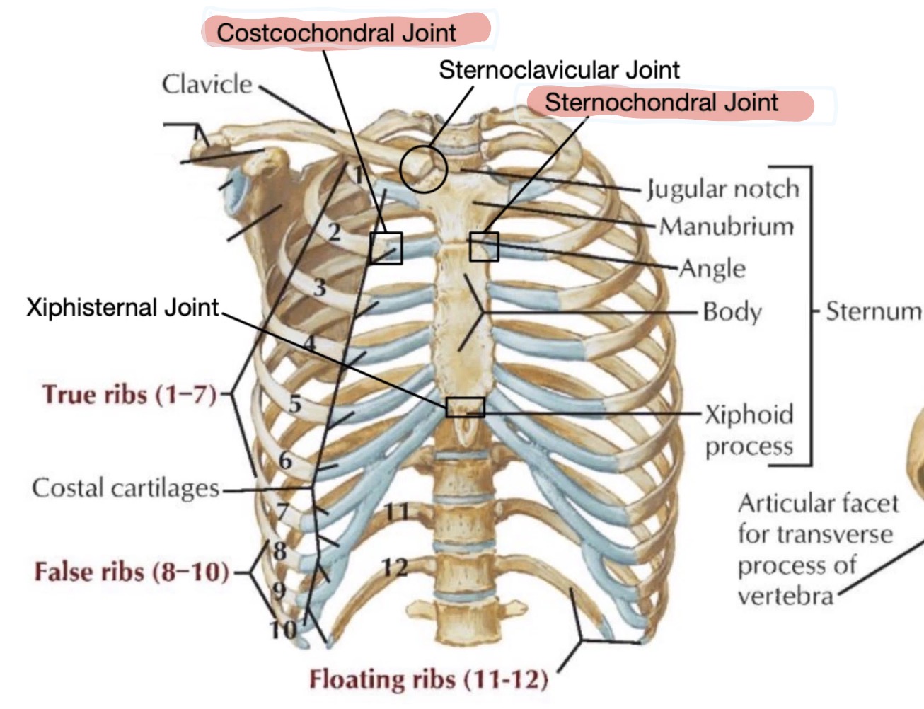
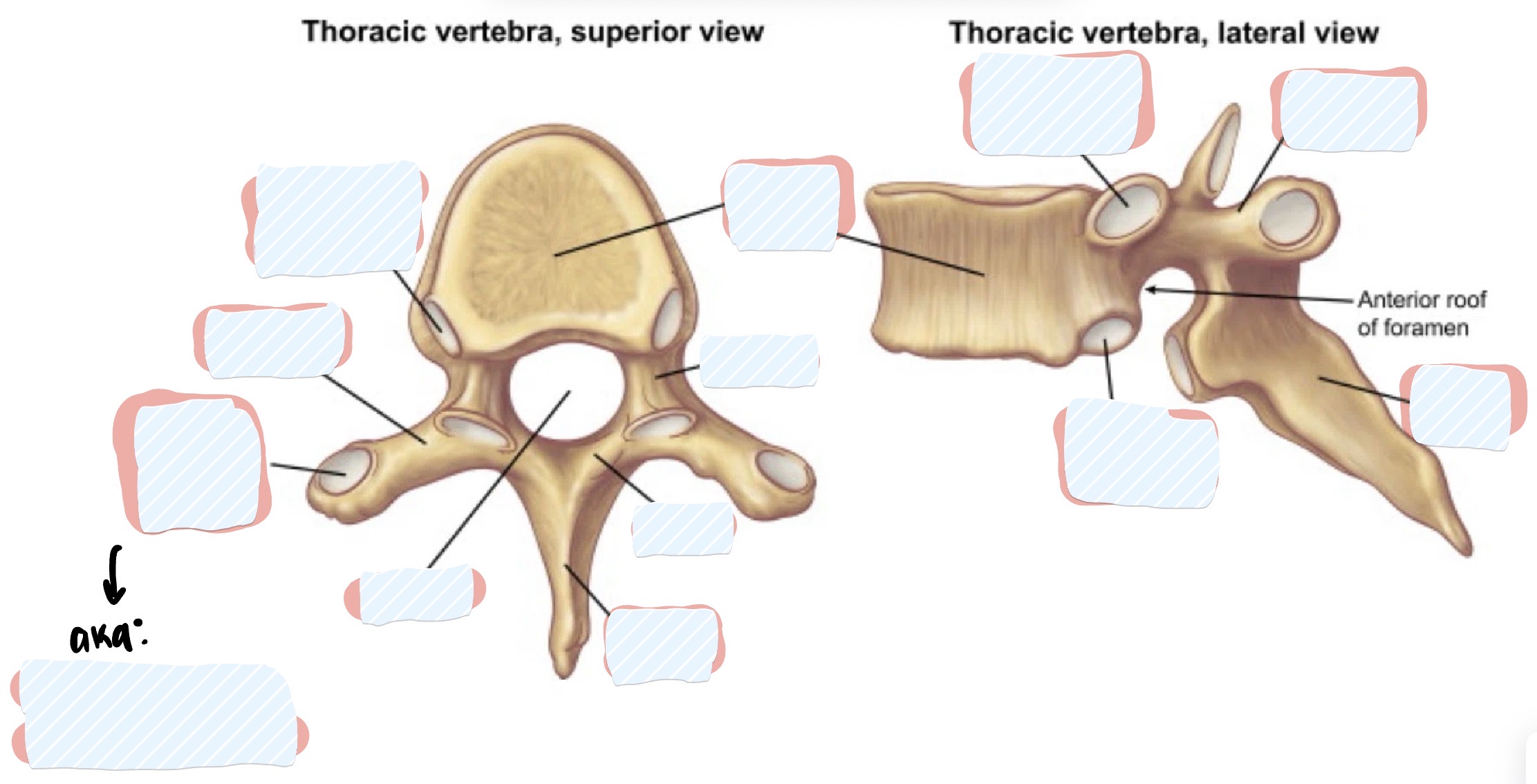
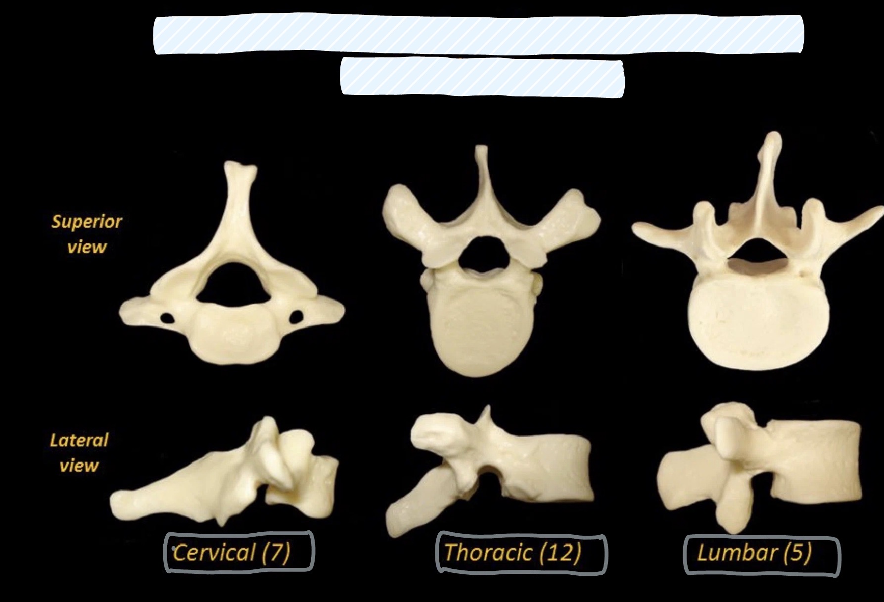
Thoracic Vertebrae
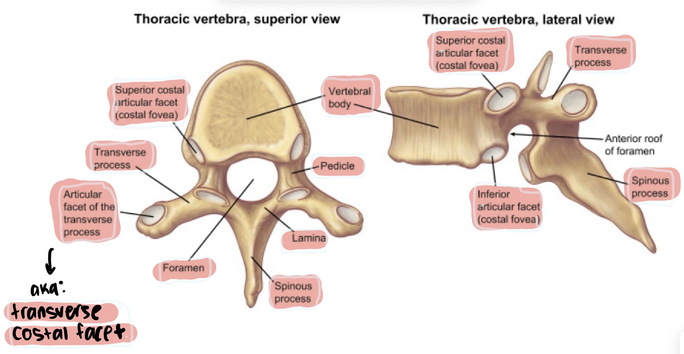
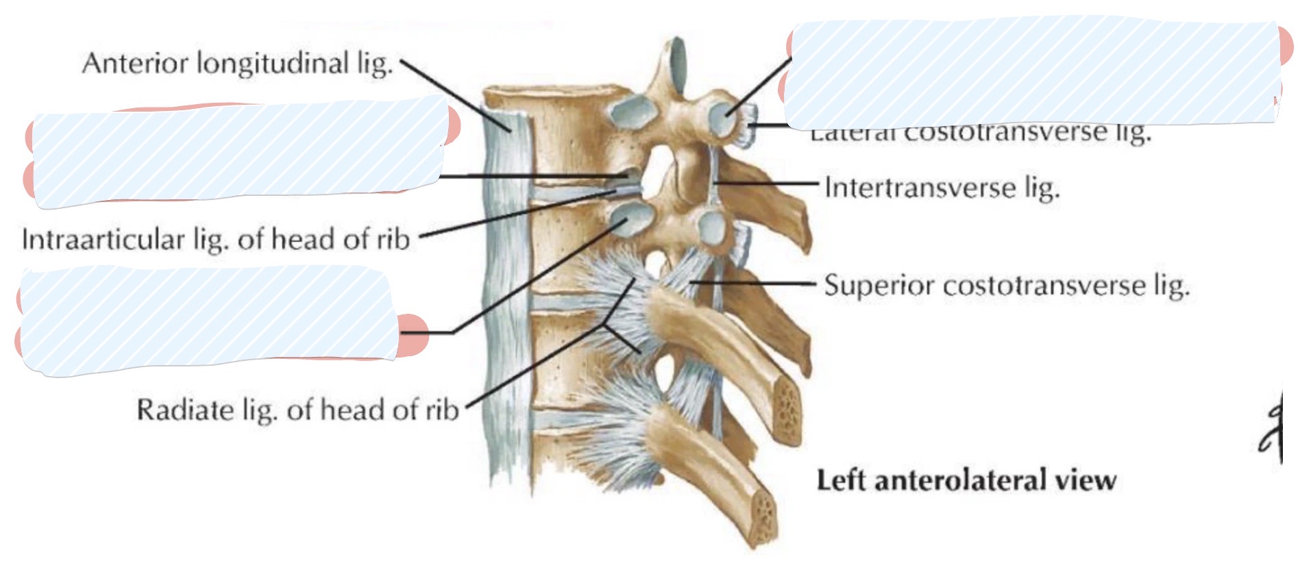
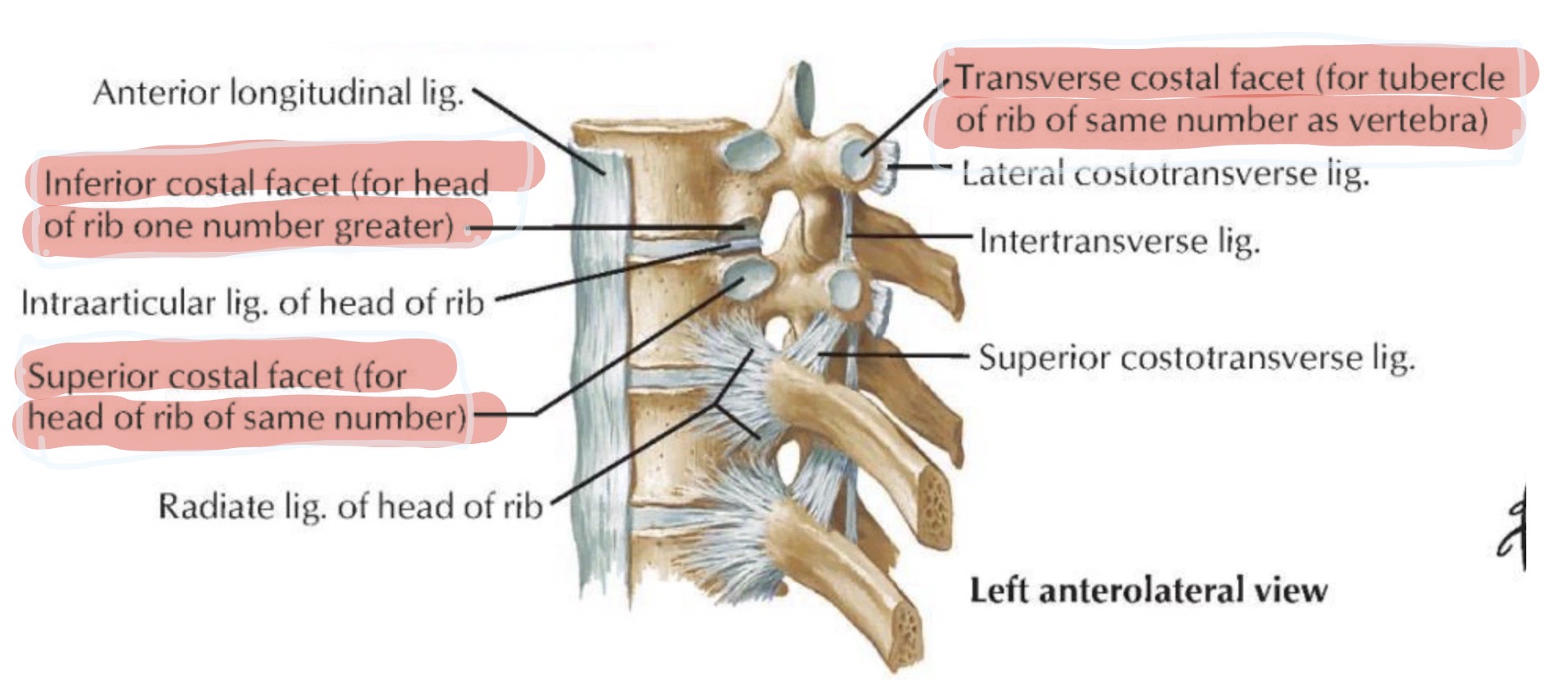
Thoracic vertebrae
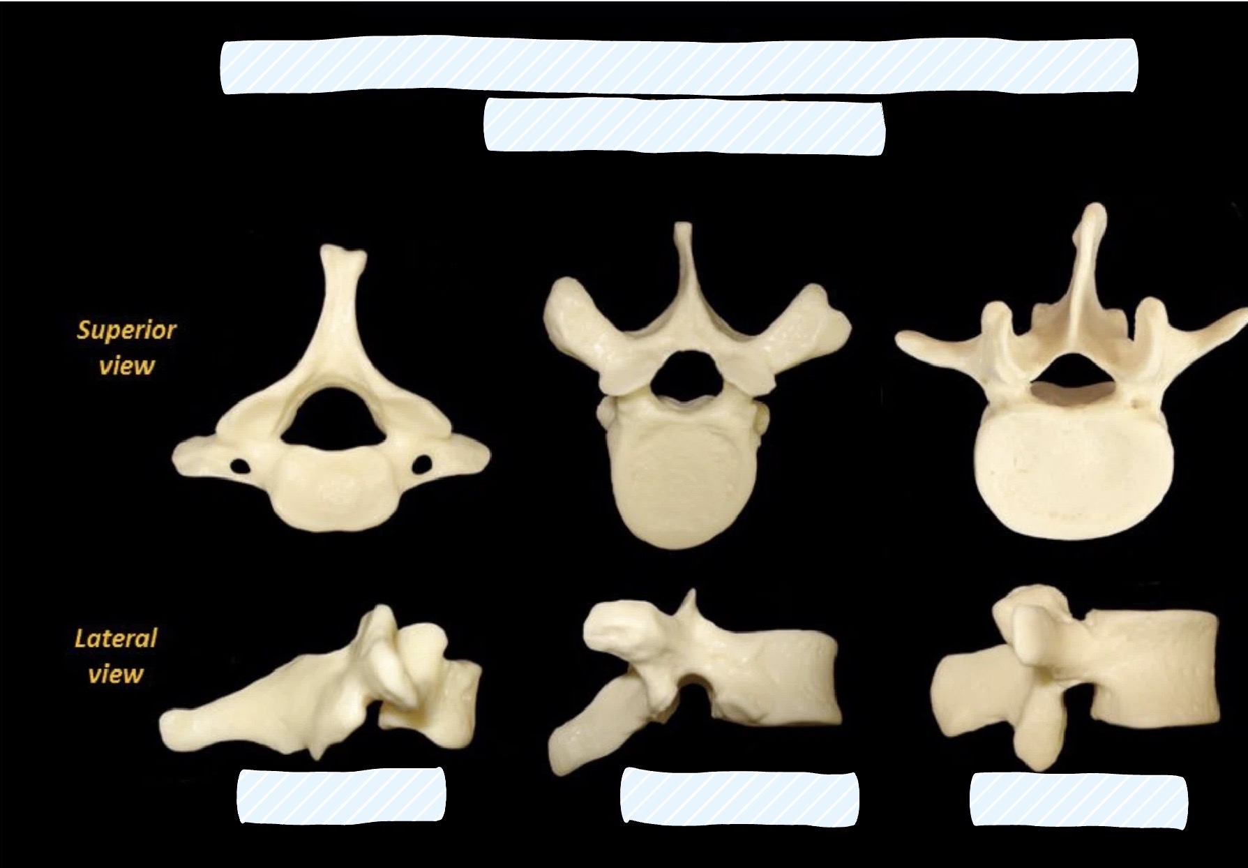
Recognize cervical vs thoracic vs lumbar vertebrae (include numbers)
LO 3: what are the muscle layers of the chest wall
intercostal muscles
external intercostals → outermost
internal intercostals → middle
innermost intercostals → deepest
diaphragm
LO 3: describe the primary muscles of respiration & innervation
diaphragm: primary muscle of respiration, separating the thoracic and abdominal cavities. Diaphragm: Innervated by the Phrenic Nerve (C3, C4, C5)
intercostal muscles: (External, Internal, and Innermost) stabilize the rib cage during the respiratory cycle
These intercostal muscles are innervated by the intercostal nerves, which are the ventral rami of spinal nerves T1 through T11
LO 3: what are the innnervations of the primary muscles of respiration
Diaphragm: Innervated by the Phrenic Nerve (C3, C4, C5).
Intercostal Muscles: Innervated by the Intercostal Nerves (T1-T11
LO 3: functions of intercostal muscles
stabilize intercostal spaces during respiration
LO 4: what are the borders of the thoracic cavity
Superior (Thoracic Inlet): Bounded by the T1 vertebra, 1st ribs, and the manubrium
Anterior border: Formed by the sternum and costal cartilages.
Lateral borders: Formed by the 12 pairs of ribs and intercostal muscles.
Posterior border: Formed by the 12 thoracic vertebrae and intervertebral discs.
Inferior border: Formed by the diaphragm
LO 4: anterior border of thoracic cavity
Formed by the sternum and costal cartilages
LO 4: lateral borders of thoracic cavities
Formed by the 12 pairs of ribs and intercostal muscles
LO 4: posterior border of thoracic cavity
Formed by the 12 thoracic vertebrae and intervertebral discs
LO 4: inferior border of thoracic cavity
Formed by the diaphragm
LO 3: Accessory muscles for inspiration & expiration
Accessory Muscles (Forced Breathing):
Inspiration: Sternocleidomastoid (elevates sternum) and Scalenes (elevate top ribs).
Expiration: Abdominal muscles (rectus abdominis, obliques) push the diaphragm up
LO 4: mediastinum & pleural cavities
Spaces Within:
Mediastinum: The central compartment housing the heart and great vessels.
Pleural Cavities: Two lateral compartments housing the lungs
LO 5: what do the intercostal nerves provide to the chest wall
The intercostal nerves provide both segmental motor and sensory innervation to the chest wall
LO 5: describe the myotomes & dermatomes of the chest wall
Dermatomes: These represent the segmental areas of skin innervated by the somatic sensory fibers of each intercostal nerve.
- area of skin supplied by one spinal nerve aka skin map (lets you feel pain, touch, temperature)
- think DERM = SKIN
Myotomes: These represent the groups of muscles (primarily intercostals) innervated by the somatic motor fibers of each intercostal nerve
- group of muscles supplied by one spinal nerve aka muscle map (lets you move muscles)
- think MYO = MUSCLE

LO 5L High yield dermatome landmarks
T4 dermatome → skin around the nipple
T10 dermatome → skin around the belly button (umbilicus)
LO 6: Identify the components and location of the intercostal bundle (on rib & intercostal muscles)
Intercostal Bundle (V.A.N) The neurovascular bundle is organized from superior to inferior as the intercostal vein, intercostal artery, and intercostal nerve.
Location: It is situated within the costal groove along the inferior border of each rib, running between the internal and innermost intercostal muscles
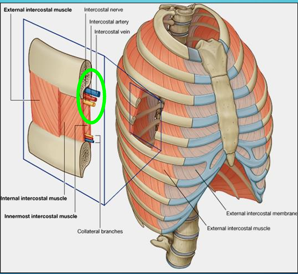
what kind of fibers are in intercostal nerves & describe them
1) Somatic motor fibers to skeletal muscle (intercostal muscles)
2) Somatic sensory fibers from skin (pain, temperature, touch) and from joints, skeletal muscles, and tendons (proprioception)
3) Sympathetic fibers to sweat glands, arrector pili muscles, and blood vessels in the skin
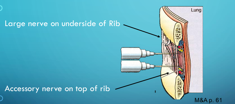
LO 6: components and location of the intercostal bundle as it relates to clinical procedures [where would you place a chest tube]
Thoracentesis and Chest Tube placement: To avoid damaging the V.A.N bundle, needles or tubes must be inserted close to the upper border of the lower rib
LO 6: components and location of the intercostal bundle as it relates to clinical procedures [where would you do an intercostal nerve block]
Intercostal nerve block: Involves injecting anesthesia to target the large nerve on the underside of the rib
where does the internal thoracic artery run
Internal thoracic artery: Runs vertically on the internal aspect of the anterior thoracic wall
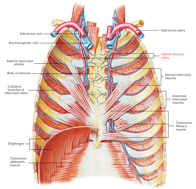
where does the internal thoracic vein drain into and where does this flow
Internal thoracic vein: Drains into the brachiocephalic veins, which then flow into the superior vena cava (SVC)

what does the azygous venous system drain and where does it empty into & what veins are involved
Azygos Venous System: This system drains the posterior chest wall and empties into the superior vena cava
azygous vein, accessory hemiazygos vein, hemiazygos vein
azygous vein what is it and which side
Azygos vein: The main vessel of the system on the right side
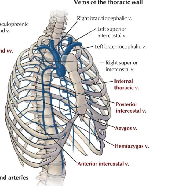
accessory hemiazygos vein where does it drain and on what side
Accessory hemiazygos vein: Drains the superior portion of the left posterior wall
Accessory = Above
“Leave heavy jewlery & accessory’s at home”
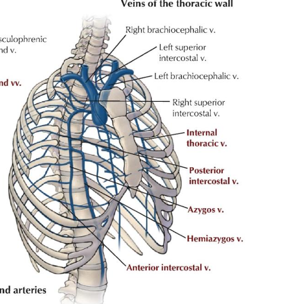
hemiazygos vein where does it drain and what side
Hemiazygos vein: Drains the inferior portion of the left posterior wall

LO 7: somatomotor pain innervation in the chest wall what is it provided by (nerves & where include thoracic vertebrae levels), what does this control
Somatomotor: Provided by somatic motor fibers in the intercostal nerves (ventral rami T1-T11) to the intercostal muscles
Control the intercostal muscles for breathing
LO 7: somatosensory pain innervation in the chest wall what is it provided by, what sensation are carries and what carries the sensations
Somatosensory pain innervation: Provided by somatic sensory fibers in the intercostal nerves, which carry sensations of pain, temperature, and touch from the skin and proprioception from joints and muscles
LO 7: sympathetic innervation of the chest wall what do the intercostal nerves carry
Sympathetic innervation: Intercostal nerves carry sympathetic fibers to targets in the skin, including sweat glands, arrector pili muscles, and blood vessels
LO 7:sympathetic innervation sympathetic pathway [where do the signal originate, where do they pass through, and where do they reach, after that where do they exit & to join what to reach where]
Sympathetic Pathway: Signals originate in the lateral horn of the spinal cord, pass through the ventral root, spinal nerve, and white ramus communicans to reach the sympathetic trunk. They then exit via the gray ramus communicans to join the ventral or dorsal rami to reach the body wall

LO 8: imaging modalities for evaluating the chest wall [projectional radiography x-ray PA vs AP describe what view they are, which view is preffered & why, and what they are used for]
Projectional radiography (X-ray): Uses radiation to create 2D images
PA (posteroanterior) is the preferred view as it prevents heart size exaggeration
AP (anteroposterior) is often used for bedbound patients

LO 8: imaging modalities for evaluating the chest wall computed tomography (CT and CTA) what does it do
Computed Tomography (CT and CTA): Obtains tomographic "slices" that can be reconstructed into 3D images for detailed diagnostic evaluation
Excellent for seeing detailed anatomy and tumors
CTA (CT Angiogram): Uses contrast dye to see blood vessels, like the aorta or pulmonary arteries
LO 8: imaging modalities for evaluating the chest wall fluoroscopy
Fluoroscopy: Provides real-time moving images, useful for procedures like esophageal exams or cardiac catheterization
Used for moving structures like the heart during catheterization
LO 8: imaging modalities for evaluating the chest wall ultrasound
Ultrasound: Can be used to visualize pleural fluid or guide procedures like thoracentesis
LO 8: what are the 2 high yield x-ray signs
Silhouette Sign: Loss of a normal border (like the heart edge) means something dense (like pneumonia) is touching it.
Pneumothorax: Shows as a dark area with no lung markings
LO 9: cartilage is an ______ tissue formed from mesoderm via ___________
General Features: Cartilage is an avascular connective tissue formed from mesoderm via chondrogenesis
LO 9: what are the 2 types of cartilage cells
Cells:
Chondroblasts: Located near the perichondrium, they secrete the extracellular matrix.
Chondrocytes: Mature cells situated in lacunae; often seen in groups called nest cell
LO 9: what are the 3 types of cartilage
hyaline cartialge
elastic cartilage
fibrocartilage
LO 9: hyaline cartilage what does it contain, what type of collagen, where is it found, what is it a precursor of
Hyaline cartilage: The most common type with a homogeneous matrix containing type II collagen and aggrecan.
Found in the ribs (costal cartilage), nose, larynx, and trachea; it has a perichondrium
it’s a precursor of bone

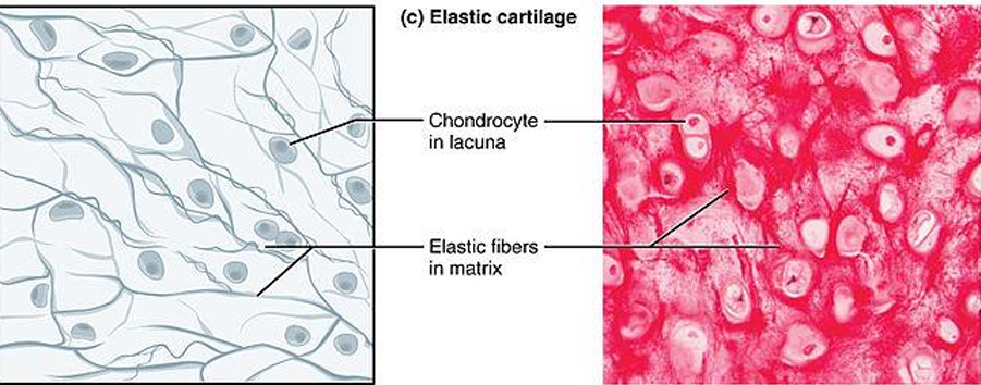
LO 9: elastic cartilage what type of collagen, what does it contain, where is it found
Elastic cartilage: Contains type II collagen and a dense network of dark-staining elastic fibers.
Found in the external ear, larynx, & epiglottis; it also has a perichondrium

LO 9: fibrocartilage what is it a mixture of, what type of collagen, what does it lack, & where is it found
Fibrocartilage: A mixture of hyaline cartilage and dense regular connective tissue (type I collagen).
It is the strongest type, lacks a perichondrium, and is found in intervertebral discs, joint capsules, and ligaments
LO 10: bone matrix what is it comprised of organic vs inorganic part
Bone Matrix: Comprised of an organic part (90-95% type I collagen) and an inorganic part (hydroxyapatite crystals of calcium and phosphate)
LO 10: what are the 4 bone cells
osteoprogenitor cells
osteoblasts
osteocytes
osteoclasts
LO 10: osteoprogenitor cells where are they found and what do they differentiate into
Osteoprogenitor cells: Stem cells found in the periosteum and endosteum stem cells that become osteoblasts

LO 10: osteoblasts what type of cells and what do they form
Osteoblasts: Cuboidal cells that form new bone by secreting the organic matrix

LO 10: osteocytes where are they found & their communication
Osteocytes: Mature bone cells found in lacunae; they communicate through thin processes called filopodia via gap junctions

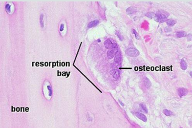
LO 10: osteoclasts what are they responsible for
Release acid, collagenase , and other lytic enzymes to break down bone matrix and release minerals (bone resorption).
Osteoclasts: Large, multinucleated cells responsible for bone resorption (breaking down matrix); they sit in Howship's lacunae
LO 10: what are the 2 types of bones
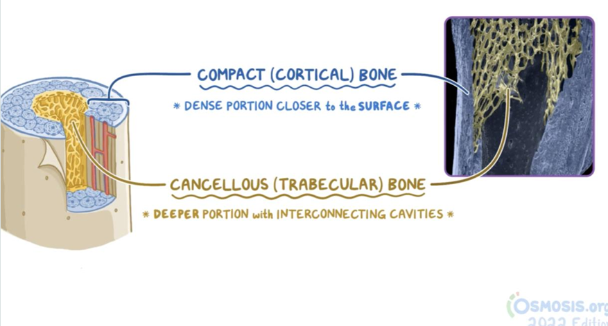
compact (cortical) bone
cancellous (spongy/trabecular) bone

LO 10: compact (cortical/dense bone) what it is, where is it found, what is it organized into
Compact (cortical) bone: Dense bone lacking large spaces, found in the diaphysis of long bones.
It is organized into osteons with concentric lamellae surrounding a Haversian cana
Made of Osteons (Haversian systems) which look like tree rings
LO 10: Cancellous (spongy/trabecular) bone what it is and what does it surround
Cancellous (spongy/trabecular) bone: A 3D lattice of bony slips called trabeculae, which surround spaces containing bone marrow
LO 10: primary (woven bone) vs secondary (lamellar) bone
Primary (woven) bone: The first randomly arranged bone tissue to appear during development or repair; it is eventually replaced.
Secondary (lamellar) bone: Mature, highly organized bone tissue
LO 10: bone development (ossification) 2 types
intramembranous
endochondral
LO 10: bone development (ossification) intramembranous
Intramembranous: Bone develops directly from membranes (e.g., flat skull bones
LO 10: bone development (ossification) endochondral
Endochondral: Bone replaces a pre-existing cartilage model (e.g., long bones of limbs)