Written Anatomy Final
 Call Kai
 Learn
Learn Practice Test
 Spaced Repetition
 Match
Match Flashcards
 Knowt Play
1/189

There's no tags or description
Looks like no tags are added yet.
Name | Mastery | Learn | Test | Matching | Spaced | Call with Kai | Chat |
|---|
No analytics yet
Send a link to your students to track their progress
190 Terms
Foregut — contents & blood supply
Esophagus, stomach, duodenum, spleen, liver, gallbladder, and pancreas; supplied by the celiac trunk.
Midgut — contents & blood supply
Jejunum, ileum, cecum, ascending colon, and proximal 2/3 of the transverse colon; supplied by the superior mesenteric artery.
Hindgut — contents & blood supply
Distal 1/3 of the transverse colon, descending colon, sigmoid colon, and rectum; supplied by the inferior mesenteric artery.
I Ate Ten Eggs at Noon — meaning
I = IVC/caval opening → T8; Ate Ten = esophageal hiatus → T10; At Noon = aortic hiatus → T12.
Peritoneum
A continuous, bilayered membrane that lines the abdominopelvic cavity and surrounds the viscera.
Parietal peritoneum
Lines the internal surface of the abdominopelvic wall.
Visceral peritoneum
Directly invests or covers organs such as the stomach and intestines.
Portal vein
Carries deoxygenated blood containing absorbed nutrients from the intestines to the liver.
Hepatic vein
Carries processed blood from the liver to the inferior vena cava.
Hepatic artery
Supplies oxygenated blood to the liver tissue.
First-pass metabolism
Processing of orally administered drugs by the liver's portal circulation before they reach systemic arterial supply.
Spleen
Depletes red blood cells and digests bacteria.
Large intestine
Primarily absorbs water and salts.
Gallbladder
Stores and concentrates bile produced by the liver and releases bile as needed into the duodenum.
Kidneys
Filter blood to remove metabolic waste and maintain fluid balance.
Ureters
Muscular ducts that carry urine from the kidneys to the urinary bladder.
Suprarenal/adrenal glands
Located between the superior aspects of the kidneys and the diaphragm; produce hormones that regulate the stress response and maintain homeostasis.
Four abdominal quadrants — planes
Established by the vertical median plane and horizontal transumbilical plane.
Diaphragm — motor innervation
Phrenic nerve.
Diaphragm — T8 opening
Inferior vena cava passes through the caval opening at T8.
Diaphragm — T10 opening
Esophagus passes through the esophageal hiatus at T10.
Diaphragm — T12 opening
Abdominal aorta passes through the aortic hiatus at T12.
Great vessels — branches of aortic arch
Brachiocephalic trunk, left common carotid artery, and left subclavian artery.
Brachiocephalic trunk — branches
Right subclavian artery and right common carotid artery.
Superior vena cava — formation
Union of the left and right brachiocephalic veins.
Superior vena cava — function
Returns blood from structures superior to the diaphragm, excluding the heart and lungs.
Pulmonary trunk
Divides into right and left pulmonary arteries, which carry deoxygenated blood to the lungs.
Pulmonary arteries
Carry poorly oxygenated/deoxygenated blood to the lungs.
Pulmonary veins
Carry well-oxygenated blood to the left atrium.
Pulmonary capillaries
Site within the lungs that facilitates gas exchange between blood and air.
Right ventricle
Pumps poorly oxygenated blood into the pulmonary trunk.
Left atrium
Receives well-oxygenated blood from the pulmonary veins.
Fossa ovalis
Shallow depression in the right atrium representing the remnant of the fetal foramen ovale.
Auricle
Increases the holding capacity of an atrium.
Coronary sinus
Drains venous blood from the heart muscle into the right atrium.
Three structures draining into right atrium
Superior vena cava, inferior vena cava, and coronary sinus.
Papillary muscles
Contract to tighten the chordae tendineae.
Chordae tendineae
Stabilize valve cusps and prevent them from prolapsing into the atria during ventricular contraction.
Tricuspid valve
Located between the right atrium and right ventricle.
Mitral/bicuspid valve
Separates the left atrium from the left ventricle.
Coronary arteries
Supply the heart muscle with oxygenated blood.
Left ventricle
Heart chamber a catheter must pass through to reach the ascending aorta most directly.
Aortic valve
Prevents backflow of blood from the aorta into the left ventricle.
Pulmonary valve
Prevents backflow of blood from the pulmonary trunk into the right ventricle.
Blood flow — systemic venous return from left subclavian
Left subclavian vein → left brachiocephalic vein → superior vena cava.
Blood flow — pulmonary circuit
Right ventricle → pulmonary trunk → pulmonary arteries → pulmonary capillaries/lungs → pulmonary veins → left atrium.
Pelvis — three bones
Ilium, ischium, and pubis.
Acetabulum — three bones
Ilium, ischium, and pubis.
Iliac crest — vertebral level
L4.
Ischial tuberosity
Bony landmark serving as an attachment point for muscles that extend the hip.
Pubic symphysis
Cartilaginous joint where a fibrocartilage disk unites the right and left pubic bones.
Acetabulofemoral joint
Triaxial synovial ball-and-socket joint formed by the acetabulum and head of the femur.
Transverse acetabular ligament
Closes the inferior portion of the bony acetabular cup.
Acetabular labrum
Fibrocartilaginous structure attached to the acetabular rim that deepens the cup and increases joint stability.
Sacroiliac joint
Synovial plane gliding joint between the sacrum and ilium that permits limited movement.
Femoral neck
Proximal femur location where fractures most frequently occur.
Linea aspera
Vertical ridge of bone on the posterior femur that serves as an attachment site for multiple muscles.
Gluteal tuberosity
Ridge of bone between the greater trochanter and linea aspera; articulates with/serves as an attachment for gluteus maximus.
Gluteus maximus — action
Extension and lateral rotation of the femur.
Gluteus maximus — innervation
Inferior gluteal nerve.
Gluteus medius & minimus — actions
Abduction of the femur.
Gluteus medius & minimus — innervation
Superior gluteal nerve.
Trendelenburg sign — muscles
Weakness of gluteus medius and gluteus minimus.
Trendelenburg sign — nerve
Superior gluteal nerve injury.
Piriformis — attachments
Sacrum to greater trochanter of the femur.
Sciatic nerve — common compression site
Lower border of piriformis.
Obturator nerve — primary hip movement affected by injury
Hip adduction.
Posterior thigh — tibial nerve muscle
Long head of biceps femoris.
Gracilis — AAI
Pubic bone → medial tibia; adducts hip and flexes knee; obturator nerve.
Semimembranosus & semitendinosus — attachment and innervation
Ischial tuberosity → medial tibia
tibial nerve.
Adductor group — AAI
Ischium/pubic bone → linea aspera & adductor tubercle; adducts femur; obturator nerve.
Anterior thigh compartment — innervation
Femoral nerve.
Medial thigh compartment — innervation
Obturator nerve.
Posterior thigh compartment — innervation
Tibial nerve.
Lumbosacral plexus — roots
Ventral rami of L1 through S5.
Lumbar plexus — roots
L1 through L5.
Sacral plexus — roots
S1 through S5.
Iliacus — innervation
Femoral nerve.
Obturator nerve — route into medial thigh
Through the obturator foramen.
Sciatic nerve — terminal branches
Tibial nerve and common fibular/common peroneal nerve.
Deep veins of lower extremity
Accompany all major arteries and their branches.
Two major deep veins
Popliteal vein and femoral vein.
Two major superficial veins
Great saphenous vein and small saphenous vein.
Small saphenous vein — drainage
Drains into the popliteal vein in the popliteal fossa.
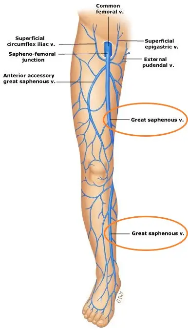
Great saphenous vein — drainage
Drains into the femoral vein in the femoral triangle.
Great saphenous vein — location
Medial aspect of the leg and thigh.
Internal iliac artery
Supplies the gluteal and medial thigh regions after branching from the common iliac artery.
External iliac artery → femoral artery
Becomes the femoral artery after passing deep to the inguinal ligament.
Femoral artery → popliteal artery
Becomes the popliteal artery after passing through the adductor hiatus.
Tibiofemoral joint
Uniaxial joint.
ACL — function
Prevents anterior displacement of the tibia.
PCL — function
Prevents posterior displacement of the tibia.
ACL — tibial insertion
Anterior border of the tibia.
PCL — tibial insertion
Posterior border of the tibia.
Tibial tuberosity fracture — major action affected
Knee extension.
Rectus femoris — attachments
Anterior inferior iliac spine (AIIS) → tibial tuberosity.
Rectus femoris — actions
Hip flexion and knee extension.
Vastus group — attachments
Linea aspera → tibial tuberosity.
Vastus group — shared action
Knee extension.
Quadriceps — function
Extension of the knee; efficiency is improved by the patella.