Benign and malignant liver neoplasms
 Call Kai
 Learn
Learn Practice Test
 Spaced Repetition
 Match
Match Flashcards
 Knowt Play
1/72

There's no tags or description
Looks like no tags are added yet.
Name | Mastery | Learn | Test | Matching | Spaced | Call with Kai | Chat |
|---|
No analytics yet
Send a link to your students to track their progress
73 Terms
Number 1 benign lesion of the liver
Hemangioma
Hemangioma is associated with
Focal nodular hyperplasia and Kasabach-Merritt syndrome
Hemangiomas are more common in ___________
Women
Hemangioma
Usually found in the right lobe
Solitary <3cm diameter
Subcapsular masses
Cat scan is helpful in determining the type of tissue in the hyperechoic mass identified on US
Contrast US is very accurate in evaluation of suspected hemangioma
Hemangioma labs
Normal LFTs
Asymptomatic
Hemangioma can increase in size with
Pregnancy and estrogen therapy
If a suspected hemangioma is accompanied by abnormal LFTs, clinical symptoms of liver disease, or an atypical sonographic appearance, it should…
Be further evaluated with contrast US, CT, MRI or NucMed to eliminate possibility of malignancy
________ ultrasound is very accurate in evaluation of suspected hemangiomas
Contrast
Contrast exam
Demonstrates enhancement of hemangioma throughout the arterial
Benign nodules demonstrate isovascular enhancement
Malignant tumors demonstrate hypervascular enhancement
USA hemangioma
Focal concentration of blood vessels
Homogeneous, hyperechoic, well defined borders, posterior enhancement
Some have areas of degeneration, necrosis, and fibrosis which are demonstrated as hypoechoic
Has mass effect on surrounding structures - Compress and displace
May appear hypoechoic in a fatty liver
Color is not useful, blood flow is too slow
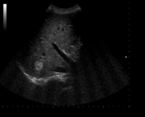
Hemangioma
Kasabach-Merritt syndrome is also called
Hemangioma thrombocytopenia syndrome
Kasabach-Merritt syndrome
Seen in infants with large hemangiomas
Sequestration and destruction of platelets within the larger cavernous hemangioma causes significant thrombocytopenia
Visible cutaneous blue or reddish lesions on extremities
Hepatomegaly or jaundice can also be identified
If the skin lesions are present with significant thrombocytopenia, the ____ and spleen should be evaluated for __________ formation
Liver; Hemangioma
Most common benign vascular liver tumor in INFANCY
Infantile hemangioma
Infantile hemangioma
Normally occurs within the first 6 months of life
Usually presents with cardiac failure early in life due to the AV malformation within the tumor
Spontaneously regresses in most patients before age 2
2nd most common benign liver mass
Focal nodular hyperplasia
Focal nodular hyperplasia (FNH)
Contains all normal parenchyma components, except normal portal venous structures
MOST COMMON IN WOMEN of childbearing years
Not encapsulated
Asymptomatic, Normal LFTS
Sulfur colloid imaging
FNH is associated with
Oral contraceptive use
Sulfur colloid imaging
Nuclear medicine exam used to detect FNH
In most pts, the FNH mass will absorb the sulfur colloid at the same rate as the normal liver tissue = warm nodule
In some pts, FNH mass will absorb the sulfur colloid at a higher rate than normal liver tissue = hot nodule
USA FNH
Variable in appearance, can be isoechoic to liver
Stealth lesion b/c it is difficult to differentiate from liver
Solitary mass <5cm
CENTRAL SCAR with radial vascularity
SPOKE WHEEL with color
May distort liver contour
Not able to diagnose on US
Contrast US demonstrates enhancement of FNH throughout arterial and portal phases
Contrast first fills central lesion and moves peripherally
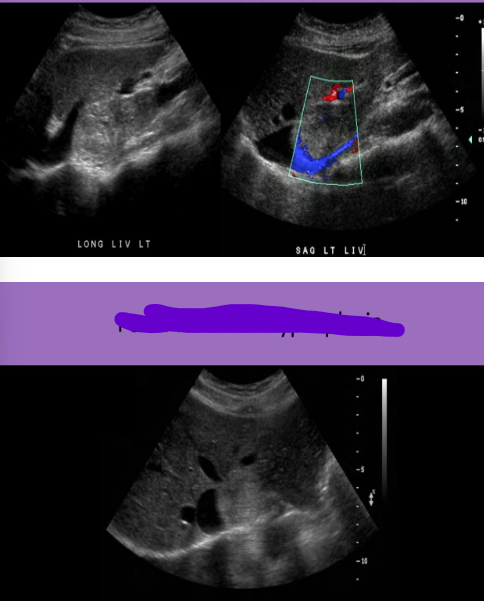
Focal nodular hyperplasia
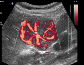
Focal nodular hyperplasia
Liver cell adenoma
Uncommon tumor
Atypical hepatocyte formation
Bile ducts and Kupffer cells are absent or few in number
Encapsulated mass
More common in women
Asymptomatic or RUQ pain
Normal LFTs
Cold nodule on sulfur imaging
Surgical intervention is recommended
Liver cell adenoma is associated with
Oral contraceptives, glycogen storage disease
USA liver cell adenoma
Hypervascular mass, with/without hemorrhage, solid and well defined
Varying echogenicity, usually hypoechoic
Increased vascularity with possible internal hemorrhage
HYPOECHOIC RIM
Cannot be differentiated from FNH on US
Contrast first pools peripherally then moves centrally
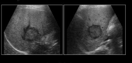
Liver cell adenoma
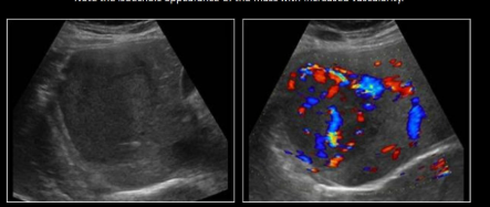
Liver cell adenoma
Hepatic lipoma
Very rare
Associated with tuberous sclerosis
Hyperechoic mass
May see propagation speed artifact due to difference in speed of sound in liver tissue and fat → broken diaphragm posterior to lesion
CT is helpful to determine type of tissue
HCC is the __th leading cause of cancer death
5
Most common primary malignancy of liver in adults
HCC
Most common risk factor for HCC
80% of pts already have this
Cirrhosis
HCC
Primary tumor of parenchymal cells that interferes with hepatocyte function
Leads to hepatoma formation
Related to formation of multiple nodules but can be solitary or diffuse
Invades vasculature causing an obstruction
3-4 month survival after detection
HCC is also assocaited with
Chronic hepatitis
70% of cases with HCC have moderate levels of ____ in blood → sign of malignancy
70
Symptoms of HCC
Fever, hepatomegaly, palpable mass, ascites, weight loss
HCC is more common in _______
Males
Lab testing for HCC
Increased AST, ALT and ALP
HCC most commonly invades what vessel
MPV
USA HCC
Single large mass or multiple masses with diffuse infiltration of cancer cells
Solid nodules <5cm
Varying echogenicity with Central Scar
Fibrosis and necrosis causes heterogenicity
HALO EFFECT → hypoechoic ring
Hypervascular
Venous invasion
Low resistance waveforms
Contrast demonstrates enhancement during arterial phase
Patient Hx of a primary cancer in the body + liver mass with halo =
Liver mets
Patient Hx of chronic liver disease and liver mass with halo =
HCC
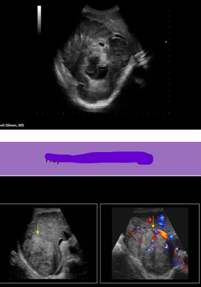
HCC
Fibrolamellar carcinoma
Subtype of HCC found in ADOLESCENTS and young adults with NO coexisting liver disease
AFP is normal
Solitary mass that can grow large (6-22cm)
Mass is well differentiated, and usually encapsulated by fibrous tissue
Punctate calcs and a central echogenic scar help to differentiate it from hepatoma
Number 1 PEDIATRIC primary liver malignancy
Hepatoblastoma
Hepatoblastoma
Genetic
Associated with Beckwith-Wiedemann syndrome or familial adenomatous polyposis
Abdominal enlargement, hepatomegaly, palpable mass
Lab testing: Abnormal LFTs, INCREASED AFP
USA Hepatoblastoma
Solid echogenic mass
May have cystic portions/septations
May contain calcs
Has tendency to invade hepatic and portal veins
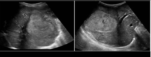
Hepatoblastoma
Liver is the #__ site for mets
1
Most common solid mass of the liver
Mets
Mets is ___x more common than primary liver cancer
20
__% of primary cancers that metastasize to liver are carried to the liver by the portal vein
50
Liver Mets
RUQ pain, weight loss, ascites, jaundice, palpable mass, N/V
Labs: Increased LFTs, Alk phos, AFP, direct bilirubin
Contrast US = rapid contrast washout in the portal venous phase
Liver Mets USA
Multiple masses that vary in size
Necrosis at the center of the lesion as the blood supply is outgrown
Hypoechoic rim USUALLY METS
Hyperechoic → GI tract primary or HCC
Hyperechoic with hypervascularity → RCC
Hypoechoic → Lymphoma, lung or breast primary
BULLS-EYE
Calcified
Abnormal lymph nodes in surrounding area
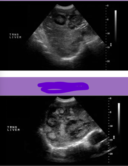
Liver mets
Kaposi Sarcoma
Malignancy of connective tissues in body
May also involve skin, lungs, GI tract and other organs
Most common liver malignancy seen with HIV/AIDS
Kaposi sarcoma
Lymphoma
Associated with HIV/AIDS
Malignancy of the lymphatic system
Solid tumor
Single or multiple
Hypoechoic and/or complex appearance
Hodgkins
Nonhodgkins
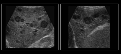
Non-Hodgkin lymphoma
Most common bloodborne chronic infection in US
Hepatitis C
A bulge in the liver capsule indicates __________, while an indented liver capsule indicates ________________
Intrahepatic; Extrahepatic
All the following describe a hemangioma except:
Most common benign mass of liver
Decreases in size with pregnancy
Most commonly seen in right lobe
Isovascular contrast enhancement
Decreases in size with pregnancy
Kasabach-merritt syndrome is associated with the formation of what benign liver mass?
Hemangioma
What benign liver masses are associated with use of oral contraceptives?
FNH and adenoma
FNH is demonstrated by US as:
Solid mass with central scar with radial vascularity
A liver adenoma is typically:
Hypoechoic
Hypervascular
Surgically removed
All of the above
All of the above
The presence of which blood testing indicated HCC is present in cases of cirrhosis?
AFP
Which of the following sonographic characteristics of a liver mass should cause a strong suspicion of malignancy?
Posterior enhancement
Hypervascularity
Calcificatinos
Hypoechoic halo
Hypoechoic halo
Focal fatty sparing vs. Hepatoma
Fatty sparing has no mass effect on the vasculature, while a hepatoma will invade vasculature
What is a sign of fibrolamellar carcinoma (FLC), but is not seen with other types of HCC?
FLC demonstrates punctate calcifications and a central echogenic scar
No coexisting liver disease
A liver mass with a target or bulls eye appearance is most likely
Liver mets
If liver mets is suspected after an upper abdominal US, the entire abdomen should be evaluated for associated:
Lymphadenopathy
What two liver malignancies are most commonly seen with AIDS?
Lymphoma and Kaposi tumor