Embryology Lecture 3
 Call Kai
 Learn
Learn Practice Test
 Spaced Repetition
 Match
Match Flashcards
 Knowt Play
1/66

There's no tags or description
Looks like no tags are added yet.
Name | Mastery | Learn | Test | Matching | Spaced | Call with Kai | Chat |
|---|
No analytics yet
Send a link to your students to track their progress
67 Terms
Cranial Placodes
thickening of the cephalic embryonic ectoderm
vison hearing and olfaction
what sensory organs does cranial placodes contribute to
Invagination
curve of the cranial placode that contains the lens, otic, olfactory, and the adenohypophyseal placode
Delamination
leads to the trigeminal and epibrachial
Adenohypophysis placode
invovled in an invagination of the oral ectoderm in the mouth
it develops directly from Rathke’s pouch
lens placode
part of cranial placode that leads to vision
invagination
in orange
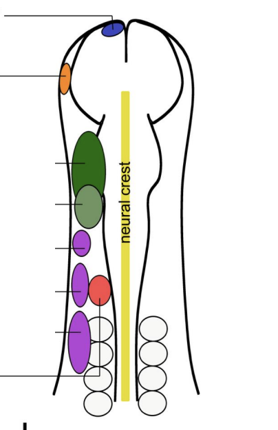
otic placode
part of cranial placode, invagination, that leads to inner ear
hearing/balance
In RED
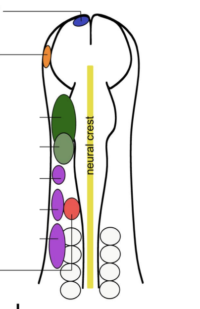
Olfactory placode
cranial placode that leads to olfactory epithelium (smell)
in blue
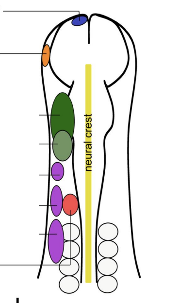
Rathke’s pouch
the embryonic precursor of the adenohypophysis
Trigeminal Placode
comes from delamination of the placode
corresponds to 2 regions
opththalmic
maxillomandibular
top two in green
Gives rise to Cranial Nerve V (5)
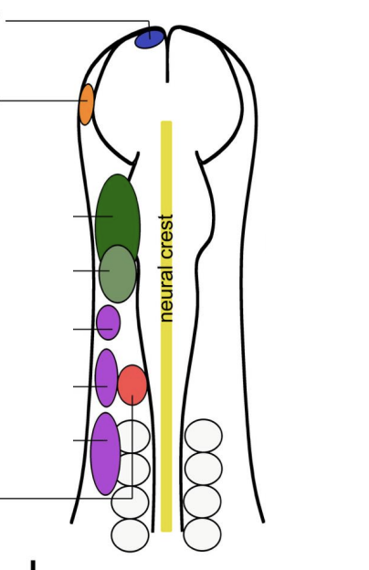
Stomodeum
future opening of the mouth
Epibranchial placode
comes from delamination of cranial placode
Corresponds to 3 regions
Geniculate
Petrosal
Nodose
In purple
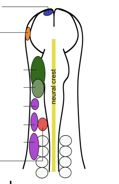
Geniculate
of the epibranchial placode that is associated with distal ganglion central nerve VII (7)
Petrosal
of the epibranchial placode that is associated with distal ganglion central nerve IX (9)
Nodose
of the epibranchial placode that is associated with distal ganglion central nerve X (10)
Gastrulation
allows the mesoderm to regionalize
Notochord
cells that migrate through the node, passing through the primitive streak
Paraxial mesoderm
the layer of mesoderm that is closes to the notochord since it is next to the primitive node
the lighter blue color
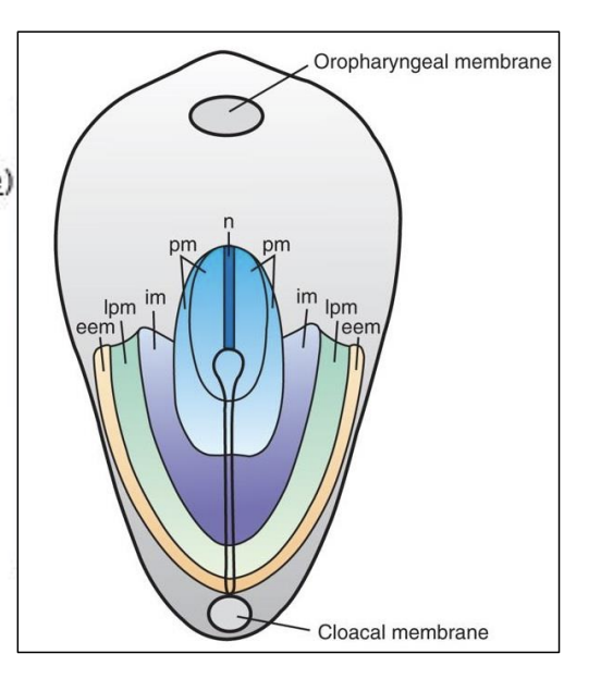
Intermediate mesoderm
the purple section
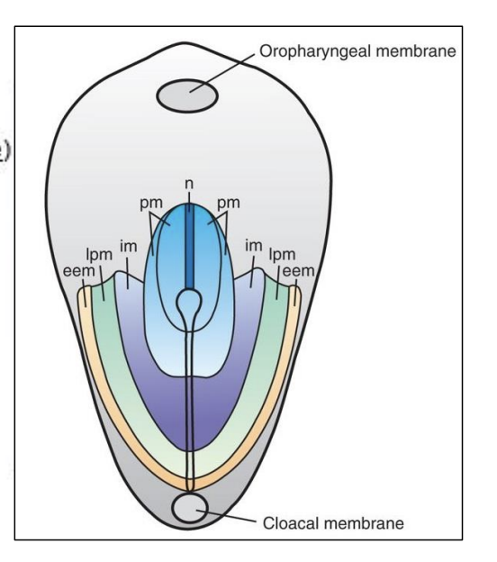
Node and Primitive Streak
Fate mapping analyses indicated that mesodermal tissue regionalization depends on the positive and the timing of cell passage through the _____ and the _______ in an anterior to posterior direction
Lateral plate mesoderm
in green
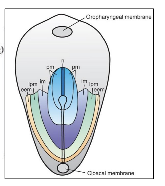
extraembryonic mesoderm
in the yellow, doesn’t directly help with the devlopment of the mesoderm proper
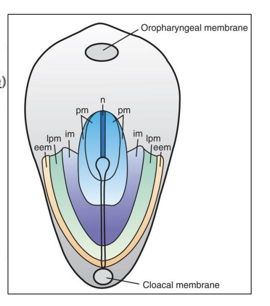
Chordamesoderm
the future notochord
Paraxial mesoderm
Gives rise to
Head mesenchyme (bones in the skull)
Somites
Sclerotome (cartilage, bones)
Myotome (skeletal muscle)
Dermatome (dermis)
Syndetome (tendons)
Large blocks in red
Sclerotome
direct derivative of the somites which are a derivative of the paraxial mesoderm
cartilage and bones, vertebrae
ventral cells migrating
Myotome
direct derivative of the somites
skeletal muscle
Dermatome
direct derivative of the somites
dermis, dorsal, located in between the primaxial and abaxial
Syndetome
direct derivative of the somites
tendon progenitors, the last compartment to form
forms at the boundary between the sclerotome and the myotome
Intermediate mesoderm
gives rise to the
kidneys
gonads
Lateral Plate Mesoderm
gives rise to
Splanchnic (visceral) mesoderm (circulatory system)
Somatic (parietal) mesoderm (body wall)
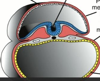
Splanchnic mesoderm
visceral, that is involved in the circulatory system
Ventral component
covering of the abdominal organs, lungs, hearts
it is a derivative of the lateral plate mesoderm
the thin red layer on the bottom half surrounding the yellow endoderm
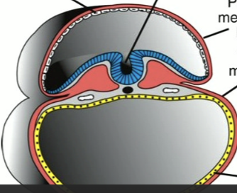
Somatic mesoderm
parietal, body wall,
Mesodermal components of the limbs, like bone(except muscles)
It is the top layer of the lateral plate mesoderm/ Dorsal
it is a derivative of the lateral plate mesoderm
the red layer on the top half that is surrounding the amnion and the the future blue neural tube
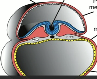
Somite
the derivative the paraxial mesoderm that has now developed
the large red blocks
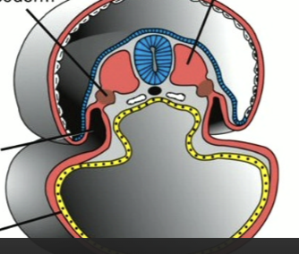
Intermediate mesoderm
the brown blocks that are surrounding the somites
This generates the urinary and genital systems
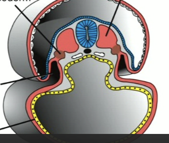
Coelom
the space between the somatic mesoderm (top) and splanchnic mesoderm
forms the body cavity
abdominal
thoracic
yolk sac
What is the area within the yellow endoderm
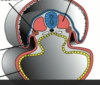
gut, parietal mesoderm, embryonic body cavity, surface ectoderm, dorsal mesentary, visceral mesoderm
what is the yellow tube
what is the outer red layer
what is the space under the yellow tube
what is the blue layer surrounding
what is the thick red column above the yellow tube
what is the red layer that is directly surrounding the yellow tube
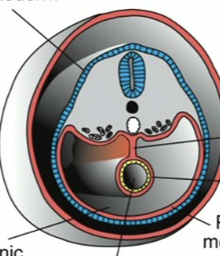
Somatopleure
Somatic (pareital) mesoderm + ectoderm
forms ventral and lateral body wall
forms the membranes that line the peritoneal, pleural and pericardial cavities
Splanchnopleure
Splanchnic (visceral) + endoderm
Form the wall of the gut and the thin serous membrane around each organ
Intermediate mesoderm
which mesoderm generates the urinary and genital systems?
Urinary system
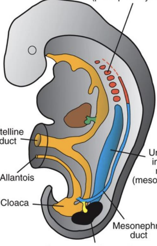
formed sequentially from 3 overlapping kidney systems
Pronephros
Mesonephros
Metanephros
Pronephros
non-functional kidney, degenerate at the end of the 4th week
the most anterior, red color on the diagram
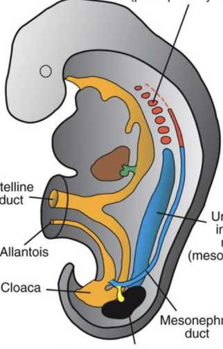
Mesonephros
functional kidney for a short time
during fetal period, then regresses
The blue on the diagram
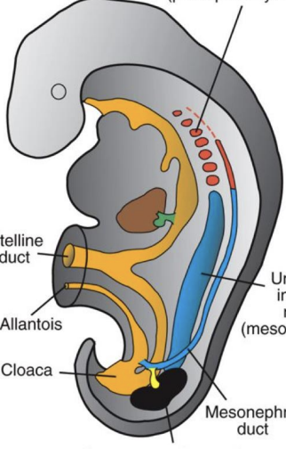
Metanephros
Permanent Kidney
appears in the 5th week and becomes functional around the 12th week
form in the most posterior region of the embryo
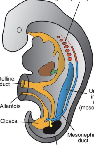
comes from the Metanephric blastema
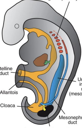
Ureteric bud
gives rise to the collecting system
the bright yellow structure in the photo
Metanephric blastema
the black structure in the image
gives rise to the excretory system
Ascent
The kidneys at the posterior region near the kidneys will go which way as a result of a decrease in the body curvature
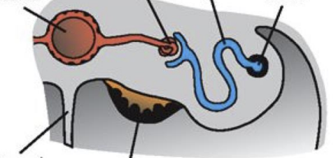
Aorta, glomerulus, dorsal mesentary, genital ridge, excretory tube, mesonephric duct
This is a cross section of the developing gonad
big red ball
small ball at the end of red
thin grey column
black thing under the orange
blue tube
end of the blue tube
Treacher collins syndrome
includes hypoplasia of the facial bones and dental malocclusion
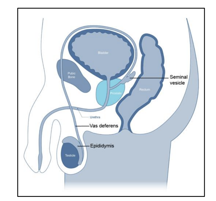
Wolffian duct
another name for the mesonephric duct
in males, this duct forms an epididymis, vas deferenes, and seminal vesicles
In females, this duct degenerates
Mullerian duct
another name for the Paramesonephric duct
in males, this ducts degenerate
In female, this duct forms the fallopian tubes, uterus, cervix, and vagina
Somites
The number of this is a good indicator of the age of an embryo
Primaxial dermamyotome
derivative of the paraxial mesoderm
closer to the neural tube that gives rise to the muscles of the back
Abaxial dermamyotome
derivative of the paraxial mesoderm
further away from the main axis of the embryo/further from the neural tube and notochord
muscles of the body wall
Location
What determine cell fate within the somite?
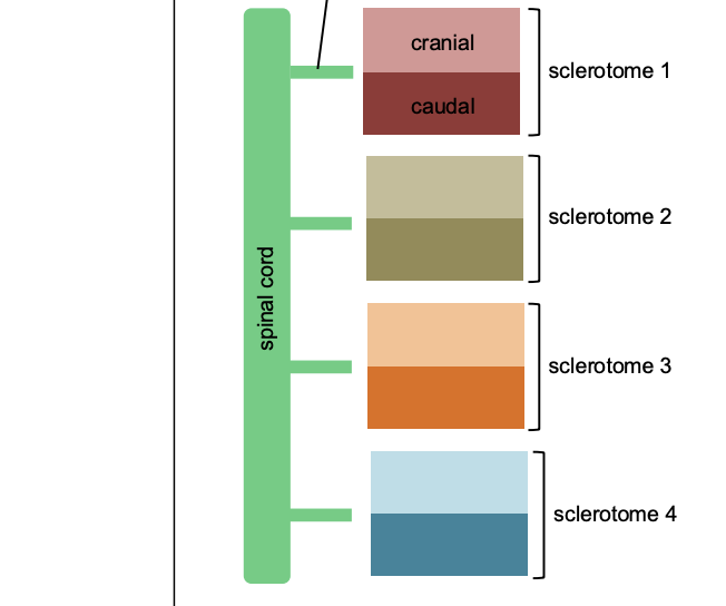
Resegmentation
the sclerotome portion of each somite undergoes this process.
A single sclerotome from one single somite will be further divided into cranial and caudal region.
Each vertebrae is formed from the fusion of the caudal half of one somite and the cranial half of its neighbor
Nucleus pulposus
what the notochord will become
surrounded by the circular fibers of the annulus fibrosus
Intervertebral disc
Annulus fibrosus + nucleus Pulposus
Annulus fibrosus
the circular fibers that surround the nucleus pulposus (what use to be the notochord)
limb bud
protrusion of the ectoderm
derived from the mesoderm of somites (muscle precursors) and somatic lateral plate (skeletal precursors)
Apical ectodermal ridge
organizing center in the limb bud
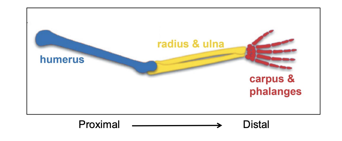
signals to the progress zone to continue to divide and multiply, once the zone gets out of the range of this, it develops into something else
Progress zone
the zone immediately adjacent to the apical ectodermal ridge
participates in the establishment of the proximal-distal axis of the limb
Phocomelia
condition in which the limbs are missing the proximal elements and you see the hand attached to the level of the shoulder
long bones are shorter than normal and the more proximal elements are lost
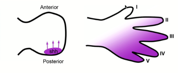
ZPA
secrets sonic hedgehog which acts as a gradient. This has an influence on the development of the digits in the hand.
ZPA is posterior (pinky side)