Cervical Spine
1/35
There's no tags or description
Looks like no tags are added yet.
Name | Mastery | Learn | Test | Matching | Spaced | Call with Kai |
|---|
No analytics yet
Send a link to your students to track their progress
36 Terms
How many C vertebrae are there?
7
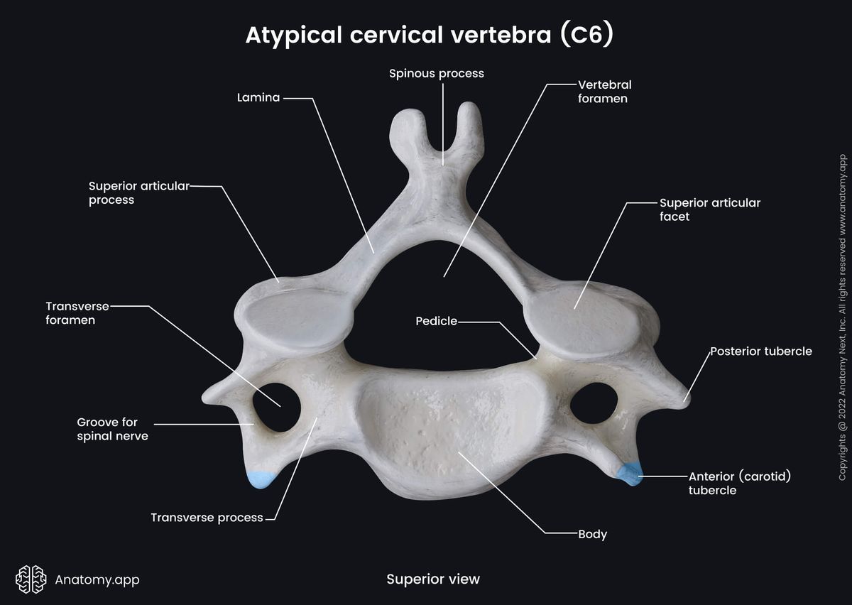
Which C vertebrae are the typical vertebrae?
C3-C6
What type of curve does the C spine have?
secondary (lordotic)
concave posterior, convex anterior
The transverse foramen of C vertebrae contain ___
an artery that goes to the brain
C3-C6 have ___ spinous processes
bifid
The area between the articulating processes of a singular vertebrae are called ___
pillars
The articulating processes (zygapophyseal joints) of C vertebrae sit at a ___ angle and are visualized in a(n) ___
90o
lateral position (for C3-C7)
AP position (for C1/C2)
C vertebrae pedicles come off of the body at a ___ angle and (intervertebral foramina) are visualized in a(n) ___
45o
oblique position (15o cephalic angle if AP, 15o caudal angle if PA)
If done as AP obliques, intervertebral foramina are demonstrated as side ___
up

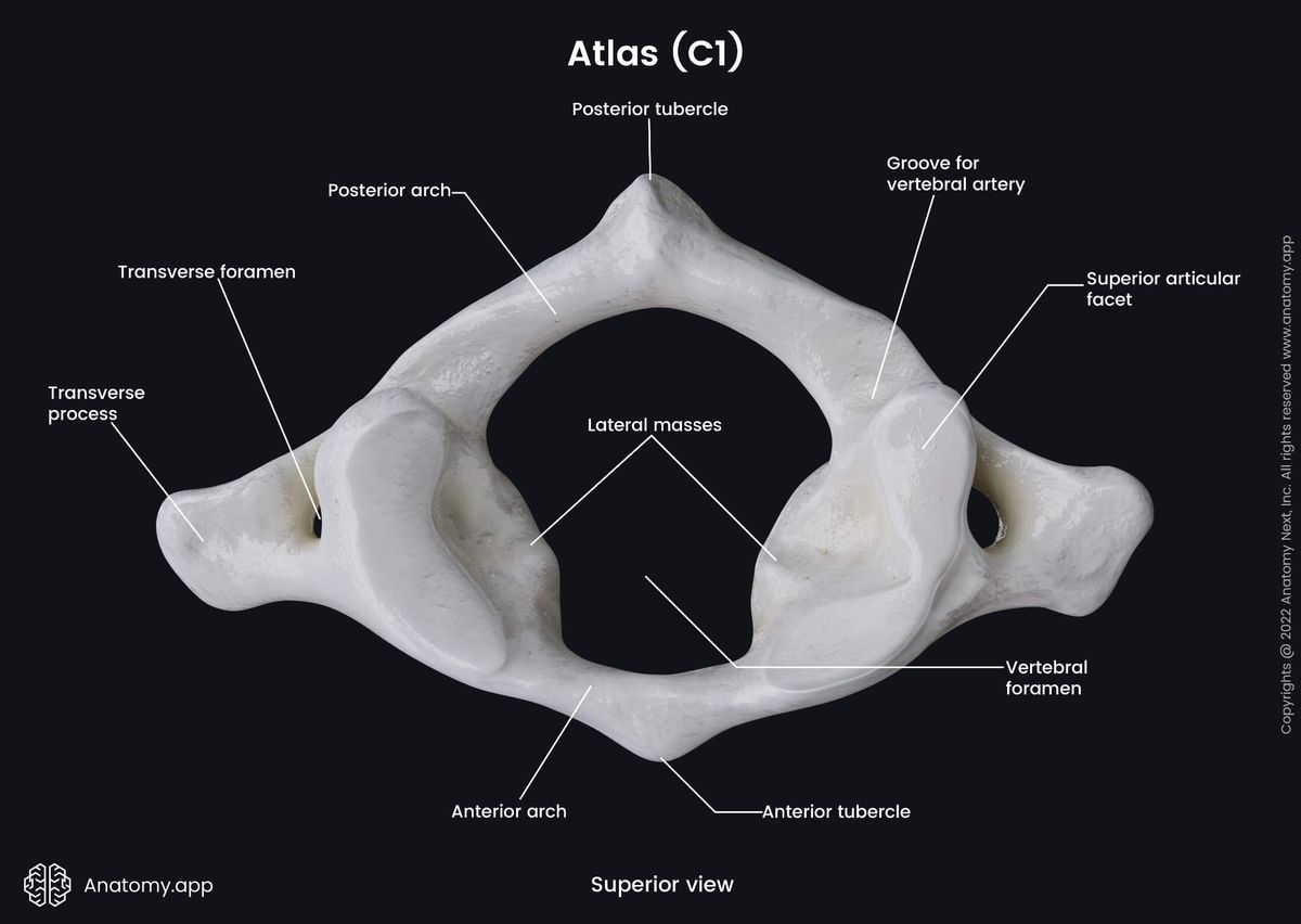
The first C vertebrae is called the ___
atlas
What are the 4 parts of the atlas?
anterior arch
posterior arch
lateral masses
transverse process
The posterior tubercle of C1 is the ___
spinous process
Which arch of C1 is bigger?
posterior
The areas of the superior articulating processes/facets of C1 are known as ___
lateral masses
Does C1 have a body?
NO
What the ligament that runs across C1 called?
transverse atlantal ligament
The posterior portion of C1 (as divided by the transverse atlantal ligament) ___
transmits the proximal end of the spinal cord
The anterior portion of C1 (as divided by the transverse atlantal ligament) ___
receives C2
What is the atlantooccipital joint?
the superior articulating facets of C1 articulating with the occipital condyles of the skull
What is the classification of the atlantooccipital joint?
diarthrodial, synovial, ellipsoidal (condylar)
allows flexion and extension (nodding your head)
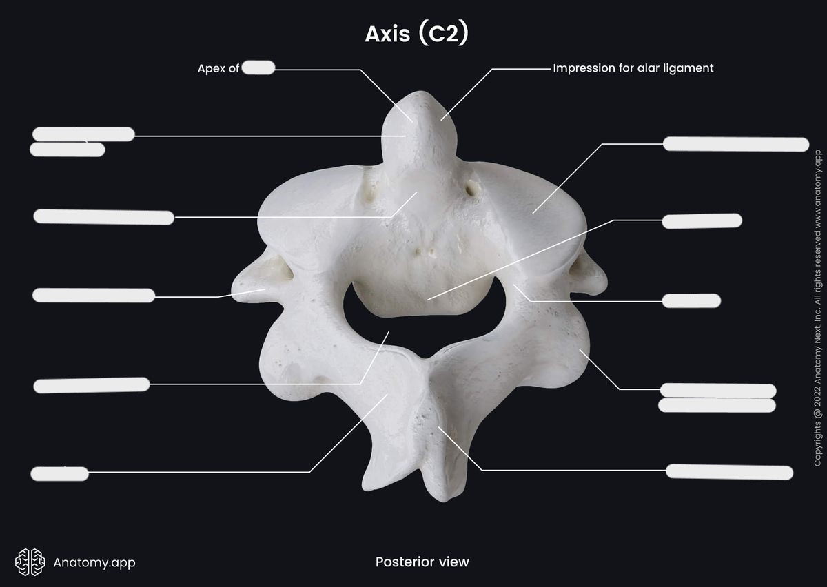
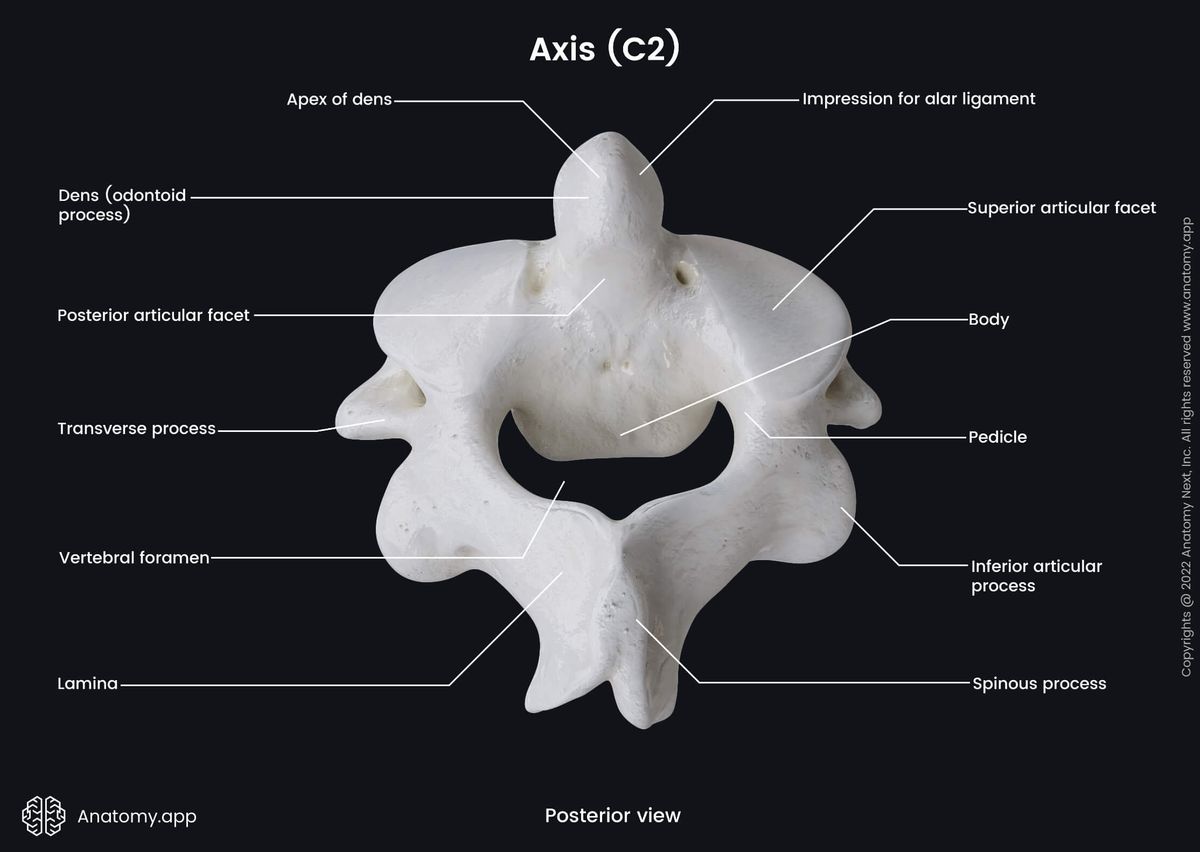
C2 is also called the ___
axis
Explain the spinous process of C2
thick and blunt
C2 has all the anatomy of a typical C vertebrae, except ___
it has the dens/odontoid process
What is another name for the atlantoaxial joint?
atlanto-epistropheal joint
What is the atlantoaxial joint?
odontoid is received into the anterior portion of C1
What is the classification of the atlantoaxial joint?
diarthrodial, synovial
Medial (dens and atlas ring): trochoid/pivot
Lateral (articulating processes/zygapophyseal): gliding/plane
The AP “open mouth” projection demonstrates ___
the incisors lined up with base of skull
odontoid tip
C1/C2 zygapophyseal joint
The C7 spinous process is called the ___
vertebral prominence
Why is C7 considered atypical?
it has a very long spinous process
Explain the Hangman’s fracture
involves C2
fracture through C2 pedicles on both sides
results from extreme hyperextension
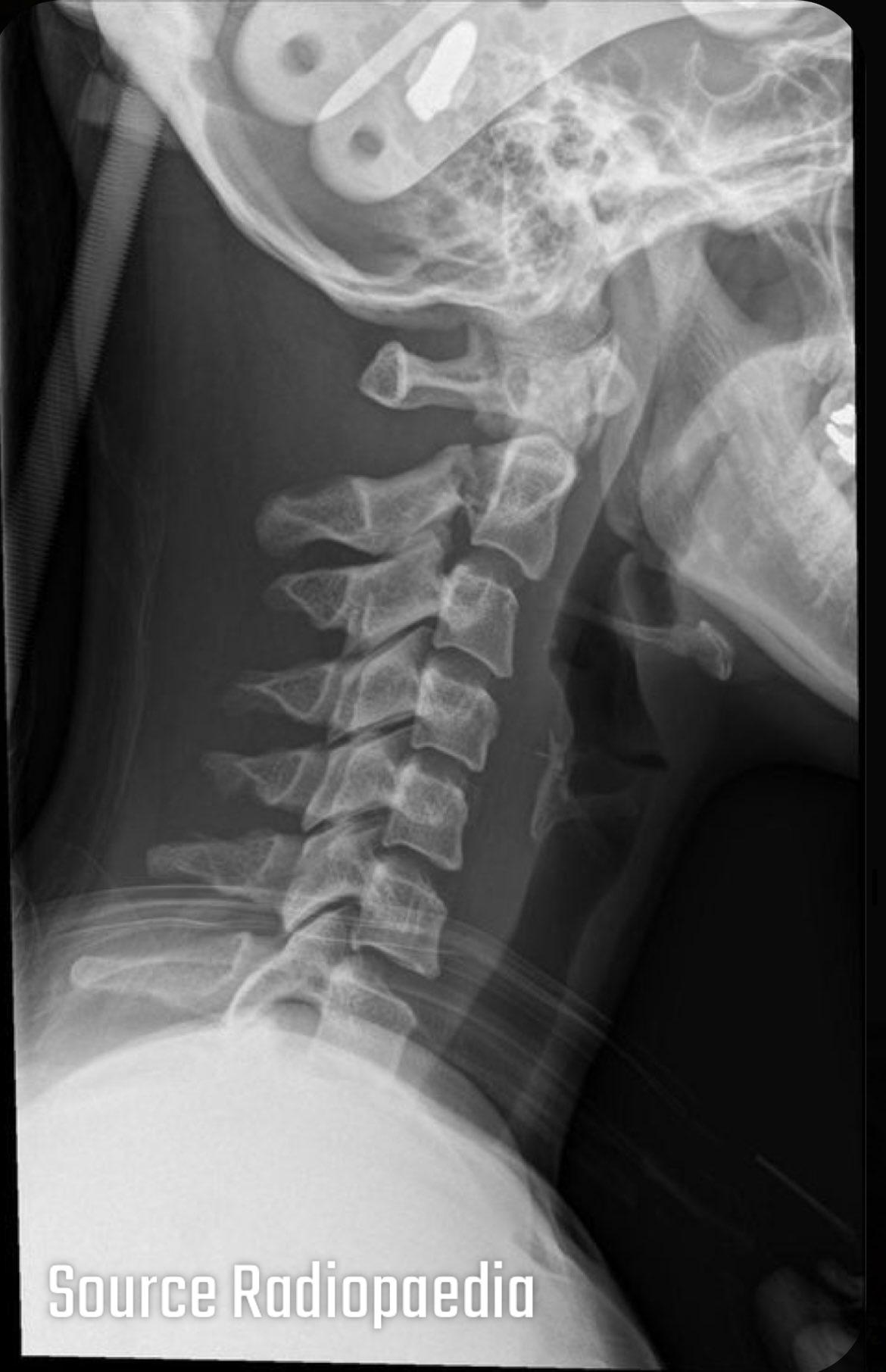
What type of fracture is shown?
Hangman’s fracture
Explain Jefferson’s fracture
involves C1
anterior and posterior arches of C1 are fractured by force (such as hitting head on bottom of shallow swimming pool)
often involves paralysis
Explain odontoid fracture (including the 3 types)
direct impact from the anterior arch of atlas during hyperextension or hyperflexion
Type 1: extends through the tip of the dens
Type 2: extends through the base of dens
Type 3: extends through the vertebral body of the axis