Cell structure and division
 Call Kai
 Learn
Learn Practice Test
 Spaced Repetition
 Match
Match Flashcards
 Knowt Play
1/93

There's no tags or description
Looks like no tags are added yet.
Name | Mastery | Learn | Test | Matching | Spaced | Call with Kai | Chat |
|---|
No analytics yet
Send a link to your students to track their progress
94 Terms
cell
the smallest, basic membrane bound unit of life, responsible for all of life’s processes
cell theory- in order to continue living a cell requires
metabolic machinery capable of obtaining energy from the environment
the ability to use this energy to support essential life processes
a set of genes to control the synthesis of all the cell components and these are passed onto the next generation through cell division
a physical boundary between itself and the environment (cell membrane)
stem cell
a cell that can produce other cells which can develop into any kind of cell in the body
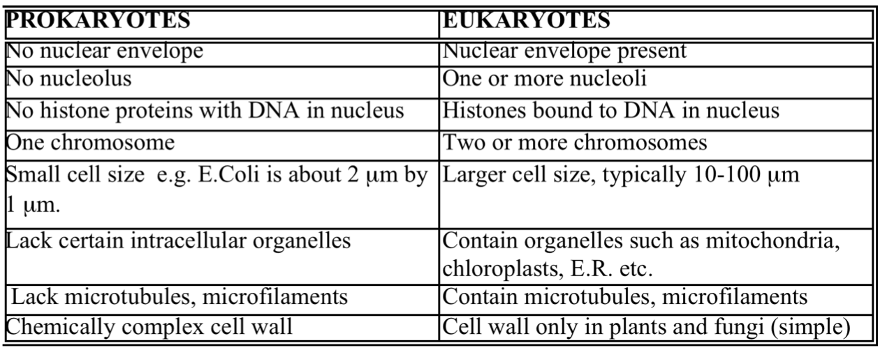
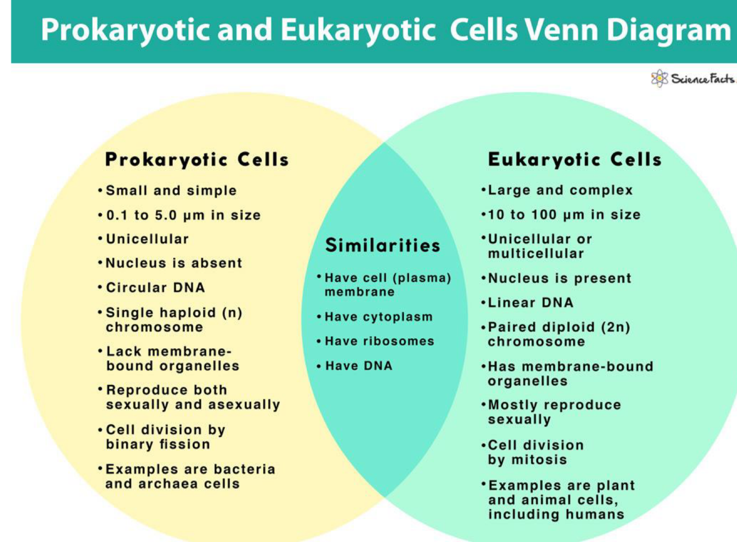
differences between prokaryotes and eukaryotes
3 shapes of bacteria
coccus (round)
bacillus (rods)
spirilla (coils)
3 parts of bacteria
external appendages and coverings
cell envelope (cell membrane and cell wall)
internal organelles
what is a cell wall made up of
peptidoglycan
what is peptidoglycan made up of
2 types of glycan chains (NAG and NAM)
what are the 2 types of glycan chain
N-acetyl glucosamine (NAG)
N-acteyl muramic acid (NAM)
Gram positive cell wall
thick (20-80nm)
porous
contains teichoic acid
stains purple
Gram negative cell wall
thin (1-3nm)
requires an extra layer of protection- outer membrane (porins)
stains pink
4 ways antibiotics can affect bacterial cells
Disrupts cell wall synthese
Inhibits DNA replication- e.g. quinolones (ciprofloxacins)
Inhibits RNA synthesis
Inhibit protein synthesis- e.g. macrolides (erythomycin)
disrupts cell wall synthesis
binds to transpeptidases and inhibits the cross-linking of peptidoglycan subunits
inhibits RNA synthesis
binds to RNA polymerase e.g. rifampicin
porins
block the entrance of harmful chemicals and antibiotics making gram-negative bacteria more difficult to treat than gram-positive cells
what is attached to the outer membrane
lipopolysaccharide
lipopolysaccharide
acts as an endotoxin because it induces fever and shock in the human host
pilus
hair like appendage required for bacterial conjugation (transfer of genetic material)
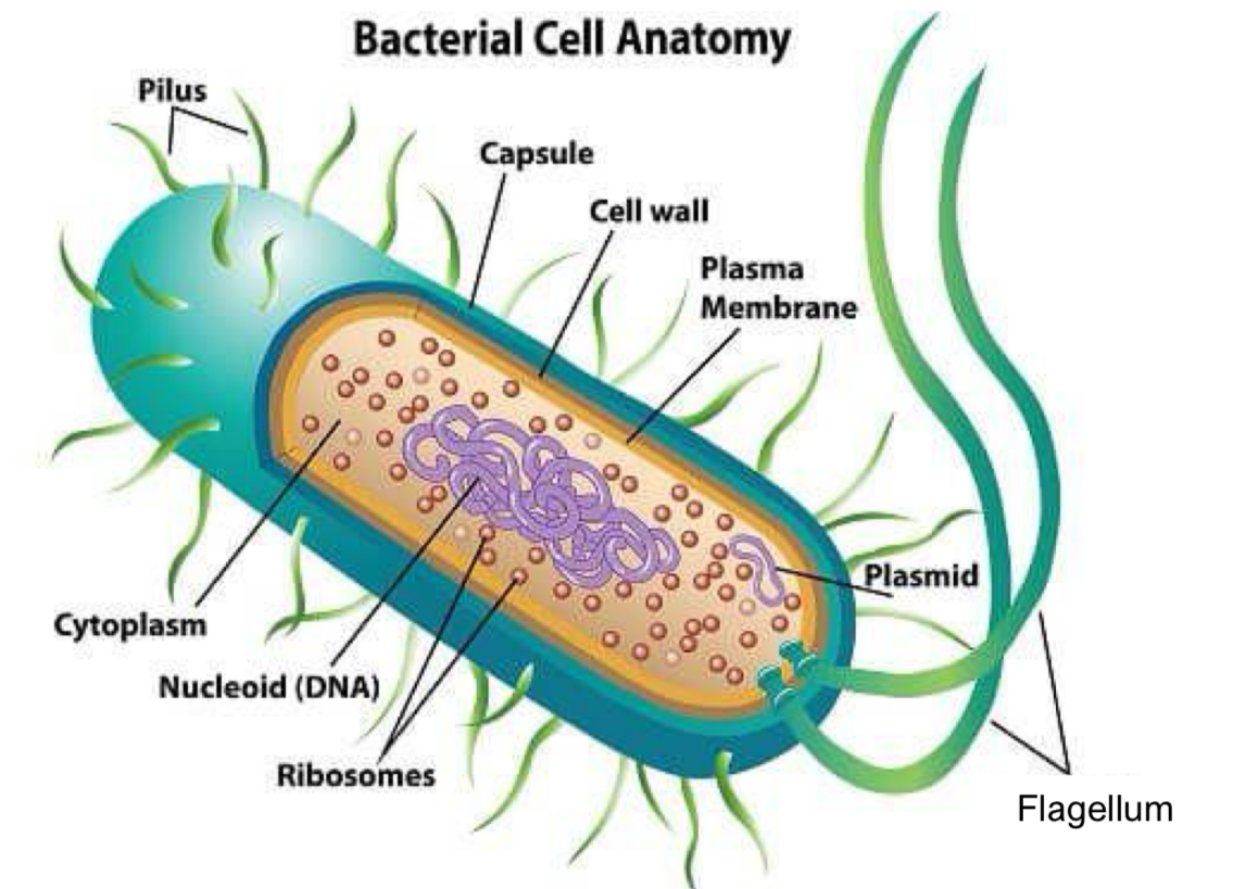
capsule
polysaccharide layer, contains water to prevent the cell from dying out, protects the cell from phagocytosis helps adhere to surfaces
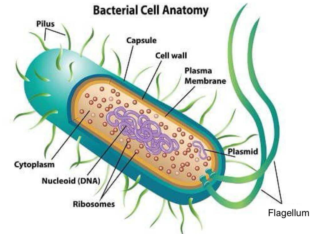
cell wall
rigid structure responsible for the characteristic shape and is made of cellulose
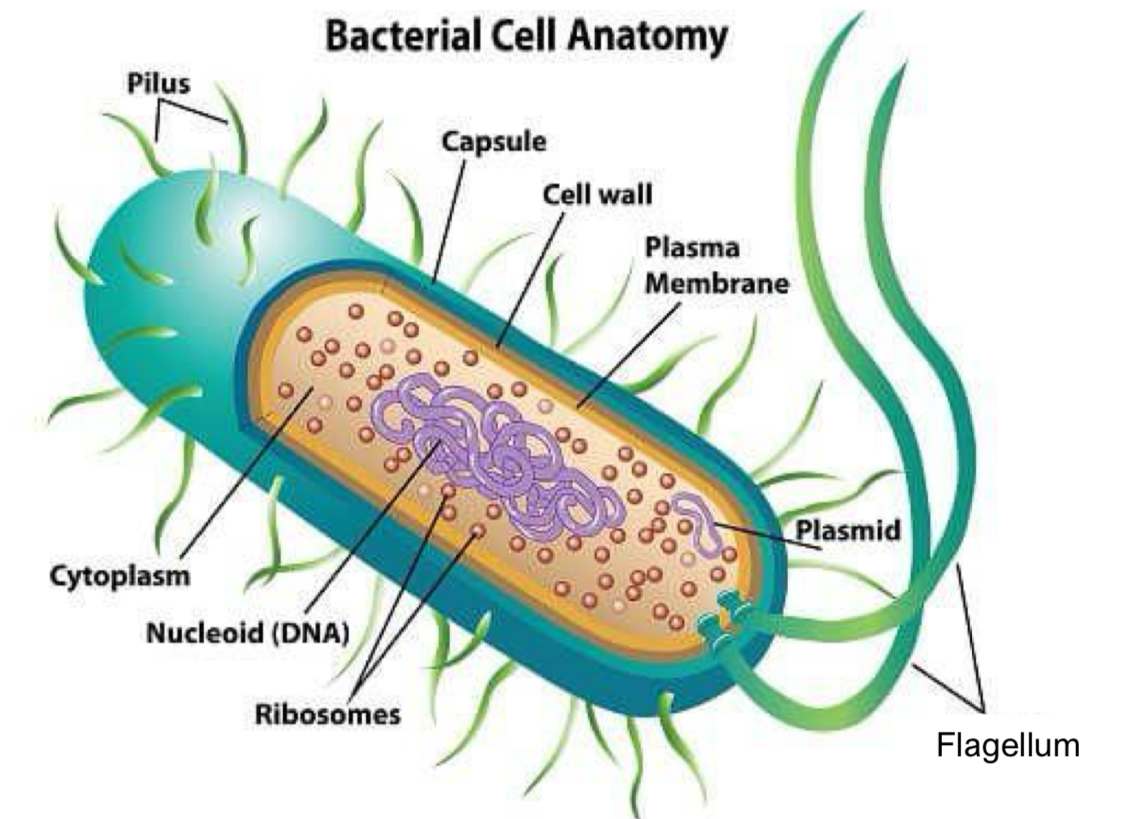
plasma membrane
consists of protein and phospholipid- transport, biosynthesis and energy transduction
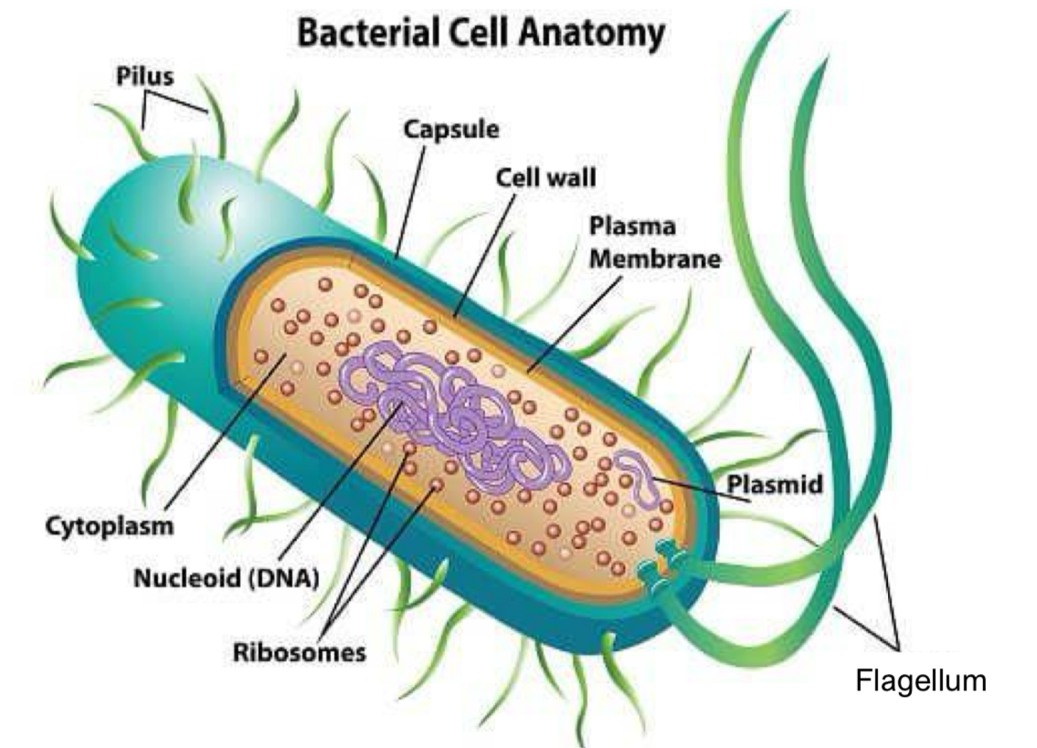
plasmid
circular DNA, bacterial virulence
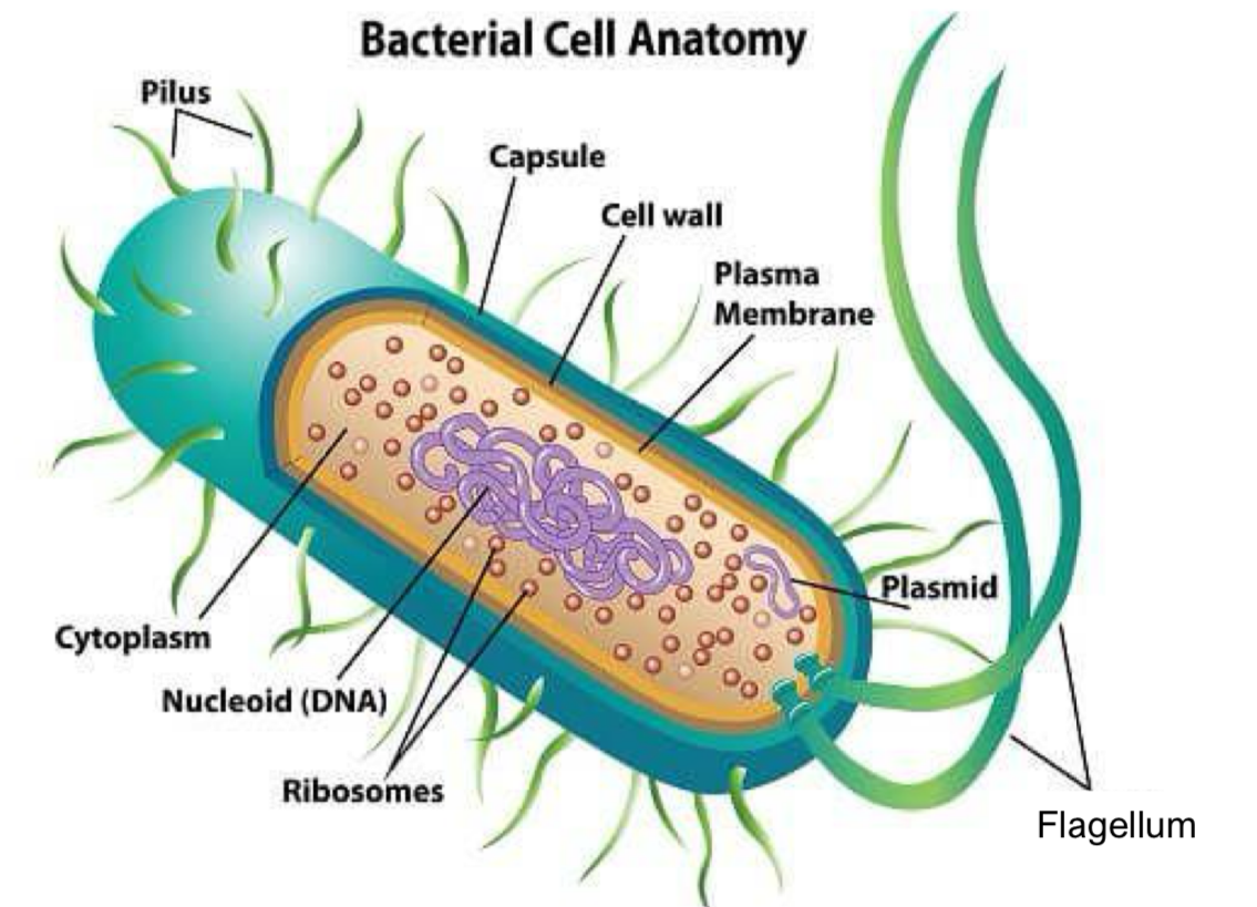
flagellum
enables movement and chemotaxis (process where cells move directionally in response to specific chemicals in the environment)
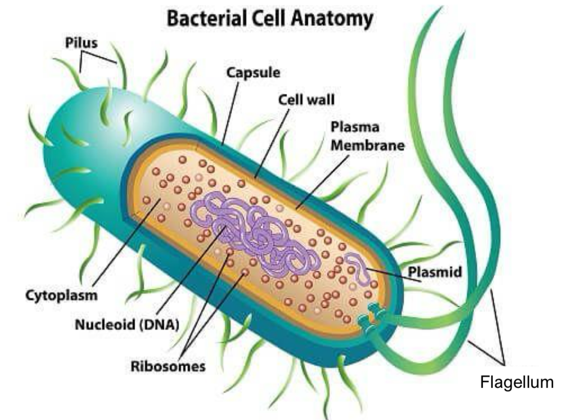
ribosomes
translate the genetic code from DNA to amino acids to produce proteins
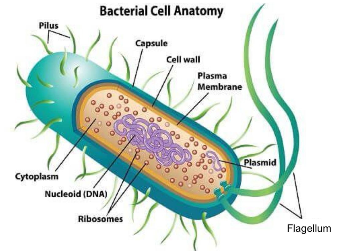
nucleoid (DNA)
regulates growth, reproduction and function of the cell
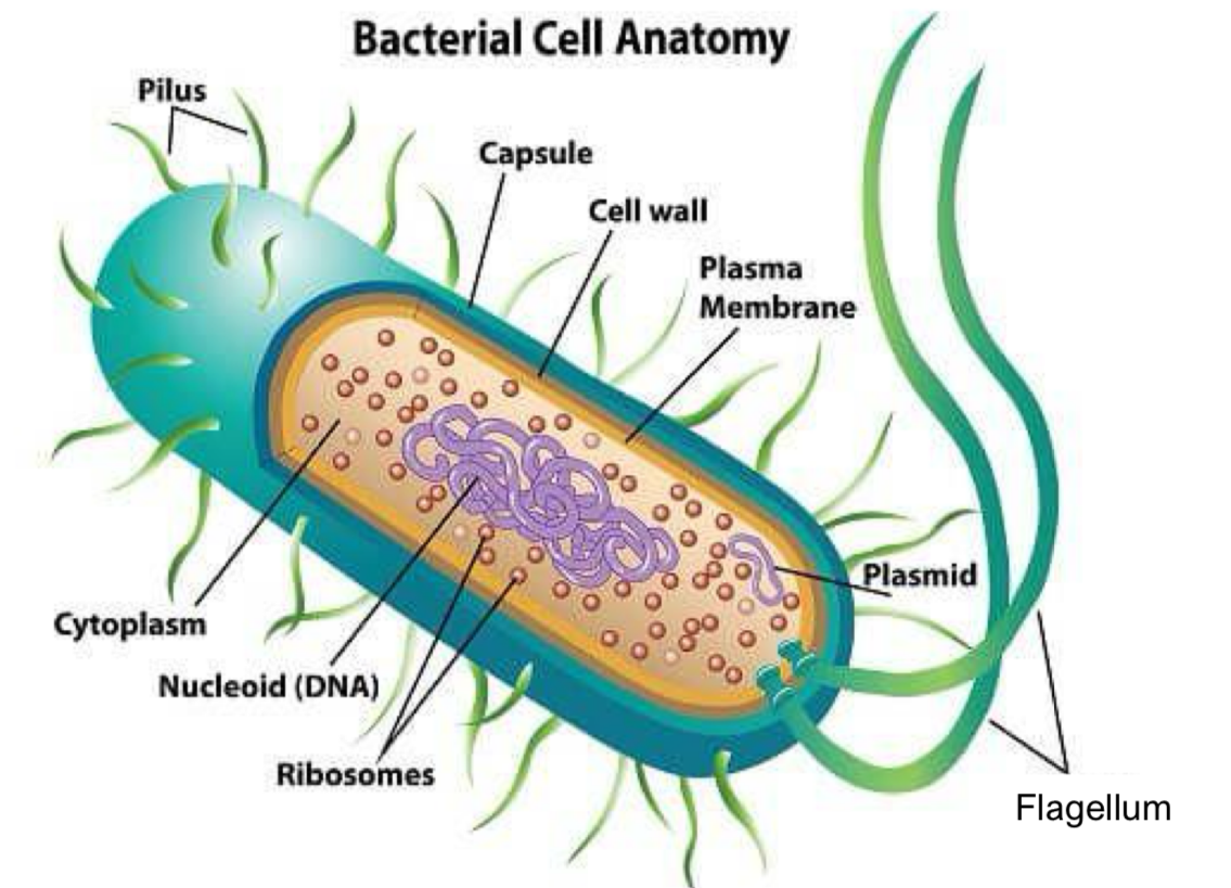
cytoplasm
fluid that fills the whole cell, house and maintain an optimal environment for the organelles. It’s where chemical reactions take place
eukaryotic cells
basic units of animals and plants
mitochondria
turns glucose and oxygen into energy
vacuole
contains cell sap, which is a weak solution of sugar and salts
green chloroplasts
contains chlorophyll for photosynthesis
what does a cell membrane consist of
a double layer of phospholipid each about 2nm thick separated by a space approximately 3.5nm
what is embedded in the fluid mosaic in the cell membrane
proteins and other lipids
what is the cell surrounded by
a plasma membrane that controls the passage of materials into and out of the cell as well as receiving messages that regulate the behaviour of the cell
nucleus
surrounded by a double membrane where pores of 40-100nm can be seen
inside nucleic acids and proteins are found
more densely packed chromatin granules appear as nucleoli (ribosomal RNA is made there)
cell organelles- mitochondria
small elongated bodies
consists of a double membrane, the outer membrane and much folded inner membrane
contains enzymes and some DNA
produces most of the cell’s adenosine triphosphate (ATP)
cristae
the folds in the inner membrane of the mitochondria
chloroplasts
have a double membrane surrounding a storm where stacks of lamellae which contain chlorophyll
performs photosynthesis
cell wall
a non-living layer found in plant cells and is derived from cellulose (a glucose polymer)
ribosome
carries out protein synthesis and is found in the rough endoplasmic reticulum
endoplasmic reticulum
can be rough- engages with protein synthesis
or smooth- involved in lipid metabolism
surface area is very large
lysosome
vesicles containing enzymes that break down proteins, lipids and nucleic acids
centriole
2 centrioles = a centrosome- attaches the cell’s cytoskeleton of microtubules
cytoskeleton
framework of tubular proteins gives cell its shape provides basis for movement
nucleus
DNA is held in the nucleus in chromatin fibres
nucleolus assembles ribosomes and communicates with cytoplasm through pores in the nuclear envelope
Golgi apparatus
flattened sacs that receive vesicles from the endoplasmic reticulum, modify, sort and package their contents and secrete it to the other organelles
plasma membrane
a bilayer of phospholipid molecules acts as a semi-permeable barrier in which proteins are embedded and acts as receptors, pumps and channels
viruses
they are tiny particles only visible in an electron microscope
2/3 parts of viruses
the genetic instructions in the form of DNA or RNA
A protein coat (capsid) that protects the DNA or RNA
in some cases an envelope of lipids that surround the protein coat when the virus is outside cells
capsid
a protein coat that surrounds and protects the DNA or RNA
how do viruses work
they replicate by taking over the machinery of a host cell and directing it to synthesis new virus particles
the newly synthesised virus particles are released from the cell and sometimes destroys the cell in the process
viruses bind to host cells in order to gain entry to the cell and incorporate their genome into the host cell DNA to achieve replication of the virus `
what do many virus cells target
glycans for cell attachment and entry
cell surfaces are especially enriched with complex carbohydrates, polysaccharide, glycoproteins and glycans
influenza
influenza virus binds to cell-surface slalic acid receptors through its surface glycoprotein hemagglutinin
cell takes up the virus by endocytosis
this vesicle fuses with a lyosome
virus takes up the lysosome and uses the acidity to break down the coat. Hemagglutinin undergoes structural changes and inserts itself into the cell membrane
membrane fusion and releases RNA into the host cell
intracellular replication
a cell surface neuraminidase cleaves sialic acid from the cell membrane allowing viral escape
viral treatment- drugs
antibiotics have no effect
antiviral drugs- inhibit or disable viral proteins that are a part of the cell entry mechanism or the viral replication and release process
viral treatment
viral infections provokes an immune response that usually eliminates the virus
the immune response can be induced artificially with vaccines
vaccines
contains an agent that could be dead virus particles or one of the viral surface proteins
how do vaccines work
the agent stimulates the body’s immune system to recognise the agent as a threat, destroy it and to further recognise and destroy any similar viruses it may encounter in the future
mitosis
replication of cells where division results in 2 identical daughter cells
meiosis
division that results in 4 gametes (Sex cells) with half the chromosome number
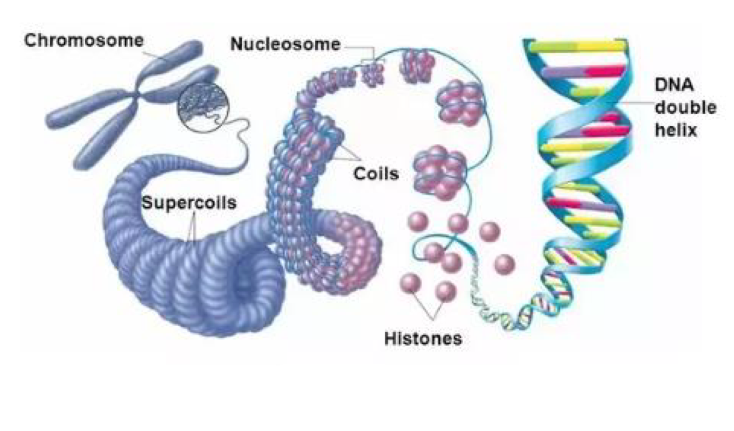
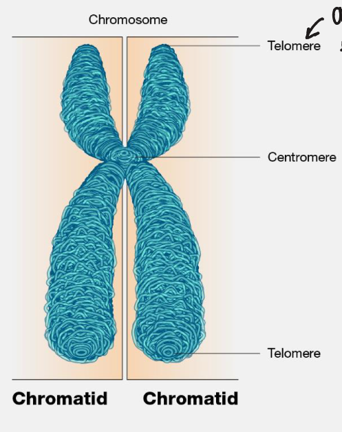
chromosomes
threadlike structure of nucleic acids and protein found in the nucleus
chromatid
2 threadlike strands that make up the chromosome
centromere
region of a chromosome where the microtubules of the chromosome join
telomere
cap at the end of each chromosome arm which maintain stability
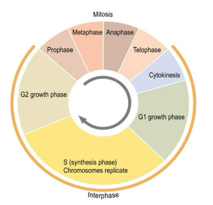
cell cycle
series of events that occur when a cell divides and grows
cell division structure
chromosome structure
stages of cell cycle
interphase
prophase
metaphase
anaphase
telophase
stages of mitosis
prophase
metaphase
anaphase
telophase
stages of interphase
G1 stage
S stage
G2 stage
G1 stage
the synthesis of RNA and protein occurs
preparing to replicate DNA by synthesising the mRNAs and proteins required to execute the future steps
cell usually grows larger and some organelles are copied
S stage
replication of DNA us complete and duplication of histone proteins occurs
the process of replication generates sister chromatids, which are identical pairs of chromosomes
these sister chromatids are attached to each other by a centromere
a centromere is a specialised sequence of DNA that links the sister chromatids and is important throughout mitosis
G2 stage
synthesis of organelles like mitochondria occurs
the cell undergoes additional growth, replenishes energy stores and prepares and reorganises the cytoplasmic components for division including duplicating some organelles ad dismantling the cytoskeleton
G2 ends when mitosis begins
how are chromosomes formed
DNA is packaged by special proteins called histones to form chromatin
chromatin further condenses to form chromosomes
chromatins is a lower order of DNA organisation whereas chromosomes are higher order of DNA organisation
prophase
chromosomes condense and become visible inside the nuclear membrane as long thin entangled threads
threads become shorter and thicker and the chromosomes care seen as separate structures
the nucleous gradually disappears
the centrioles migrate to opposite ends of the cell
the spindle of microtubules begins to form
proteins catalyse the condensation process (the chromosomes become compact) including cohesion and condensin
cohesion forms rings that hold the sister chromatids together
condensin forms rings that coil the chromosomes into highly compact forms
pro-metaphase
phosphorylation of nuclear lamins by M-CDK causes the nuclear membrane to break down into numerous small vesicles
the spindle microtubules now have direct access to the genetic material of the cell
each microtubule is highly dynamic, growing outward from the centrosome and collapsing backward as it tries to locate a chromosome
the microtubules find their targets and connect to each chromosome at its kinetochore, a complex of proteins positioned at the centromere
the actual number of microtubules that attach to the kinetochore varies between species
at the end of pro-metaphase…
kinetochore microtubules attach the chromosomes to the spindle pole
interpolar microtubules extend from the spindle pole across the equator
astral microtubules extend from the spindle pole to the cell membrane
metaphase
nuclear membrane disappears and each chromosome is seen as 2 chromatids joined at the centromere
the chromosomes become attached by their centromeres to the equator of the spindle
anaphase
the centromere splits
enzymatic breakdown of cohesion- which linked the sister chromatids together during prophase- causes this separation to occur
the spindle microtubules to which centromeres are attached shorten and drag chromatids away from the equator to opposite ends of the cell
anaphase A
the kinetochore microtubules shorten and draw chromosomes toward the spindle poles
anaphase B
the astral microtubules that are anchored to the cell membrane pull the poles further apart and the interpolar microtubules slide past each other, exerting additional pull on the chromosomes
telophase
the chromatids assemble at the poles and a nuclear membrane forms around each group
inside the nuclear envelope chromatids become uncoiled, nucleoli reform and the nucleus takes on a granular appearance
cell division is completed by cytoplasmic cleavage (cytokinesis)
cytokinesis
the cell membrane between the 2 nuclei separated to form 2 daughter cells each of which is a copy of the original cell
why do people get Down syndrome
they have an extra chromosome
prophase I
the chromosomes condense and the nuclear envelope breaks down- crossing over occurs
metaphase I
pairs of homologous chromosomes move to the equator of the cell
anaphase I
homologous chromosomes move to the opposite poles of the cell
telophase I & cytokinesis
chromosomes gather at the poles of the cells
the cytoplasm divides
synapsis
pairing of homologous chromosomes
how does crossing over work
spindle fibres will pull the homologous chromosomes together creating tetrads leading to crossing over
this creates genetic diversity
prophase II
a new spindle forms around the chromosomes
metaphase II
chromosomes line up at the equator
anaphase II
centromere divides and chromatids move to the opposite poles of the cells
telophase II and cytokinesis
a nuclear envelope forms around each set of chromosomes and the cytoplasm divides
what happens when fertilisation occurs
the 2 haploid cells fuse to form one cell zygote
because of recombination and reassortment the gametes have a varied combination of genes