Study Guide + Quizzes: 9 - Joints and Articulations
1/20
There's no tags or description
Looks like no tags are added yet.
Name | Mastery | Learn | Test | Matching | Spaced | Call with Kai | Chat |
|---|
No analytics yet
Send a link to your students to track their progress
21 Terms
What is a joint/articulation?
site where two bones come together

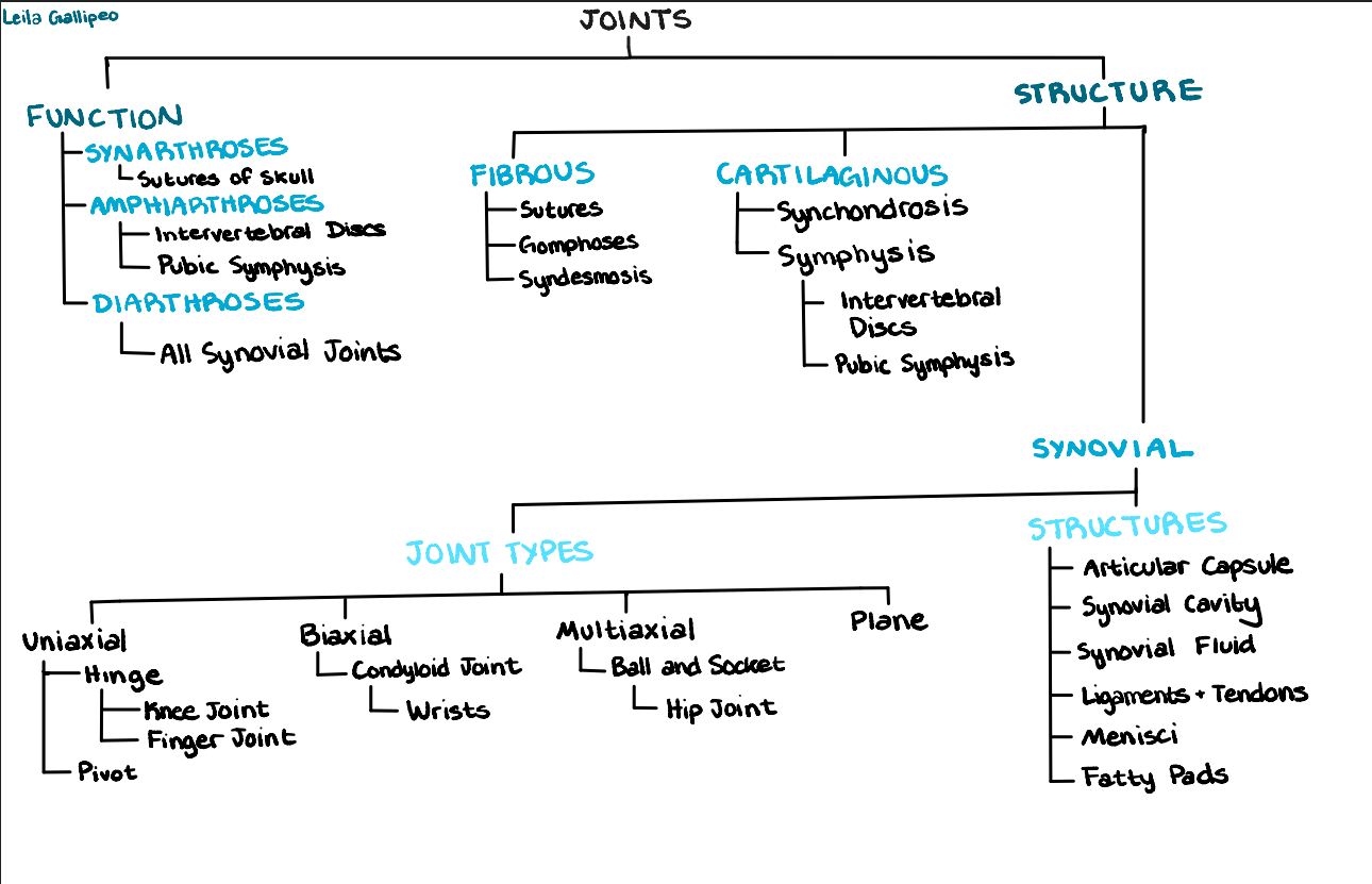
Functional Classification of Joints
functional: based on the amount of movement allowed
synarthroses: immobile joints
sutures of skull
amphiarthroses: slightly moveable
intervertebral discs between vertebrae
diarthroses: freely moveable joints
joints of appendicular skeleton (all synovial joints - hip, elbow, etc.)


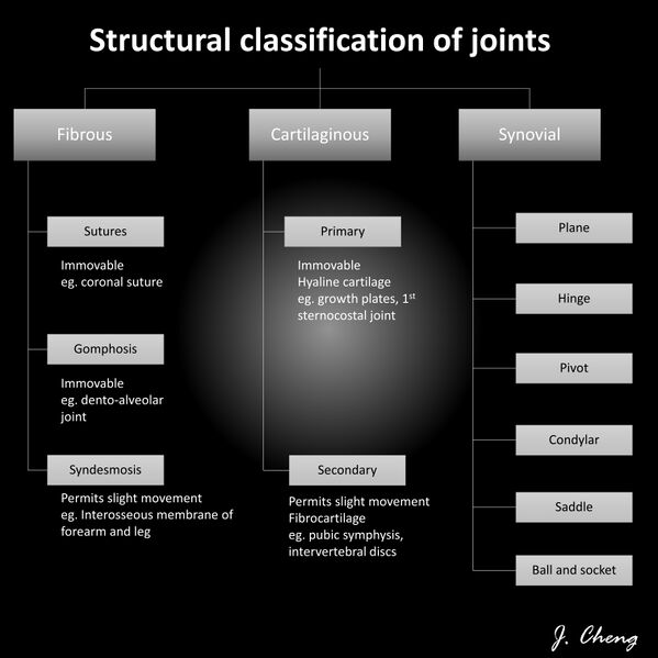
Structural Classification of Joints
structural: based on material, which joins bones
fibrous joints: joints composed of fibrous tissue, no joint cavity present
sutures: short fibrous fibers, found in skull, immovable
syndesmosis: cord of fibrous tissue (ligament); amphiarthrosis with “give” but no actual movement
ex: distal tibiofibular joint
gomphosis: tooth within its bony socket; short ligament
cartilaginous joints: joints composed of cartilage, no joint cavity
synchondrosis: plate of hyaline cartilage; sites of bone growth during youth; eventually ossify = synarthrotic
ex: joint between first rib and manubrium; epiphyseal plate
symphysis: pad or plate of fibrocartilage; compressible “shock absorber”; limited movement = amphiarthroses
ex: intervertebral discs, pubis symphysis
synovial joints: fluid-filled joint cavity; freely movable = diarthrosis

General Structure of a Synovial Joint
there are 5 distinct features of a synovial joint
articular cartilage: hyaline cartilage covers the surface of each bone
joint cavity: a potential space between the two bones, filled with synovial fluid
articular capsule: double layered capsule surrounding cavity
external, tough flexible fibrous capsule
synovial membrane: loose CT lining of fibrous capsule, also covers all internal joint surfaces excluding hyaline cartilage
synovial fluid: lubricating fluid within cavity; reduces friction between bones, provides “weeping lubrication” nourishes cartilage, contains phagocytes
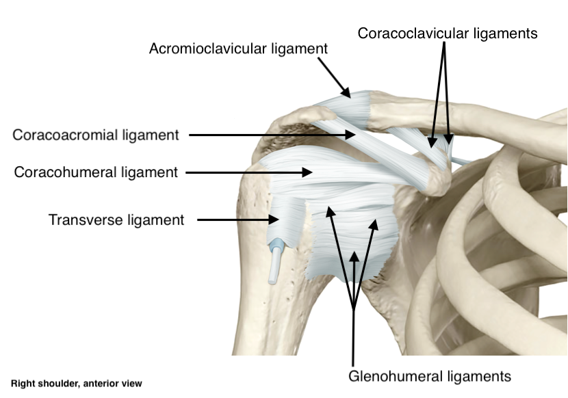
reinforcing ligaments: ligaments that strengthen joint
ligament: joins bone to bone
extra features:
fatty pads: hip and knee
menisci or articular discs: separate cavity into 2 compartments
bursa: flattened sacs with synovial membrane + fluid inside = prevent friction, cushion. it is located between skin and bone, muscles, tendons, and ligaments
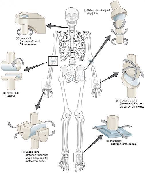
Types of Synovial Joints
ball and socket joints: most freely movable joints, all angular movement, the head of one bone fits into socket of another
ex: hip and shoulder
condyloid joints: permit all angular motion, except rotation
ex: wrists and knuckles
gliding joints: cartilaginous joints, flat bones glide/slide over on another
ex: intervertebral discs
hinge joints: permit flexion and extension only
ex: elbow and knee
pivot joints: permit rotation
ex: first intervertebral joint (atlantoaxial joint)
saddle joints: thumb

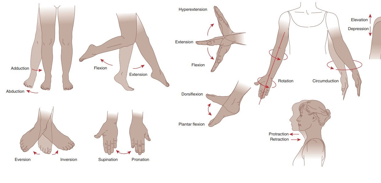
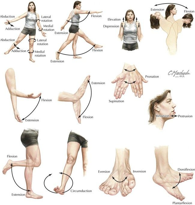
Types of Joint Movements
flexion: decreasing angle between 2 bones
extension: increasing angle between 2 bones
dorsiflexion: bringing foot closer to shin, rolling back on heel
plantar flexion: pointing one’s toe, tippy toes
hyperextension: increasing angle greater than normal (180 degrees)
abduction: moving limb AWAY from midline (raising arm)
adduction: moving a limb back towards midline
circumduction: moving limb in circular (cone-shaped) manner
rotation: turning movement of a bone along axis (atlas over axis, shoulder/hip)
supination/pronation: movements between radius and ulna at radioulnar joint (palm up = supination, palm down = pronation)
inversion/eversion: movement of foot (sole inward = inversion, sole out = eversion)
elevation/depression: moving up or down (shoulder shrug = elevation, opening mouth = depression)
protraction/retraction: going forward or back (thrust forward = protraction, pull back = retraction)
Concept Map


Match the joints to their locations
synarthroses: sutures of skull, gomphoses of teeth
amphiarthroses: intervertebral disc and pubic symphysis
diarthroses: finger joints, ankle and wrist joints
multiaxial synovial joint: shoulder
biaxial synovial joint: metacarpal, phalangeal joints
uniaxial synovial joint: elbow
cartilaginous joints (3 answers):
lack a synovial cavity
have a synovial cavity
articulating bones are held together with cartilage connective tissue
permit little or no movement
permits gliding movement
1, 3, and 4
lack a synovial cavity
articulating bones are held together with cartilage connective tissue
permit little or no movement
fibrous joints (3 answers):
Lack a synovial cavity
Have a synovial cavity
Articulating bones are held together by dense irregular connective tissue
Permitted movement is free and unrestricted
Permit little or no movement
Permit rotation around a central axis
1, 3, and 5 are correct
lack a synovial cavity
articulating bones are held together by dense irregular connective tissue
permit little or no movement
Which of the following are specific structural types of fibrous joints? (Select 3 answers)
Sutures
Syndesmoses
Synchondroses
Symphyses
Interosseous membranes
Planar joints
Sutures, syndesmoses, and interosseous membranes
synovial joints (Select 3 answers)
Lack a synovial cavity
Have a synovial cavity
Articulating bones are covered by a layer of hyaline cartilage called articular cartilage
Provide a range of motion from slightly movable to freely movable
Permit no movement whatsoever
Bones are bound exclusively by a solid sheet of fibrocartilage
2, 3, and 4 are correct
have a synovial cavity
articulating bones are covered by a layer of hyaline cartilage called articular cartilage
provide a range of motion from slightly moveable to freely movable
Which of the following are functional or structural components found in synovial joints? (Select 3 answers)
Articular capsule
Synovial fluid
Periodontal ligament
Sutural ligament
Accessory ligaments (such as extracapsular and intracapsular ligaments)
Epiphyseal cartilage plate
articular capsule
synovial fluid
accessory ligaments


All of the following are freely moving joints, EXCEPT______
gomphosis
hinge
ball and socket
sutures
pivot
gomphosis
sutures
Match the following terms with their functions and/or definitions.
Match the following terms with their functions and/or definitions.
flexor: The muscle that closes the joint
extensor: The muscle that opens the joint
orbicularis: round
trapezius: triangular
deltoid: Greek letter delta
pectoralis: chest /breast
origin: a bone that remains immobile for an action
insertion: bone that moves during the action
flexion ________, adduction _________
Decreases the size of the angle; moves the arm or leg towards the body
Decreases the size of the angle; moves the arm or leg away from the body
Increases the size of the angle; moves the arm or leg towards the body
Increases the size of the angle; moves the arm or leg away from the body
Decreases the size of the angle; moves the arm or leg towards the body
Muscle nomenclature is based on ____________
location of origin and insertion
direction of muscle fibers
appearance
Muscle action
muscle shape
Number of origins
location of origin and insertion
direction of muscle fibers
appearance
Muscle action
muscle shape
Number of origins