Microbiology - Ch. 4 Prokaryotic and Eukaryotic Cell Anatomy - flashcards (CONDENSED)
 Call Kai
 Learn
Learn Practice Test
 Spaced Repetition
 Match
Match Flashcards
 Knowt Play
1/60

There's no tags or description
Looks like no tags are added yet.
Name | Mastery | Learn | Test | Matching | Spaced | Call with Kai | Chat |
|---|
No analytics yet
Send a link to your students to track their progress
61 Terms
Why do we need to know the average size of bacterial cells, and what unit is it measured in?
For identification purposes. Measured in microns (µm).
What is an example average size for a bacterial cell?
~2.0 µm diameter × ~8 µm length.
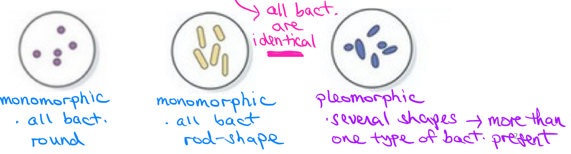
What is the difference between monomorphic and pleomorphic bacteria?
Monomorphic: one shape only (all cells identical)
—
Pleomorphic: multiple shapes present, more than 1 type of bact.
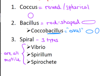
What are the 3 basic bacterial cell shapes?
Coccus = round/spherical
Bacillus = rod-shaped (coccobacillus = "oval" shaped)
Spiral = 3 types (vibrio, spirillum, spirochete)
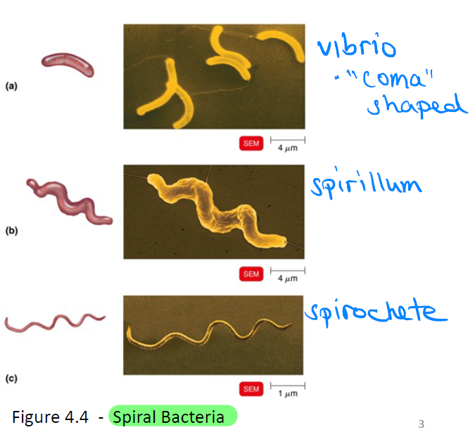
What are the 3 types of spiral bacteria, and what do they have in common?
Vibrio = "comma" shaped
Spirillum
Spirochete
All spiral types are motile
What is the difference between spirilla (singular: spirillum) and spirochetes?
Spirilla: rigid, thick, loosely coiled; move via external flagella
—
Spirochetes: flexible, thin, tightly coiled; move via internal endoflagella, located inside bacteria.

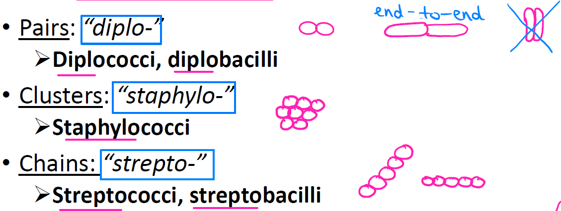
What are the cell arrangement prefixes for pairs, clusters, and chains, and what are they called?
Pairs = "diplo-" → diplococci, diplobacilli
Clusters = "staphylo-" → staphylococci
Chains = "strepto-" → streptococci, streptobacilli
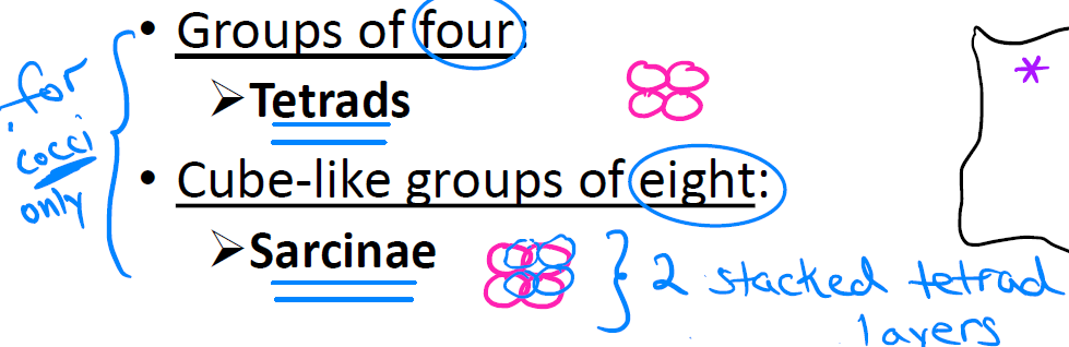
What are tetrads and Sarcinae?
Tetrads = groups of 4 (cocci only)
Sarcinae = cube-like groups of 8 (2 stacked tetrad layers)
For some bacteria, how is the genus name formed, and what's an example?
Genus naming (shape + arrangement bacteria):
Genus = arrangement + shape.
Ex: Streptococcus = "strepto" (chain) + "coccus" (round).
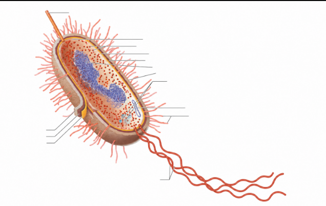
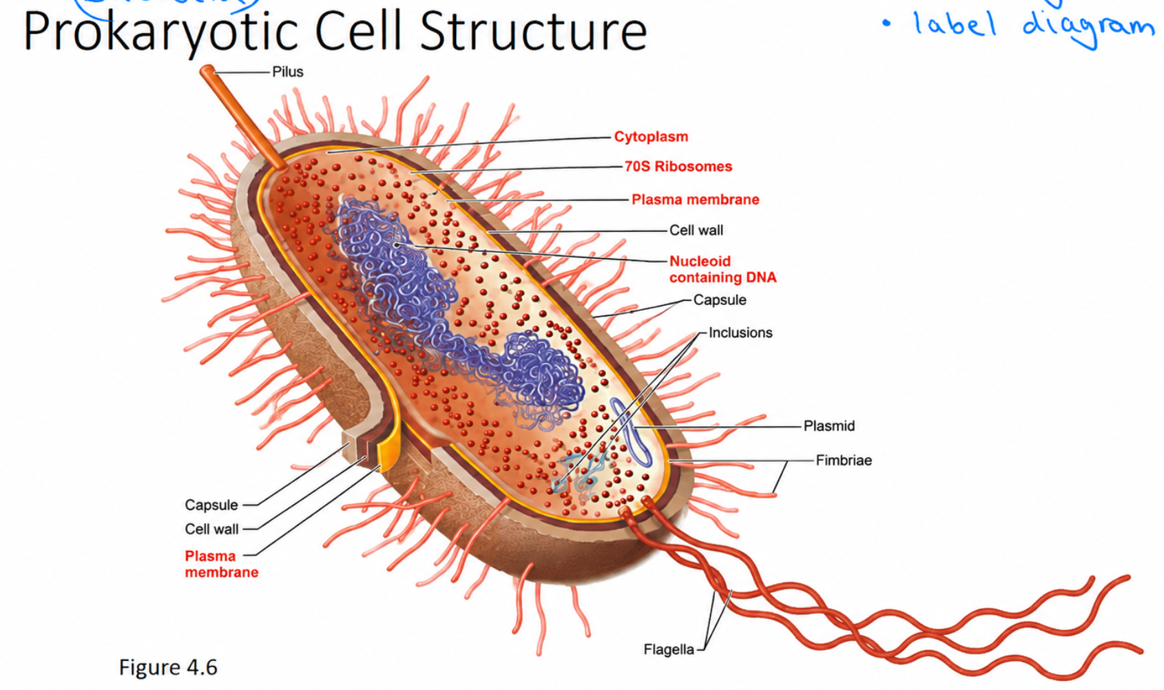
Glycocalyx — location, description, composition, and types?
Location: outside cell wall (surrounds it); not always present.
—
Description: viscous, gelatinous, sticky
—
Composition: polysaccharide or polypeptide
—
Capsule: thick, organized, tightly attached
—
Slime layer: thin, unorganized, loosely attached
—
What is the function of a capsule?
Adhesion: sticky (carbohydrate) → attaches to host tissue
—
Protection: makes bacterium look bigger → evades phagocytosis
What are two examples of encapsulated bacteria, and what's notable about each?
Streptococcus mutans — present in mouth/teeth
—
Streptococcus pneumoniae — causes pneumonia; has huge capsules
Flagella — location, function, and composition?
Location: external filaments; not always present
—
Function: movement → finds nutrients, escapes phagocytosis
—
Composition: protein flagellin → forms filaments → flagellum
—
H antigen: flagellin used to ID strains (ex: E. coli O157:H7)
What exactly is an "H antigen," and what does the E. coli O157:H7 example show?
H antigen
Flagellin protein = surface marker ("H antigen")
Flagellin → filaments → flagellum
Ex: E. coli O157:H7 (pathogenic strain)
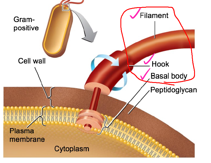
What are the three parts of a flagellum, and their functions?
Filament — outermost region
Hook — attaches to (joins) the filament to the basal body
Basal body — anchors the flagellum
Which bacteria may have flagella?
Found in some bacilli, and in all spirilla-type bacteria (unlike spirochetes, which move via endoflagella instead).
Axial filaments (endoflagella) — which species found in, location, composition, and function?
Axial filaments (endoflagella)
Found in: spirochetes (vs. flagella in spirilla)
—
Location: internal, within cell wall, anchored at one end
—
Composition: protein
—
Function: rotation → corkscrew/spiral motility
—
Fimbriae — location, composition, description/function?
Location: external to cell
Composition: made of proteins
Description/Function: hair-like appendages that allow for attachment
Pili — location, composition, function?
Location: external to cell
Composition: made of proteins
Function: involved in "twitching" motility, and DNA transfer from one cell to another (ex: conjugation pilus — used for DNA/gene transfer from one cell to another)
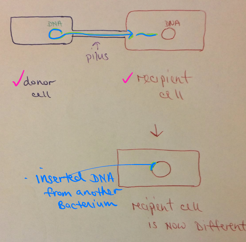
How does conjugation via pilus work?
Conjugation via pilus:
Pilus connects donor → recipient cell; transfers DNA; recipient becomes genetically altered.
Cell wall — location, function, composition?
Location: outermost layer if no glycocalyx present; most bacteria have a cell wall
Function: prevents osmotic lysis and protects the cell membrane
Composition: contains peptidoglycan (rows of carbohydrates connected to proteins)
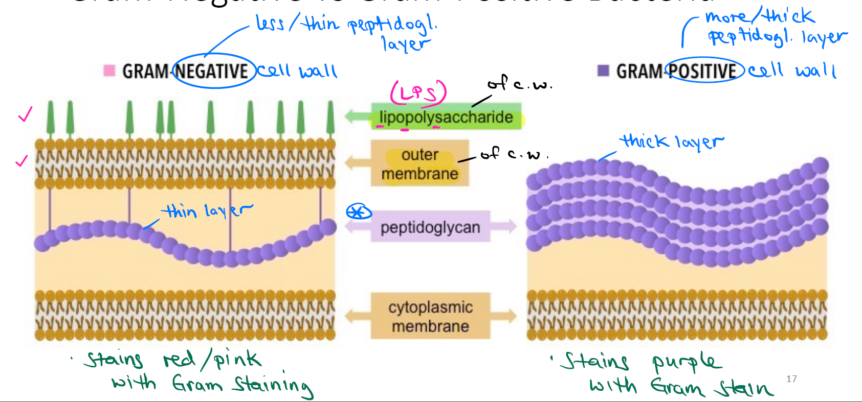
What is the difference between Gram-positive and Gram-negative bacteria (peptidoglycan amount and stain color)?
Gram-positive: more peptidoglycan (thick layer) → stains purple
==
Gram-negative: less peptidoglycan (thin layer) → stains red/pink
What are the structural differences between Gram-negative and Gram-positive cell walls (layers, from outside in)?
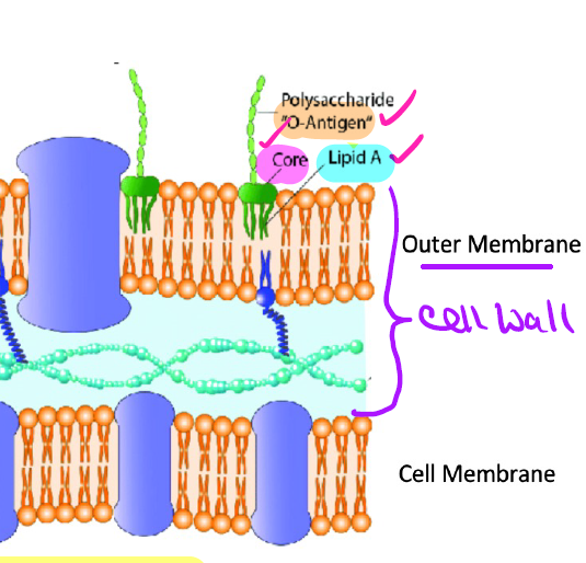
Gram-negative: has an outer membrane with lipopolysaccharide (LPS), then a thin peptidoglycan layer, then cytoplasmic membrane. Stains red/pink.
—
Gram-positive: no outer membrane/LPS — has a thick peptidoglycan layer directly outside the cytoplasmic membrane. Stains purple.
Compare Gram-positive vs Gram-negative cell walls in terms of teichoic acids and Penicillin sensitivity.
Gram-positive: Has teichoic acids; no outer membrane; sensitive to Penicillin (PCN destroys Gram+ bacteria)
==
Gram-negative: No teichoic acids; has outer membrane with LPS; resistant to Penicillin (not destroyed by PCN)

What is LPS, where is it located, and what does it contain?
LPS (lipopolysaccharide) is the major component of the outer membrane of the Gram-negative cell wall, attached to the cell wall's outer membrane. Contains lipids & carbohydrates.
What are the 3 components of LPS, and their functions?
Lipid A — embedded into cell wall's outer membrane; functions as a toxin
Core polysaccharide — joins Lipid A & O polysaccharide
O polysaccharide — sticks out; functions as an antigen ("O antigen," acts as a surface marker); helps distinguish between subspecies/strains (ex: E. coli O157:H7 — "O157" = O polysaccharide)
What is Gram staining, and what does it target?
A differential stain used to classify/identify bacteria; targets the cell wall containing peptidoglycan.
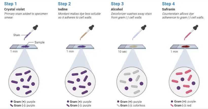
What are the 4 steps of the Gram stain procedure?
Crystal violet (purple dye) — all cells start off purple/violet
Iodine (mordant) — rinse with H₂O
Alcohol wash (decolorization) — rinse; Gram-positive cell walls keep the purple dye, Gram-negative do not
Safranin (counterstain) — rinse; Gram-positive appears purple/violet, Gram-negative appears red/pink
Gram staining is used for identifying bacteria based on what 4 characteristics?
Gram reaction (+/-)
Shape
Arrangement
Size
What is atypical about the genus Mycobacterium's (mycolic) cell wall, and how is it stained?
Mycobacterium cell wall:
Mycobacterium cell walls require acid-fast staining instead of Gram staining because they are rich in mycolic acid.
a waxy lipid that makes the cell wall impermeable to standard Gram stain dyes like crystal violet
What is atypical about genus Mycoplasma & Domain Archaea cell walls?
Mycoplasma: lacks cell walls entirely
Domain Archaea: HAS cell wall but NO peptidoglycan present in cell wall
Plasma (cell) membrane — location, composition, and characteristics?
Location: deep to cell wall
Composition: phospholipid bilayer, encloses cytoplasm
Characteristics: peripheral (surface) vs. integral/transmembrane proteins (span membrane)
What are the 3 functions of the plasma (cell) membrane in bacteria?
Transport — allows passage of some molecules but not others across the cell membrane ("selective permeability")
Site of ATP production — because bacteria have no mitochondria
Site of photosynthesis — because bacteria have no chloroplasts (only possible in certain bacteria)
What is passive transport, and does it require energy?
Movement of particles from an area of high → low concentration (includes simple diffusion and facilitated diffusion). Does not require energy.
What is active transport, and does it require energy?
Movement of particles from an area of low → high concentration. Requires energy (ATP).
What is the difference between simple diffusion and facilitated diffusion?
Simple diffusion: movement of small, uncharged particles across the cell membrane (no protein needed)
—
Facilitated diffusion: movement of large or charged particles across the cell membrane; particles require a protein channel or carrier
What is osmosis, and what direction does water move?
Movement of H₂O across a membrane; requires a concentration difference. Water moves toward the area of high concentration of particles.
Define isotonic, hypotonic, and hypertonic solutions, and their effect on the cell.
Isotonic: same concentration of particles inside & outside the cell → no net H₂O movement
Hypotonic: low particle concentration outside the cell → H₂O enters → cell swells
Hypertonic: high particle concentration outside the cell → H₂O leaves → cell shrinks
In osmosis, which side is the water moving?
Osmosis direction
Water moves from hypotonic (low solute / high water) → hypertonic (high solute / low water) across a semi-permeable membrane.
Cytoplasm — location and composition?
The substance inside the plasma membrane; 80% water plus proteins, carbohydrates, lipids & ions. No organelles present, except for 70S ribosomes. (BACTERIA)
Bacterial ribosomes — description, function, composition?
Called 70S ribosomes (vs. Eukaryotic ribosomes, which are 80S)
Description: not surrounded by a membrane (non-membrane bound)
Function: sites of protein synthesis
Composition: composed of 2 subunits — large subunit & small subunit
Nucleoid — description and composition?
Nucleoid
"Nucleus-like" region, NOT membrane-bound; contains circular, supercoiled bacterial chromosome (most of cell's DNA).
Plasmid — description and function?
Small, circular, extrachromosomal genetic material, in addition to the nucleoid. Carries "non-crucial" genes that play a role in adaptability & survival (ex: genes for antibiotic resistance & production of toxins).
Inclusion bodies — description/location?
AKA reserve deposits; structures that store reserve materials in the cytoplasm.
What are the 4 examples of inclusion bodies and what does each store?
Metachromatic granules —
Polysaccharide granules —
Lipid inclusions —
Carboxysomes —
Metachromatic granules — phosphate reserves/storage
Polysaccharide granules — energy reserves of glycogen (polysaccharide)
Lipid inclusions — energy reserves of fat
Carboxysomes — enzyme reserves involved in photosynthesis
Endospores — location, description, and function?
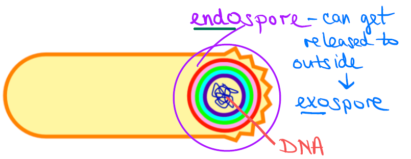
Location: formed inside cell ("endospore"); released = "exospore"
—
Formed by: Bacillus, Clostridium — dormant survival form under nutrient stress
—
Function: protects DNA
—
Resistant to: heat, chemicals, desiccation, radiation — very hardy once activated
What is the difference between sporulation and germination?
Low nutrients/harsh conditions → triggers sporulation (active cell → dormant endospore)
—
Favorable conditions return (nutrients available, right temp/moisture) → triggers germination (dormant endospore → active cell again)

Compare Prokaryotic vs. Eukaryotic cells: chromosome, organelles, ribosomes, cell wall, and division.
Prokaryote: one circular chromosome (not in a membrane); no organelles (except ribosomes); 70S ribosomes; peptidoglycan in cell walls; unicellular; divides by binary fission
—
Eukaryote: paired chromosomes in a nuclear membrane; membrane-bound organelles; 80S ribosomes; polysaccharide in cell walls (if present); unicellular & multicellular; divides by mitosis
Eukaryotic cell wall — where found, composition, and function?
Found in: plants, algae (Kingdom Protista), and fungi
Composition: made of carbohydrates — cellulose (in plants), chitin (in fungi)
Function: structure and protection of the cell
Eukaryotic cell membrane — key features?
Has sterols (a type of steroid) bound to the cell membrane
Has carbohydrates — for attachment and cell-to-cell recognition
Capable of endocytosis
What is the difference between phagocytosis and pinocytosis?
Phagocytosis: engulfing particles
Pinocytosis: engulfing fluids and dissolved substances
What are Eukaryotic ribosomes called, and what is their function?
80S ribosomes; site of protein synthesis.
Nucleus — structure and function?
Double membrane structure (nuclear envelope) that contains the cell's DNA.
What is the difference between Rough ER and Smooth ER?
Rough ER: studded with ribosomes; sites of protein synthesis
Smooth ER: no ribosomes; site of cell membrane, fat, and hormone synthesis
Both form a folded transport network
What is the function of the Golgi Complex?
Modifies, sorts & packages proteins from the ER.
Lysosomes — origin and function?
Vesicles formed by the Golgi Complex; contain digestive enzymes.
Vacuoles — origin and function?
Cavities in the cell formed by the Golgi complex; bring food into cells and provide storage.
Mitochondria — structure and function?
Double membrane; contain inner folds (cristae) and fluid (matrix); involved in cellular respiration (ATP production).
Chloroplasts — structure and function?
Locations for photosynthesis; contain flattened membranes that contain chlorophyll pigment.
Centrosomes — function?
Form the mitotic spindle; critical role in cell division.