Sensation & Perception Quiz 2
1/92
There's no tags or description
Looks like no tags are added yet.
Name | Mastery | Learn | Test | Matching | Spaced |
|---|
No study sessions yet.
93 Terms
Vision at birth
20/500: What a person with normal vision can see in 500 ft, they can see it in only at 20 ft away.
Vision at 6 months
20/40: what a person with normal vision can see at 40 ft, they can see it in only from 20 ft away
Infants mostly rely on __________ to see
rods
Low Acuity
Low vision
Cell elongation
Outer segment of cones are short and cannot pick up as much information. They are less sensitive Explains low acuity in babies. Outer segment of cones will get longer with age and visual acuity and sensitivity will increase. Inner segment gets thinner
Cell Migration
There are big spaces in between the cones of an infant. Explains low acuity in infants. As the outer segment elongates, cones migrate to make more room in the fovea.
Photopigments in infants
There are less photopigments in the infants eyes-explains low sensitivity and acuity
Neuron connections in infants
Explains low acuity. Infants are born with few neural connections. As they age, they make thousands of connections which help them to see better.
Contrast Similarity
Explains low acuity in infants-they have a harder time seeing small differences in hue and brightness
Phototopic Period
Day. Mostly cones are activated. More sensitive to red.
Mesopic
Dusk/Dawn. Cones and rods are active and more sensitive to green.
Scotopic Period
Night/Dark. Mostly rods are activated. More sensitive to blue.
Purkinje shift
When we switch from cones to rods (light to dark from day to dusk/dawn), we are more sensitive to short wavelengths (blue, green)
Action Potential
Charges in the neuron become more positive. When they reach a certain charge (surpass the threshold), they fire.
Synapse
Gaps between neurons where communication occurs between the axon of one neuron and the dentrite of another.
Types of Neurotransmitters
excitatory or inhibitory
Inhibitory signals
Slow the firing rate down, makes it less likely for a cell to fire. Lateral (perpendicular)signals.
Excitatory signals
Increases the firing rate, makes it more likely for a cell to fire. Occurs in the direction/line of sight.
Neural Convergence
Because we have 126 million receptors, it would be difficult for them all to have their own cell to communicate to. Convergence is where multiple receptors communicate to one bipolar cell, and then that cell communicates to a ganglion cell.
Spatial Summation
When one cell fires, the signal may come from many different inputs. You are sampling large areas of the eye when just looking at one fired cell. (More receptors picking up light = more excitatory signals; increases the chance of firing)
Receptor with less convergence
Cones: Cones are more likely to not have convergence-more on a 1:1 ratio from receptors to cells.
Receptor with more convergence
Rods: Because there are so many rods, they are more likely to converge.
Effect of limited cone convergence
If there is no convergence, it is easy to pinpoint which receptor is receiving light when the bipolar cell fires. As a result, we can pick up more detail. less convergence, however, makes it less likely for a cell to fire (think working by yourself vs on a team, you get more done on a team)
Effect of more convergence on rods.
Because 1 bipolar cell receives a lot of rods, they are more sensitive (Having more cells converge, increases the chance of a cell firing). However, when bipolar cells fire, figuring out which cone picked up light is harder.
Receptor to the bipolar cell
The Receptor sends an excitatory signal directly (in line of sight) to the bipolar cell.
Photoreceptor to the Horizontal cell
The photoreceptor sends an excitatory signal straight to the horizontal cell (tells horizontal cell to fire).
Horizontal to Bipolar
The horizontal cell sends an inhibitory signal to a neighboring receptor’s bipolar cell.
Bipolar cell to Ganglion Cell
The bipolar cell sends and excitatory signal to the ganglion cell.
Bipolar cell to Amacrine cell
Bipolar cell sends an excitatory signal to amacrine cell (tells the amacrine cell to fire).
Amacrine Cell to Ganglion Cell
Amacrine cell of neighboring receptor sends an inhibitory signal to the ganglion cell
Neural Integration
Photoreceptors send an excitatory signal directly to a bipolar cell. However, they also send an excitatory signal to a horizontal cell. The signals of the horizontal cells of its neighbor receptor send an inhibitory signal to the bipolar cell. Then, the bipolar cell sends an excitatory signal to the ganglion cell. However, it also sends excitatory signals to amacrine cells. Then, the amacrine signals from neighboring receptors send an inhibitory signal to the ganglion cell.
Lateral Inhibition
horizontal/inhibitory signals help highlight edges. when these inhibitory signals combine with excitatory signals, the two firing rates combine, creating differences in shading.

A on the diagram
Photo receptor: sends excitatory signals


D on the Diagram
Horizontal cells

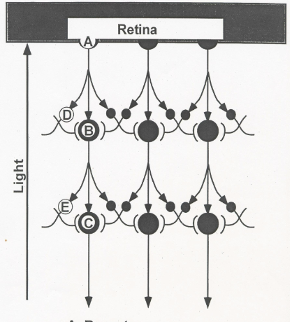
B on the Diagram
Bipolar cells


E on the Diagram
Amacrine cells


C on the Diagram
Ganglian cells

Define Ganglion cells
Cells in the eye that convey information from retinal neurons to the rest of the brain
Less Light =
Less inhibition. Darker subjects have a slower firing rate.
More Light =
More Inhibition. Lighter subjects have a greater firing rate.
Explain the illusion of simultaneous contrast (boxes with same shade of gray)
The lighter big box has more inhibition. Because the small gray box is surrounded by these strong signals, it appears to be darker. The darker big box has less inhibition. Because the small gray box is surrounded by the weaker more inhibited signals, it appears lighter.
Temporal Retina
Side of each eye/retina that faces outward towards the rest of the brain
Nasal Retina
Side of each eye/retina that faces inward towards the nose
The side of the eyes where the right visual field is located.
The left side of each eye is where the right visual field lies. Temporal retina side of left, nasal retina of right eye.
The side of each eye where the left visual field is located.
On the right side of each eye. Nasal retina on the left, temporal retina on the right.
The right visual field is ________
Processed by the left primary visual cortex
The left visual field is ________
on the right side of each eye and processed on the right side of the primary visual cortex
Optic Nerve
Where nerve fibers exit the retina
Optic Chiasm
Where signals from the eye cross over if needed (Left visual field-nasal retina crosses over to right side of brain. Right visual field-nasal crosses over to left side of brain)
Contralateral processing
Information from the right visual field is processed on the left side of the visual cortex.
Ipsilateral side
(inside the brain) eye transmits signals to the same side of the LGN (think temporal retina) does not cross over optic chiasm.
Lateral Geniculate Nucleus
Exists on both sides of the brain. Where the signals from each visual field meet before moving on to the visual cortex. 90% synapse here
Has 6 layers
Magnocellular layers (1-2): Inner layers. Rod processing. Good for motion.
Parvocellular layers (3-6): Outer layers. Processes cones. Detects details, texture.
Layers of the Lateral Geniculate Nucleus
Magnocellular layers (1-2): Inner layers. Rod processing. Good for motion.
Parvo cellular layers (3-6): Outer layers. Processes cones. Detects details, texture.
Superior colliculus
10% of signals from the retina synapse here. The information sent here does not feed back. It is lost. However, the information here is good for coordination and motor of the eye. Reflexes
Primary Visual Cortex
Where cells process the simple properties of vision: lines, edges, orientation, motion
6 layers. Information is sent to layer 4, then the projects to 3 and 5.

A on the Diagram
Left visual Field


B on the Diagram
Temporal Retina


C on the Diagram
Superior Colliculus


D on the Diagram
Right Visual Field


E on the Diagram
Temporal Retina


F on the Diagram
Optic Nerve


G on the Diagram
Optic Chiasm


H on the Diagram
Lateral Geniculate Nucleus (LGN)


I on the Diagram
Primary Visual Cortex
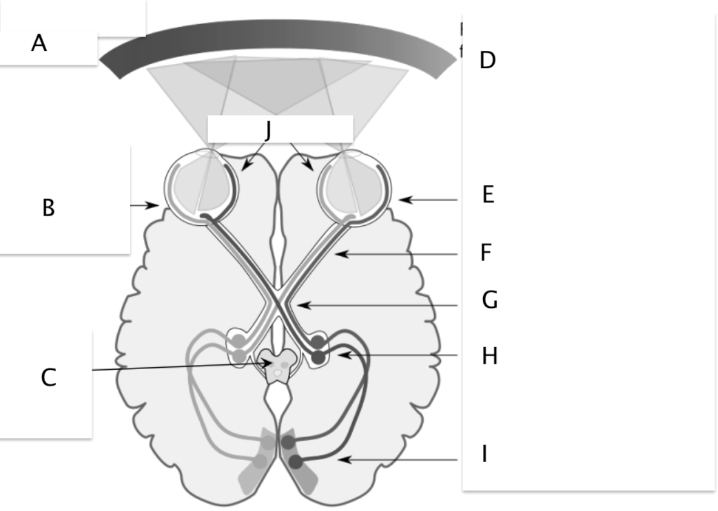

J on the Diagram
Nasal Retina

Left of eye, ________
Processed on the right side of the brain, right visual field
Right of eye, ________
left visual field, left of brain
Retinotopic mapping
Signals that in the retina to the Lateral Geniculate Gyrus to the visual cortex keep the same spatial relationships. (cones activated together stay together) We know this through single-cell recordings (electrophysiology)
Electrophysiology
Single cell recordings, measures the activity of one/multiple neurons. Can damage them!
Receptive field
Area on the retina that when stimulated, changes the activity of a cell. Measures a cell and how it responds to light in different visual fields. Records a signal from an optic nerve fiber. Receptive fields characterize what a cell does. We can study what causes cells to fire the most.
-Tells you where on the retina the firing rate changes
-Tells you he type of stimuli that causes the most change
Light that projects to your right visual field will be detected by receptors in the ______________
side of your eye, and the ______________ side of your brain.
Left, Left
Temporal lobe
Smell, hearing (recognition)
Occipital lobe
Vision
Frontal lobe
Taste
Parietal lobe
Touch (movement, coordination, balance)
The types of cells in frogs
Edge detector, Bug Detector, Moving Edge Detector, Dimming Detector (brightness)ctcor
At what two structures do ganglion cells synapse through the optic nerve?
Through the Superior colliculus and the Lateral Geniculate Gyrus
Ganglion/LGN receptive fields
Have a center surround receptive field: circle with a smaller circle in the middle. One circle has Excitatory signals (positive) while the other has inhibitory (negative).
-If light hits the area where excitatory, firing rate increases
-If light hits area where inhibitory, firing rate decreases
-If full cell is hit, resorts to base rate of firing
Ganglion/LGN cells
Respond to whether there is a dot of light.
-Parvocellular: cones, in the fovea, detail, detects a sustained (nonmoving) stimulus
-Magnocellular: rods, respond quickly, transient stimulus (sensitive to motion), evenly distributed across retina
Primary Visual Cortex receptive fields
Cells do not have center-surround, instead they have lines or spots for excitatory and inhibitory signals. Detects bands of light.
3 types: simple cell, complex cell, and hypercomplex cell
Simple cell
In visual cortex. There are different areas where excitatory and inhibitory signals lie. Detects bands of light.
Detects location, orientation. P cells from the LGN-comes from fovea
Complex cell:
No simple excitatory/inhibitory patterns
M cells. Bar of light must move for detection to occur: responds to moving lines/edges. Sensitive to direction of motion, but not position.
Objective: we can say a cell is responding the most when light moves in a certain way across the cell (ex: orientation from x to Y)
Hypercomplex
M cells. Sensitive to moving line/edge of a specific length-bar of light must move into receptive field and stop to be detected.
Organization of visual cortex
Retinotopic mapping: More of the cortex is given to fovea, so more detail can be processed. neurons are organized by location on the retina, orientation, and ocular dominance (L or R)
Cortical Blindness
Damage to visual cortex
Scotoma: Blindness due to lack of awareness of the visual field. Not due to eye issues.
Blindsight
(blindly seeing) Those with cortical blindness cannot consciously perceive stimuli but can guess what stimuli is or what motion occurs.
Dorsal stream
Goes from V1 up to parietal lobe. Tells us the where, and how (where objects are located, where and how to move). Guides action.
Ventral Stream
Goes down from V1 to temporal lobe. Tells us the what (what are we looking at, what is happening). Helps us recognize objects and faces
Damage to parietal lobe
We do not know where to move to pick objects up (almost as if coordination is off). I can tell you what an object is but not distance between two objects.
Damage to temporal lobe
Cannot recognize a stimulus. I can tell you two objects are close together, but cannot name them.
Monkey study
Monkeys were asked to do an object discrimination task and a landmark task (distance). When their temporal lobe was lesioned they could not complete the object discrimination task, but could complete landmark test. When their parietal lobe was lesioned instead, they could do the discrimination task but not the landmark.
On center cell
Excitatory signals are in the inner circle and inhibitory on the outside
off center cell
inhibitory signals in inner circle and exhibitory on outside