mamary gland
 Call Kai
 Learn
Learn Practice Test
 Spaced Repetition
 Match
Match Flashcards
 Knowt Play
1/25

There's no tags or description
Looks like no tags are added yet.
Name | Mastery | Learn | Test | Matching | Spaced | Call with Kai | Chat |
|---|
No analytics yet
Send a link to your students to track their progress
26 Terms
internal anatomy
3 structure
streak canal- functions to keep milk in udder and bacteria out of udder
teat cistern- duct in teat with capacity of 30-45ml, separated from streak canal by folds of tissue called Furstenbergs rosette
gland cistern- separated from teat cistern by cricoid fold, holds up ot 45 ml of milkand is the collecting area for hte mammary ducts. from this branches the mammary duct
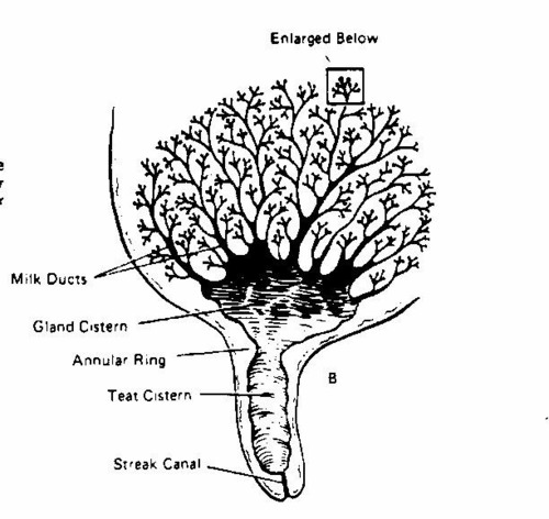
stroma- what does it contain
contains fibroblasts, apidocytes, pasma cells and blood vessles
alveoli
structure
what is it lined with and what is secreted
what it it surrounded with
what is stimulated and how
where do they empty and what does this form
what does this now empty into and then where does that empty to
➢Alveoli- secreting epithelial cells
• Small bulb-shaped structure with hollow centre
• Lined with epithelial cells that secrete milk
• Each alveoli is surrounded by network of capillaries and myoepithelial cells
• Contraction of myoepithelial cell stimulates milk ejection
➢Groups of alveoli empty into a duct forming a unit called a lobule
• Several lobules create a lobe
• Ducts of lobe empty into a galatophore, which empties into the gland cistern
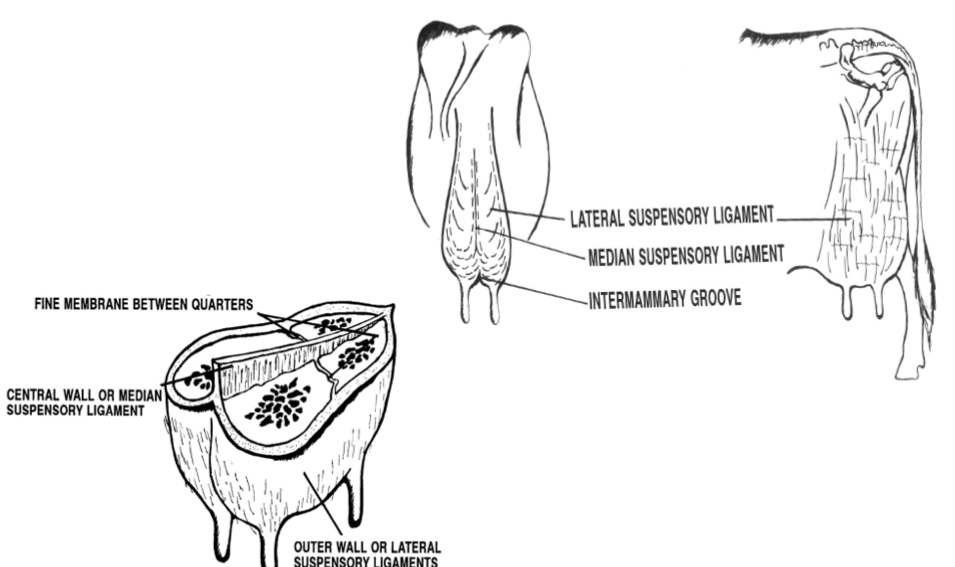
mammary gland suspension
skin- minor role in support
median suspensory ligament- separates r and l udder half, connects udder to abdominal wall by lamellae, elastic tissue which responds to weight of milk in udder
lateral suspensory ligament- inflexible, surround the outer wall of udder, attached to the prepubic an dsuboubic tendons
intermammary groove- formed where lateral suspensry ligament and median suspensory ligament meets
circulation
how much blood does one gallon of milk need
where does blood enter udder
what does the mammary artery branch into
where does blood exit
• One gallon of milk requires 400 gallons of blood being passed through udder- Ratio may increase in low producing cows
• Blood enters the udder through two external pudic arteries, one for each half of the udder;
• The mammary artery branches into cranial mammary artery and the caudal mammary artery
• Blood exiting udder from veins at the base of udder, blood can travel through two routes Via external pudic veins Via subcutaneous abdominal veins
mammogenesis
what is it
where does it penetrate
what bud does this form
how is the secondary bud form
what happens to the secondary branch and what surrounds the newly formed structure
when does it begin
what is expressed in the mesenchyme and what receptors does it singla through and what does this do
what does pthlh signals increase
wh
it is the development of mammary tissue from paired mammary ridges (thickened epidermal tissue) on ventral surface of developing embryo
ridges begin to develop inwards and penetrate into the mesoderm
primary bud begins to send out branches that further penetrate dermis this is the secondary
canalisation- secodnary branch out and form canals that form the duct system of the gland. myoepithelial cells surround the terminal portions of the developing gland
begins during 7-8 wks gestation when primary and secondary ducts develop
• Bone morphogenetic protein 4 (BMP4) expressed in the mesenchyme signals through its receptor BMPR1A to MSX2 and inhibits hair follicle formation at the developing nipple.
• Parathyroid hormone-related protein (PTHLH) signals from the epithelium to the mesenchyme to increase the expression of BMPR1A
lactogenesis
when is it
what are the 2 stages
onset of milk secretion
▪ During the second half of pregnancy, secretory activity accelerates, and colostrum is produced.
▪ The capacity of the breast to secrete milk during later pregnancy is called lactogenesis stage 1, or lactogenesis 1.
▪ Lactogenesis stage 2 occurs after birth (days two or three to eight postpartum) with the onset of copious milk secretion.
▪ During lactogenesis 2, milk volume increases rapidly from 36 to 96 hours postpartum and then abruptly levels off.
galactopoeisis
what is it
what happens when milk is not removed
what controls breast milk synthesis
what is this reposnecalled
▪ Galactopoiesis, or the maintenance of a milk supply, requires removal of milk from the breast.
▪ When milk is not removed or not removed adequately, capillary blood flow decreases, and the lactation process can be inhibited.
▪ It is the quantity and quality of infant suckling or milk removals that controls breast milk synthesis. This can be mimicked by the use of milking machines.
▪ As long as milk is regularly removed, the alveolar cells will continue to secrete milk.
▪ This phenomenon, called the supply-demand response, is a feedback control that regulates the production of milk to match the infant of the infant.
oestrogen- purpose in
udder
growth of
breast
milk
development of stromal tissue of udder
growth of ductal system
fat in breast
inhibits milk secretion
progesterone
role
what does the concurrent elevation of e2 and p4 during later pregnancy do
promotes dev of lobules and alveoli
alveoli cell roliferation, enlarge and become seretory
do not cause alveoli to secrete milk but inhibits, milk only secreted after prepared udder is further stimulated by prolatin
concurrent elevation of e2 and p4 during later pregannacy establish conditions needed for geometri cell multiplication to occur resulting in lobuloalveolar growth
prolactin
function
what does it signal through
milk production
signals throug JAK/Stat to stimulate prolif of lobuloalveolar system in mammary growht
what is prolactin inhibited by
PIH - dopamine (binding decreases cAMP levels)
suckling reposne inhibits PIH release
prolactin levels
lactogenesis triggered following expulsion of placenta by fall in prog and estrogen and continued presence of prolactin
as p ande drop anteriro pituitary is no longer inhibited and releases lots of prolactin
they rise and fall in proportion to freq intesnity and duration of nipple stimulation
if mother does not breastfeed prlactin levelsusually reach non pregnant levels by 7 days postpartum
what are other regulatory factors
GH
acts synergically with oestrogen to develop mammary gland ducts
IGF 1
primary mediator of GH
during foetal development levels are low and graudally increase from birth o puberty where they reach to their highest levels
Glucocortcoids and ACTH
mintaining tigh tjunction in breast parenhcyma
milk ejection reflex
what does it ultimately lead to
what do the nerve pathways lead to and what does this cause
wher is this released from and what does this cause
causes alveoli to reelase milk they have nade
when the newborn suckles and stimulates a message is sent up the nerve pathways to the paraventricular and supraoptic nuclei in the hypothalamus causing the production of oxytocin
oxytocin is relased from posterior pg and causes myoepithelia (musckes around the alveoli) to contract and push stored milk down the ducts through the collecting sinuses and out the nippple pores
cow involution
mammary epithelial cells de differentiate during dry period
become non secretory within 7 days of drying off
ageing cells are lost by apoptosis and replaced by division of remaining cells
involution in rodents
defoliation occurs- mammary epithelial cells fall off the basement membrane
requires more extensive regeneration at the start of next lactation
what are mechanisms for secertion
exocytosis
lipid synthesis and secretion
transmembrane secretion of ions and water
trasncytosis of extra alveolar proteins from intersistial spae
paracellular pathway - transfer of materials between milk space and intersitiial space
amino acid absorption
through where
what happens once they reach inside the cell and examples
through basal membrane by transport systems
once inside the cell they are covalently bound together to form proteins in the RER
eg milk proteins, casein, beta lactoglobulin and alpha lactoalbumin
these proteins are transferred from the RER to the golgi
casein secretion
what form and what is this formed from
where is it transported and how
as a micelle which is formed in the golgi from the casein molecules, calcium and phosphorus
transported to apical membranes of cell via secretory vessels budding off of the golgi which are bounded by a lipid bilayer membrane
glucose to lactose
where does glucose enter the cell from
what is some converted into
how is lactose formed
what does this lead to
glucose enters the cell via the basolateral membrane
some converted into galactose
both glucose and glactose enter golgi and enter a reaction forming lactose
formation of lactose in the golgi results in drawing water into the cell, into th golgi and then becoming prt of milk
milk fat precursors to milk fat
taken up by the epithelial cells at the basolateral membrane
acetate and beta hydroxybutyrate
preformed fatty acids, glycerol and MAG are absorbed at the basolateral membrane
all enter into the synthesis of triglycerides of milk
milk fat triglycerides are synthesised on SER and form small droplets
small droplets fuse together as moving towards the apical membrane
then forces out
transport of milk components not synthesised in the epithelial cells
an example
how are they taken into the cell
where are they transported
what happens once they are transported here
where is it released
eg immunoglobulins which bind to specific receptors on the basolaeral surface of cells
taken into the cell in endocytic vesicles
transported to the apical side of the cell via the vesicles
membrane of transport vesicles fuses with inner surface of apical membrane of cell
releases immunoglobulin into the lumen of the alveolus
paracellular pathway
in which situations is it used
what does this allow
what does this lead to and what can this be used for
when udder is inflamed eg mastitis or involution, or when oxytocin is causing milk ejection, the tight junctions open or some become leaky
allows lactose and potassium to move from the lumen into the extracellular space
and sodium and chlorine to move into the lumen from the extracellular space
changes in electrical conductivity of the milk and this can be used for mastitis detection
another example is leukocytes whuch pass between the epithelial cells breaking open the junctions and enter the paracellular
this allows other extracellular components to diffuse into lumen and milk components to diffuse out
stages of milk
colostrum
1-3 days
first child immunisation
protein, minerals, vitamins, immunoglobulins
transitional milk 2-4 days after birth
breast milk with some colostrum
mature milk
7+ days
90 water, 10 carbs, proteins and fats
within species what does milk production vary with
breed/genotype
stage of lactation
nutrition
health/disease
interval since last milk withdrawal
litter size
age