Family Medicine: Neurology
 Call Kai
 Learn
Learn Practice Test
 Spaced Repetition
 Match
Match Flashcards
 Knowt Play
1/78

There's no tags or description
Looks like no tags are added yet.
Name | Mastery | Learn | Test | Matching | Spaced | Call with Kai | Chat |
|---|
No analytics yet
Send a link to your students to track their progress
79 Terms
Ischemic Stroke
Acute focal neurologic deficit resulting from diminished blood flow to an area of the brain due to thrombosis, embolism, or systemic hypoperfusion
-Presentation: contralateral paralysis, can wrinkle forehead (vs Bell’s palsy)
-Dx: non-contrast head CT to rule out hemorrhage
-Tx: IV thrombotics (tPA or Tenecteplase) for eligible patients within 3-4.5 hours of symptom onset, mechanical thrombectomy for large vessel occlusion, lower BP if > 220/120, start ASA + clopidogrel x 21 days, statin therapy, BP control
Carotid
What artery is implicated in this ischemic stroke?
-Amaurosis fugax
MCA
What artery is implicated in this ischemic stroke?
-Aphasia (dominant hemisphere), neglect, hemiparesis, gaze preference, homonymous hemianopsia
ACA
What artery is implicated in this ischemic stroke?
-Leg paresis, hemiplegia, urinary incontinence, abulia
PCA
What artery is implicated in this ischemic stroke?
-Homonymous hemianopsia, visual agnosia, alexia without agraphia
Basilar
What artery is implicated in this ischemic stroke?
-Coma, cranial nerve palsies, apnea, drop attack, vertigo
Lacunar Infarct
What type of ischemic stroke is being described?
-Pure motor stroke, pure sensory stroke, dysarthria, ataxic hemiparesis
Hemorrhagic Stroke
~13% of strokes, further divided into intracerebral hemorrhage and subarachnoid hemorrhage
-Causes: HTN, cerebral amyloid angiopathy, anticoagulation, AVM, trauma, brain tumor, drugs
-Presentation: sudden severe headache, vomiting, and decreased LOC alongside focal neurologic deficits. Markedly elevated BP, bradycardia, irregular respirations, papilledema, fixed pupils
-Dx: non-contrast CT of the head
-Tx: stop bleeding, lower BP, treat ICP
TIA
Transient episode of neurologic dysfunction due to focal brain, retinal, or spinal cord ischemia without acute infarction on MRI. The blockage does not last long enough to cause permanent damage
-Presentation: mirror stroke symptoms but last minutes to < 1 hour with complete resolution of symptoms within 24 hours
-Dx: CT without contrast, MRI with diffusion-weighted imaging is more sensitive for excluding small infarcts, carotid doppler US to evaluate for stenosis, EKG to rule out AFIB, ABCD score
-Tx: admit to hospital, ASA 325 immediately + clopidogrel x 21 days, statin, carotid endarterectomy if > 70% stenosis
Carotid Endarterectomy
What is indicated when a patient has symptomatic internal/common carotid artery stenosis > 70%?
-Associated with amaurosis fugax in TIAs and strokes
Bell’s Palsy
Cranial nerve VII palsy, can be due to HSV or URIs
-Presentation: acute onset of unilateral facial weakness where both the upper and lower parts of the face are affected, unable to wrinkle forehead, decreased taste, hyperacusis
-Dx: clinical, consider Lyme disease
-Tx: oral prednisone within 72 hours of onset, antivirals if herpes zoster is suspected
CN III
What cranial nerve is affected?
-”Down and out” eye, ptosis, mydriasis
-If painful and involving the pupil, think PCA aneurysm
-If painless and pupil sparing, think ischemic/microvascular
CN IV
What cranial nerve is affected?
-Vertical diplopia, worse when looking down (trouble going down the stairs), head tilt away from the affected side. Often post traumatic
CN VI
What cranial nerve is affected?
-Inability to abduct the affected eye, leading to horizontal diplopia worse at distance
-Often the first sign of elevated ICP
CN V
What cranial nerve is affected?
-Paroxysmal lancinating facial pain
Alzheimer Disease
Neurodegenerative disease characterized by beta-amyloid plaque and neurofibrillary tangle formation, leading to impaired neuronal signaling and neuronal apoptosis
-Most common form of dementia and a leading cause of death in older adults
-Presentation: starts with short-term memory loss and progresses to loss of motor skills, long-term memory, disorientation, and death (5-10 years)
-Dx: clinical, definitive is by brain autopsy. Can diagnose by intellectual decline in 2+ areas of cognition documented by the MMSE. CT/MRI can show diffuse cortical atrophy with enlargement of the ventricles
-Tx: cholinesterase inhibitors (rivastigmine, donepezil) and memantine, avoid anticholinergics
Rivastigmine + Memantine
What is the dual therapy for Alzheimer’s?
Dementia
Significant or moderate impairment of cognition or memory that represents a marked deterioration from a previous level of function, associated with increasing age
-Dx: MMSE/Mini-Cog, labs to rule out reversible causes (B12 deficiency, hypothyroidism, normal pressure hydrocephalus)
-Tx: tx reversible causes if possible
Vascular Dementia
Second most common type of dementia, which is associated with arteriosclerotic small-vessel disease
-Correlates with cerebrovascular events
-Stepwise deterioration with periods of clinical plateau, which may cause a sudden decline after each event
-Tx: aggressive vascular risk factor control
Frontotemporal Dementia
Form of dementia where personality changes precede memory changes
Lewy Body Dementia
Form of dementia characterized by Parkinsonian motor symptoms, visual hallucinations, rapid eye movement sleep behavior disorder
-Severe neuroleptic sensitivity, avoid antipsychotics
Delirium
Acute cognitive dysfunction secondary to an underlying medical condition and is usually reversible
-Presentation: acute, rapid deterioration in mental status with a fluctuating level of awareness and disorientation. Inattention and visual hallucinations are most common symptoms. Seen after surgeries and in alcohol withdrawal
-Dx: MMSE, labs to rule out reversible causes
-Tx: address underlying cause, nonpharmacologic reorientation, low-dose haloperidol for severe agitation
Multiple Sclerosis
Immune-mediated, chronic inflammatory demyelinating disease of the CNS characterized by lesions disseminated in time and space
-Peak onset in women aged 20-40, MC is relapsing-remitting
-Presentation: episodes of focal neurologic dysfunction lasting > 24 hours such as optic neuritis, internuclear ophthalmoplegia, paresthesias, electric sensation down spine with neck flexion, hyperreflexia, Babinski sign, cerebellar signs, urgency, transient worsening of symptoms with heat, fatigue
-Dx: MRI of the brain and spinal cord with and without contrast, LP shows oligoclonal bands
-Tx: high dose IV methylprednisolone, disease modifying therapy to reduce relapse rate
Optic Neuritis
Unilateral painful vision loss, central scotoma, decreased color vision, afferent pupillary defect
-Classic first presentation of MS
Parkinson Disease
Loss of dopamine-containing neurons in the substantia nigra and locus coeruleus, which can as a result of an acetylcholine and dopamine imbalance + show Lewy bodies on pathology
-Presentation: resting tremor, cogwheel rigidity, and bradykinesia. Can also present with masked facies, loss of postural reflexes, shuffling, stooped posture, anosmia, REM sleep behavior disorder, constipation, depression
-Dx: clinical, Levodopa response, MRI, gold standard is autopsy
-Tx: symptomatic care, levodopa/carbidopa if > 65 or bromocriptine if < 65 years to delay levodopa dyskinesia
Essential tremor
Bilateral postural/action tremor, which worsens with voluntary movement, of the hands, forearms, and/or head without a resting component
-Shaking occurs with simple tasks like tying shoelaces, handwriting, shaving, holding cups, or holding the hands against gravity. Improves with alcohol
-Autosomal dominant
Propranolol
What is the first line treatment for an essential tremor?
Tourette Syndrome
Multiple motor tics + > 1 vocal tic, onset before age 18 with a duration > 1 year
-Strongly comorbid with OCD and ADHD
-Tx: behavioral therapy, clonidine or aripiprazole for refractory cases
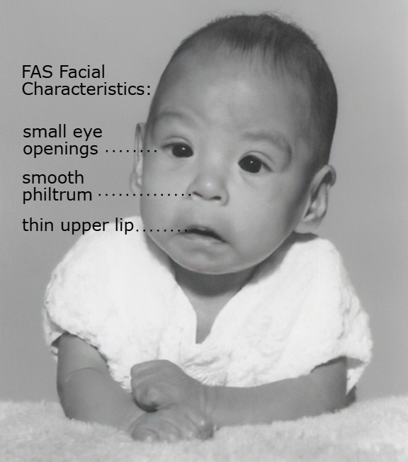
Fetal Alcohol Syndrome
Condition affecting children of mothers who consumed alcohol during pregnancy
-Presentation: Must have all three
Characteristic facial features → smooth philtrum, thin vermilion border of the upper lip, short palpebral fissures
Pre- and/or postnatal growth restriction
Central nervous system abnormalities → microcephaly, intellectual disability, learning disabilities, hyperactive and impulsive behaviors, executive dysfunction
-Can also have congenital heart defects like VSD and ASD
-Dx: clinical diagnosis
-Tx: prevention is the only cure
Antalgic Gait
Shortened stance phase on the painful side
-Associated with osteoarthritis, hip/knee injury
Parkinsonian Gait
Stooped posture, narrow base, shuffling steps, decreased arm swing, festination, and freezing
Hemiparetic Gait
Arm flexed, leg extended/circumducted. History of prior stroke
Apraxic Gait
Feet appear glued to the floor, wide base, small steps. Associated with normal pressure hydrocephalus
Ataxic Gait
Wide based, lurching, unsteady, worse with eyes closed
-Associated with alcohol intoxication, cerebellar stroke, multiple sclerosis, B12 deficiency
Shunt
What is the treatment of choice for normal pressure hydrocephalus?
Wet, wobbly, wacky
What is the triad of normal pressure hydrocephalus?
-Urinary incontinence, gait apraxia, dementia
Cluster Headache
Unilateral, excruciating, sharp, searing, or piercing pain with ipsilateral autonomic features like lacrimation, nasal congestion, conjunctival injection, ptosis, miosis, and eyelid edema
-Seen most often in males, patients are restless
-Tx: 100% oxygen at 12-15 L/min x 15 minutes via non-rebreathing mask + sumatriptan
Verapamil
What is the first line prophylactic treatment for cluster headaches?
Migraine
A headache of varying intensity, often unilateral, pulsating, with nausea and sensitivity to light and sound
-Seen most often in teenage to middle age females
-Presentation: pulsating, lasts 4-72 hours, unilateral, disabling with photophobia and phonophobia. Without aura is MC. Triggered by stress, sleep deprivation, menstruation, red wine, aged cheese, MSG, nitrates, and missed meals
-Dx: clinical
-Tx: sumatriptan, antiemetics, beta-blockers/antiepileptics as preventatives
Tension Headache
Bilateral, mild-to-moderate, dull pain that is not worsened by routine activity
-Tx: NSAIDs, acetaminophen, ASA + caffeine
Increased ICP
Sustained rise in pressure inside the rigid skull above the normal ~ 7-15 mmHg in adults. Sustained elevation induces cerebral perfusion and can cause herniation and death
-Causes: TBI, hemorrhage, tumor, hydrocephalus, meningitis, encephalopathy, IIH
-Presentation: headache that is worse in the morning, N/V, papilledema on fundoscopy, visual changes, AMS, herniation symptoms
-Dx: non-contrast CT of the head, LP for opening pressure, MRI, fundoscopy
-Tx: address underlying cause, elevate head of bed to 30 degrees, mannitol or hypertonic, sedation/analgesia, dexamethasone
> 60
In a patient with elevated ICP, you need to maintain a cerebral perfusion pressure (MAP - ICP) of what?
Idiopathic Intracranial Hypertension
Headache, transient visual obscurations, papilledema, pulsatile tinnitus in a young, obese, female
-Dx: normal imaging, elevated opening pressure on LP with normal CSF composition
-Tx: weight loss + Acetazolamide
Glioblastoma
Most common primary malignant brain tumor in adults
-”Butterfly” lesion crossing the corpus callosum on MRI
-Has a poor prognosis
Meningioma
Most common benign adult brain tumor
-Dural based, extra-axial mass with a “dural tail” and homogenous enhancement
-More common in women
-Only surgical if symptomatic
Pituitary Adenoma
Pituitary tumor associated with bitemporal hemianopsia, hormone hypersecretion, acromegaly, Cushing disease
Acoustic Neuroma
Cranial nerve VIII tumor at the cerebellopontine angle, leading to unilateral sensorineural hearing loss + tinnitus + imbalance
Pilocytic Astrocytoma
Most common pediatric brain tumor
-Cystic cerebellar lesion with a mural nodule
Medulloblastoma
Highly malignant cerebellar tumor of childhood
-”Drop metastases” via CSF
Ependymoma
Pediatric brain tumor found in the fourth ventricle
Brain Tumor
Benign or malignant masses of the brain
-Presentation: progressive headache that is worse in the morning or with Valsalva, new-onset focal seizures, personality changes, N/V, papilledema, signs of increased ICP, constitutional symptoms
-Dx: MRI of the brain with/without contrast, biopsy
-Tx: surgical resection, radiation or chemo
Polyneuropathy
Loss of nerve function in multiple areas
-MCC in the US is diabetes mellitus (think feet here) but can also be seen with alcohol use disorder, B12 deficiency, and chemo
-Presentation: stocking-glove burning/tingling/numbness, length-dependent, decreased vibration/proprioception, loss of ankle reflexes. Leads to foot ulcers, Charcot joint, autonomic dysfunction
Carpal Tunnel
Numbness/tingling in the thumb, index, middle, and radial half of the ring finger due to compression of the median nerve
-Dx: positive Tinel/Phalen
-Tx: night splints, NSAIDs, steroid injection, surgical release if refractory
Cubital Tunnel
Numbness of the 4th/5th digits, weak grip, claw-hand deformity due to ulnar neuropathy of the elbow
Peroneal Nerve Palsy
Lateral knee compression leading to foot drop, sensory loss over the dorsum of the foot
Guillain-Barre Syndrome
Acute, ascending symmetric weakness + areflexia 1-3 weeks after an infection (Campylobacter, CMV, EPV, Zika, COVID)
-CSF shows albuminocytologic dissociation
-Tx: IVIG or plasmapharesis
Carbamazepine
What is the treatment of choice for trigeminal neuralgia?
Trigeminal Neuralgia
Brief, severe, lancinating, “electric shock-like” facial pain in the trigeminal distribution that is triggered by light touch, chewing, brushing teeth, or cold air
Postherpetic Neuralgia
Persistent burning pain > 90 days after a herpes zoster rash in the same dermatome.
-Tx: gabapentin
Generalized Seizure
Seizures that occur when there is widespread seizure activity in the left and right hemispheres of the brain, starting in the midbrain or brainstem and spreading to both cortices
-Dx: witness history, labs, EEG, MRI brain
-Tx: valproate, levetiracetam, lamotrigine
Absence Seizures
Brief impairment of consciousness with an abrupt beginning and ending
-The patient has no recollection and witnesses often miss them
-EEG shows 3-Hz spike-and-wave activity
-Tx: ethosuximide
Tonic-Clonic Seizure
Bilaterally symmetric seizures without focal onset
-Presentation: sudden loss of consciousness with a fall to the ground. Stiff and rigid phase (10-60 seconds) followed by generalized convulsions and limb jerking. Associated with a postictal phase
-Tx: valproate, lamotrigine, levetiracetam
Atonic Seizure
Sudden loss of muscle tone that can mimic syncope
Myoclonic Seizure
Brief muscle jerks, which often occur in the morning
-Tx: valproate or levetiracetam
Febrile Seizure
Convulsion associated with fever > 38 C, age 6 months to 5 years, no CNS infection associated
-Simple = generalized, < 15 min, once in 24 hours
-Complex: focal features, > 15 min, or recurrent in 24 hours
Infantile Spasms
Clusters of brief tonic spasms in infants
-EEG shows hypsarrhythmia
-Tx: ACTH
PNES
Not due to epilepsy but resemble seizures clinically
-Dx: video EEG monitoring
-Tx: therapy
Status Epilepticus
A single seizure lasting > 5 minutes or two or more seizures within 5 minutes without return to baseline between them, which can present in convulsive and nonconvulsive forms alike
-Tx: IV lorazepam, intubation
-Dx: witness history, EEG, MRI brain
Valproate
What antiepileptic medication should be avoided in pregnancy due to its teratogenic nature?
Subarachnoid Hemorrhage
Bleeding into the subarachnoid space between the arachnoid and pia mater, which is typically caused by the rupture of a saccular aneurysm
-Causes: traumatic SAH, AVM, vasculitis, cocaine, HTN, smoking, heavy alcohol use
-Presentation: sudden, severe “thunderclap” headache, nausea, vomiting, photophobia, neck stiffness, focal deficits
-Dx: non-contrast CT of the head, LP shows xanthochromia
-Tx: secure aneurysm, BP control, Nimodipine to prevent vasospasm, pain control, antiemetics, stool softeners, seizure prophylaxis
Syncope
Transient loss of consciousness and postural tone secondary to an acute decrease in cerebral blood flow, characterized by rapid recovery of consciousness without resuscitation
-Causes: vasovagal, orthostatic hypotension, cardiac, cerebrovascular disease
-Dx: H&P, EKG, orthostatic vital signs, labs
-Tx: treat the underlying cause
Vasovagal Syncope
MCC of syncope
-Classic prodrome of warmth, nausea, diaphoresis, tunnel vision, then brief LOC after standing, pain, fear, micturition, or coughing
Orthostatic Hypotension
Defect in vasomotor reflexes, which is common in elderly, diabetics, and patients on diuretics, vasodilators, alpha blockers
Cardiac Syncope
Syncope due to arrhythmias, structural disease
-Red Flag Sx: exertional syncope, syncope without prodrome, palpitations, family history of sudden cardiac death, abnormal EKG
TBI
Traumatic brain injury on the spectrum from concussion to intracranial hemorrhage, which is listed as the leading cause of death and disability in patients < 45.
-Dx: ABCs + cervical spine immobilization, GCS, non-contrast head CT
-Tx: depends on severity, ranging from observation to sedation/intubation
Concussion
Brief alteration of mental status + loss of consciousness
-Presentation: headache, dizziness, nausea, photophobia, difficulty concentrating, irritability, sleep disturbance
-Risk of second impact syndrome
-Tx: brief physical and cognitive rest, then gradual return. Tylenol for headache, avoid NSAIDs/ASA
Epidural Hematoma
Arterial bleed above the dura (middle meningeal)
-CT: biconvex/lens-shaped hyperdensity that does not cross suture lines
-Presentation: classic “lucid interval” followed by rapid deterioration
-Tx: surgery
Subdural Hematoma
Venous (bridging veins) bleed below the dura mater
-CT: crescent-shaped hyperdensity that can cross suture lines but not the midline
-Common in elderly, alcoholics, and anticoagulated patients
Skull Fracture
Linear, depressed, or basilar
-Basilar → raccoon eyes, Battle sign, hemotympanum, cerebrospinal fluid rhinorrhea