Wrist/Hand Examination and Common Conditions
 Call Kai
 Learn
Learn Practice Test
 Spaced Repetition
 Match
Match Flashcards
 Knowt Play
1/23

Earn XP
Description and Tags
09/07/2026
Name | Mastery | Learn | Test | Matching | Spaced | Call with Kai | Chat |
|---|
No analytics yet
Send a link to your students to track their progress
24 Terms
Compartment 1 Muscles
Extensor Pollicis Brevis
Abductor Pollicis Longus
Which compartment of the wrist is involved with DeQuervain’s Tenosynovitis?
Compartment 1
Triangular Fibrocartilage Complex (TFCC) Defn
Approximates the distal radius and ulna with the carpal bones
Supported by the Ulnolunate/Ulnotriquetral, Ulnar Collateral, and Radioulnar Ligaments
Contains an articular disc homologous to the meniscus of the knee that is concave on both the superior and inferior surfaces
Triangular Fibrocartilage Complex (TFCC) MOI
Functionally absorbs 20% of compression/loading at the wrist
FOOSH/FOOA with supination or chronic rotational loading (racquet sports)
Triangular Fibrocartilage Complex (TFCC) Signs and Symptoms
P1 = medial wrist at distal ulna; TTP @ dorsal depression
Agg. = forceful gripping, end-range sup/pon; passive supination with ulnar dev.
May report painful clicking that may be recreated with movement of carpals into ulnar dev.
Imaging may reveal positive ulnar variance
Ulnar Variance
The measurable difference between the lengths of the radius and ulna at the Distal Radioulnar Joint (DRUJ)
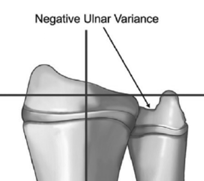
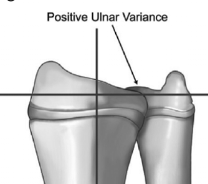
Plain-film radiograph paired with lines crossing at the base of the articular surface of the radius, with one line following the long axis of the radius and the other as the reference for radial and ulnar length.
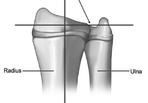
Neutral Ulnar Variance
Radius = Ulna

Negative Ulnar Variance
Radius > Ulna
Positive Ulnar Variance
Radius < Ulna
May predispose an individual to TFCC injury due to increased compressive load with ulnar deviation.
Triangular Fibrocartilage Complex (TFCC) Management
Goal: reduce stress on TFCC for recovery
Interventions:
long arm cast or splint for 6 weeks
elbow flex 90 deg
wrist UD and ext
Proximal strengthening
After cast removal, begin A/AAROM for flex/ext » pron/sup » RD/UD
~2wks post-cast may begin progressive strengthening if asymptomatic
Caution: torsion forces are the most stressful on the TFCC
Types of Wrist Instability
Dissociative
dorsal intercalated segmental instability (DISI)
ventral intercalated segmental instability (VISI)Non-dissociative
Dorsal Intercalated Segmental Instability (DISI) Defn
Most common dissociated instability = scapholunate instability, also known as scapholunate advanced collapse (SLAC wrist)
Lunate migrates dorsally as scaphoid flexes with wrist.
Dorsal Intercalated Segmental Instability (DISI) MOI
FOOSH/FOOA with wrist ext and UD
Injury to the scapholunate ligament
Dorsal Intercalated Segmental Instability (DISI) Signs and Symptoms
P1 = chronic, vague pain and weakness with grip
TTP at scaphoid and/or lunate
Laxity with mobility testing at scapholunate articulation
(+) Wason’s Scaphoid Shift = “clunk” » moving between ext/RD and flex/UD
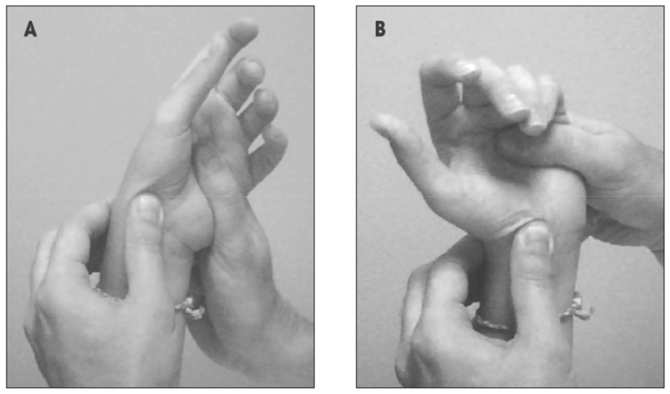
Watson’s Scaphoid Shift Test
Palpate the scaphoid tubercle with your thumb.
Passively ulnarly deviate the wrist.
Maintain scaphoid pressure and move into radial deviation.
(+) clunk and concordant pain; may also produce click with release of pressure
Dorsal Intercalated Segmental Instability (DISI) Management
Conservative: 2wks immobilization with splint or cast
If conservative fails or ligament injury too severe: scapholunate ligament repair via dorsal capsulodesis (takes flap of dorsal capsule and immobilizes the scaphoid and lunate to prevent instability; flap attached with Tacit anchors)
Ventral Intercalated Segmental Instability (VISI) Defn
Second most common dissociated instability = lunotriquetral instability
however, MUCH less common than DISI
Ventral tilt of the distal lunate; lunate is tethered to scaphoid
Ventral Intercalated Segmental Instability (VISI) MOI
FOOSH/FOOA with wrist ext and RD
Injury to the lunotriquetral ligament
Ventral Intercalated Segmental Instability (VISI) Signs and Symptoms
Similar to DISI:
P1 = chronic, vague pain and weakness with grip
TTP at lunate or triquetrum; may reproduce P1 with shear forces at lunotriquetral articulation
Laxity with mobility testing at lunotriquetral articulation
Non-dissociative Instability Defn
Most common dynamic instability at the wrist
Insufficiency of dorsal intercarpal ligaments
Common in gymnasts due to repetitive, heavy closed-chain load
Non-dissociative Instability Signs and Symptoms
May be asymptomatic; volitional subluxation and reduction
“clunk” may be present with reduction
Non-dissociative Instability Management
Conservative: splint or cast for 2 weeks
If conservative fails: surgical repair
Carpal Tunnel Syndrome Defn
Ischemic compression of the median nerve due to increased carpal tunnel pressure from synovial fluid, tendon tension, or stenosis
Often associated with OA of the wrist
Prevalence: 3.7%
Men = 0.6%
Women = 9.2%
Carpal Tunnel Syndrome MOI
May be related to:
pregnancy, renal dysfunction, infection
gout, pseudogout, acromegaly, amyotrophy