Axial Skeleton (pt.2)
 Call Kai
 Learn
Learn Practice Test
 Spaced Repetition
 Match
Match Flashcards
 Knowt Play
1/49

Earn XP
Description and Tags
facial bones, vertebrae, etc.
Name | Mastery | Learn | Test | Matching | Spaced | Call with Kai | Chat |
|---|
No analytics yet
Send a link to your students to track their progress
50 Terms

Nasal bones (2)
form the bridge of the nose
protect the upper entry to the nasal canal
structure of the tip of the nose is cartilage and not bone
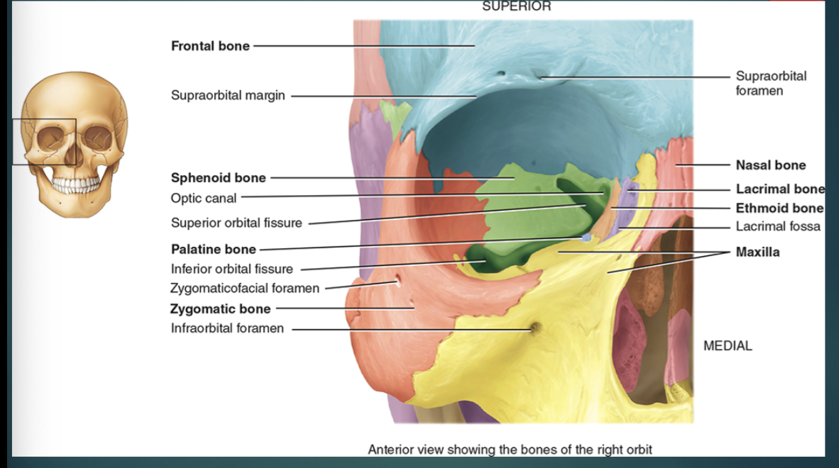
Lacrimal bones (2)
smallest facial bones
part of the medial wall of the orbit
Lacrimal fossa

Lacrimal fossa
Lacrimal bones;
houses the lacrimal sac where tears collect and pass into the nasal cavity
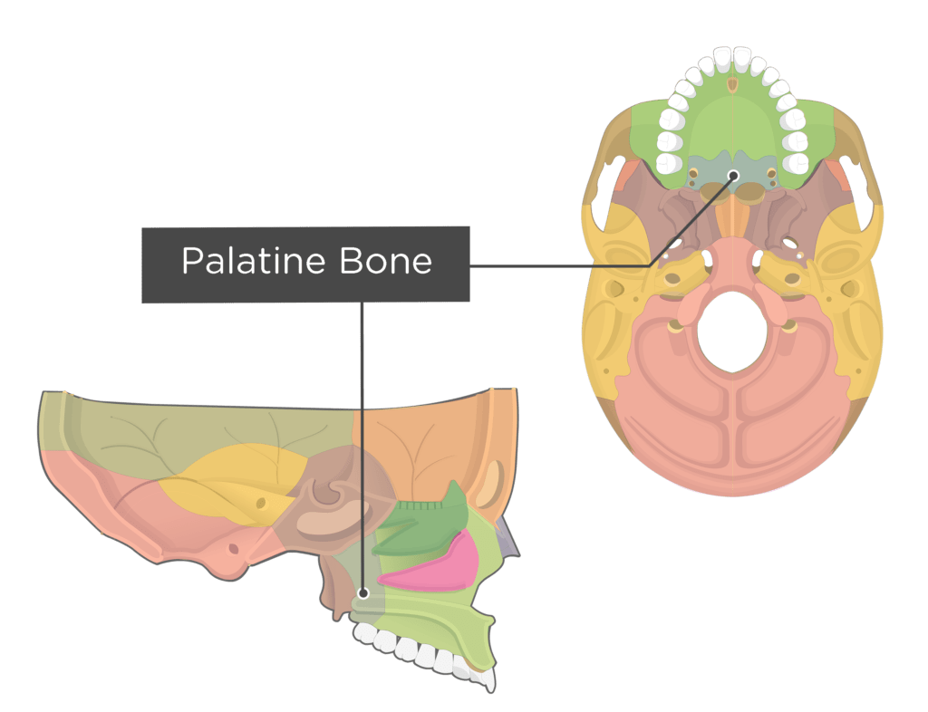
Palatine bones (2)
Posterior hard palate
Posterior lateral wall of nasal cavity
Part of orbit floor

Inferior nasal concha (2)
Inferior lateral wall of the nasal cavity
Increases surface area of the nasal cavity and swirls the air

Vomer
Inferior portion of the nasal septum


Zygomatic bones (2)
“cheek bones”
Lateral wall and floor of the orbit
Temporal processes form part of the zygomatic arch
Zygomaticofacial foramen

Zygomaticofacial foramen
Zygomatic bones;
maxillary division of the trigeminal nerve CN V and blood vessels

Maxillae (2)
Articulates with all other facial bones except the mandible
Forms part of the floor of the orbit, lateral walls, floor of nasal cavity and most of the hard palate
Separate the nasal cavity from the oral cavity
Maxillary sinus
Alveolar processes
Dental alveoli
Infraorbital foramen
Incisive foramen
Inferior orbital fissure
Maxillary sinus


Alveolar processes
Maxillae;
ridgelike arches that contain dental alveoli

Dental alveoli
Maxillae;
sockets for teeth
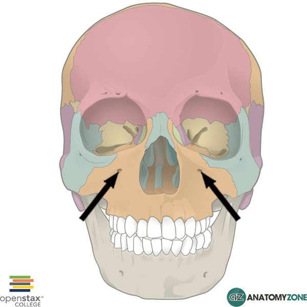
Infraorbital foramen
Maxillae;
blood vessels and maxillary division of the trigeminal nerve CN V pass through

Incisive foramen
Maxillae;
greater palatine blood vessels and nasopalatine nerve pass through

Inferior orbital fissure
Maxillae;
located between the sphenoid bone and maxilla
contains infraorbital nerve branch of trigeminal CN V and blood vessels

Mandible
Largest, strongest facial bone
One of the only movable skull bone
Anterior coronoid process
Mental foramen
Mandibular foramen

Anterior coronoid process
Mandible;
area where muscles attach

Mental foramen
Mandible;
mental nerve passes through
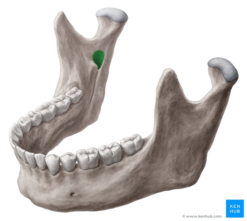
Mandibular foramen
Mandible;
inferior alveolar nerve, a branch of the trigeminal nerve CN V and blood vessels pass through

Nasal septum
divides the nasal cavity into right and left sides
made of the vomer, perpendicular plate of the ethmoid bone, and septal nasal cartilage
Anterior portion = hyaline cartilage
Posterior articulation = perpendicular plate of ethmoid bone
Posterior-inferior articulation = formed by vomer
Orbits
pyramid-shaped cavities housing the eyes
formed by: frontal, sphenoid, ethmoid, palatine, zygomatic, lacrimal maxilla
Superior orbital fissure
Supraorbital foramen/notch
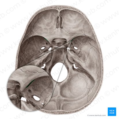
Superior orbital fissure
Orbits;
located at the superior lateral angle of the apex

Supraorbital foramen/notch
Orbits;
located at the supraorbital margin
Foramens and canals
structures passing through these openings are important for bloods vessels, nerves, or ligaments
Sutures
immovable fibrous joint in adults
moveable in young children and act as growth centers
holds most bones of the cranium and face together
most sutures are named for the bones they unite
Coronal
Saggital
Lambdoid
Squamous

Coronal suture
Sutures;
joint of frontal and 2 parietal bones
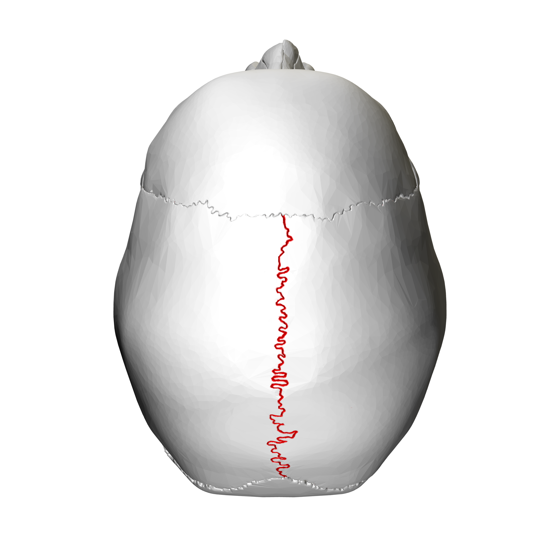
Sagittal suture
Sutures;
unite the two parietal bones superiorly

Lambdoid suture
Sutures;
unites the 2 parietal bones to the occipital bone

Squamous suture
Sutures;
unite the parietal bones to the temporal bones laterally

Paranasal sinuses
cavities within certain skull bones (frontal, sphenoid, ethmoid, maxillae)
lined by mucous membranes continuous with the nasal cavity
secrete mucous which drains into the lateral wall of the nasal cavity
very small at birth and when young
allows skull to increase in size with little change to mass
increase surface area of nasal mucosa
resonating chambers that improve voice quality
Fontanels
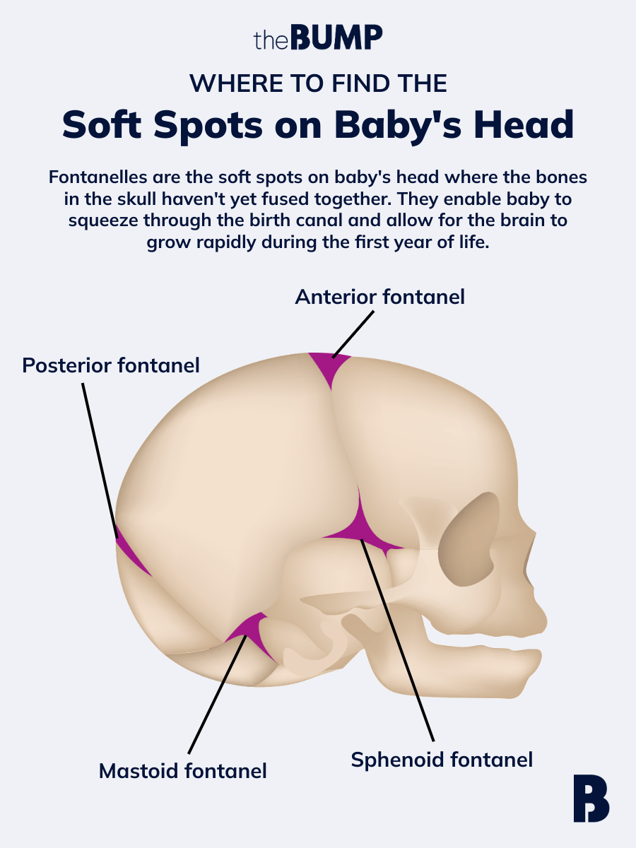
Areas of unossified mesenchyme (dense connective tissue) located between incompletely ossified cranial bones at birth
Skull initially consists of thin sheets of mesenchyme surrounding the developing brain
Undergo intramembranous ossification over time and are eventually completely replaced by bone
Provide flexibility to the fetal skull and allow space for rapid brain and skull growth

Anterior fontanel
Fontanels;
largest, located midline between the parietal bones and frontal bones
closes at 18-24 months

Posterior fontanel
Fontanels;
midline between the parietal and occipital bones
closes at about 2 months

Anterolateral fontanels
Fontanels;
between the frontal, parietal, temporal, and sphenoid bones
close at about 3 months
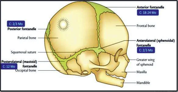
Posterolateral fontanels
Fontanels;
between the occipital, parietal, and temporal bones
close at about 12 months

Hyoid bone
does not articulate with any other bone
located between mandible and larynx
suspended from styloid process of the temporal bone by ligaments and muscles
provides attachments for some tongue, neck and pharynx muscles
consist of a body and paired lesser horns and greater horns
often fracture due to strangulation
Vertebral column
flexible rod that can move forward, backward, sideways, and rotate
encloses and protects spinal cord
supports the head
attachment points for ribs, pelvic girdle, muscle of back and upper limbs
7 cervical
12 thoracic
5 lumbar
sacrum
coccyx
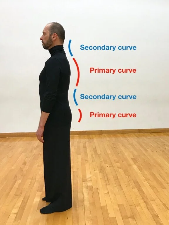
Primary curves
Vertebral column;
thoracic and sacral
retain the original curve of the fetus
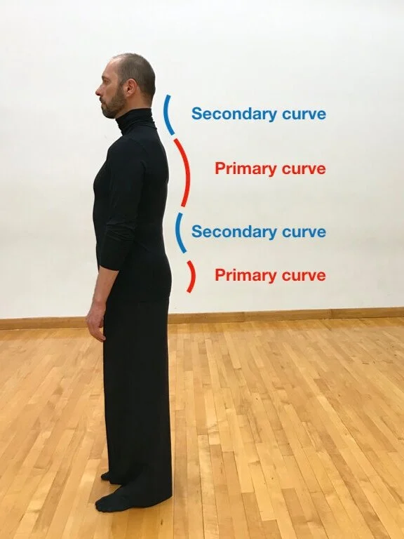
Secondary curves
Vertebral column;
cervical and lumbar
develop after birth
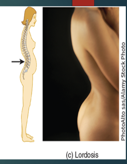
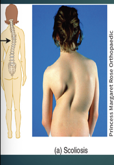
Abnormal curves
Kyphosis
Lordosis
Scoliosis
Kyphosis

Lordosis
Scoliosis
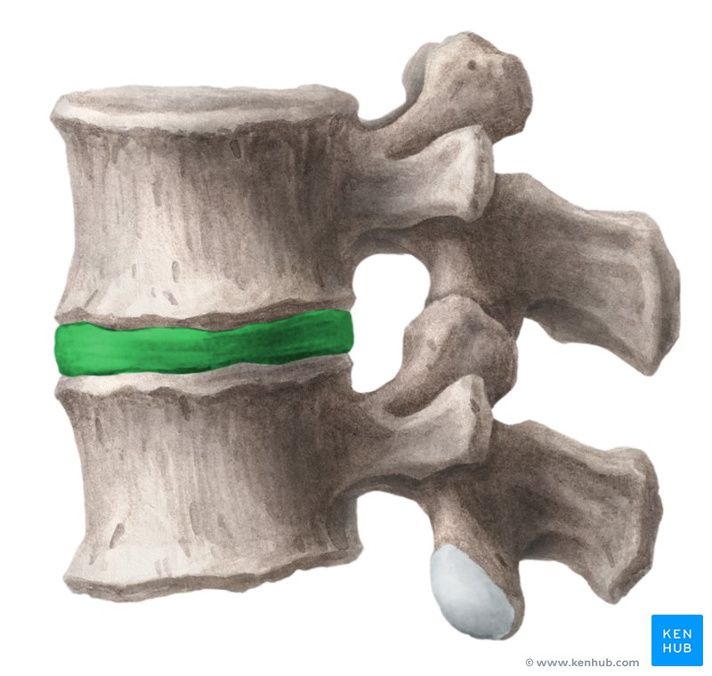
Spinal column: Intervertebral discs
adjacent vertebral bodies from inferior to the 2nd vertebra to the sacrum
form strong joints, permit movement of the vertebral column, absorb shock
can compress
avascular
rely of blood flow of the adjacent vertebrae for oxygen, nutrients, and waste removal
with age, the nucleus pulposus hardens and becomes less elastic
Superior and inferior surfaces of thin layers of hyaline cartilage

Annulus fibrosus
Spinal column: Intervertebral discs;
outer ring of fibrous cartilage

Nucleus pulposus
Spinal column: Intervertebral discs;
inner soft, pulpy, highly elastic substance

Sacrum
Composed of five “sacral vertebrae”
Begins fusing between 16-18 years old.
Completed by age 30.
Serves as a strong foundation for the pelvic girdle
The sacral canal is a continuation of the vertebral canal
The adult female sacrum is wider and shorter than the male sacrum

Coccyx
Four “Coccygeal vertebrae” fuse between ages 20-30
Female coccyx points inferiorly
Male coccyx points more anteriorly

Thorax
Made up of the sternum, costal cartilage, ribs, and thoracic vertebrae
Encloses and protects the organs of the thoracic and superior abdominal cavities
Support for the bones of the upper limb
Plays a role in breathing

Ribs
12 pairs of ribs
Numbered 1-12 from superior to inferior
Articulates with the corresponding thoracic vertebra
First-seventh ribs have direct anterior attachment to the sternum by costal cartilage
Eighth-tenth ribs attach indirectly to the sternum by the costal cartilage of the seventh rib
Eleventh and twelfth ribs do not attach (floating ribs)
Blood vessels and nerves follow the inferior aspect of the ribs
Costal cartilage contributes to the elasticity of the thoracic cage