Bioengineering - Human Movement
1/25
There's no tags or description
Looks like no tags are added yet.
Name | Mastery | Learn | Test | Matching | Spaced | Call with Kai |
|---|
No analytics yet
Send a link to your students to track their progress
26 Terms
What is the requirement of human movement
neurological control of muscle activation
Cause and treatment of arthritis
Caused by thinning or damage to the cartilage
treatment such as knee braces, footwear to reduce mechanical loading
Consequence of diabetes
Reduced healing ability causes diabetic foot ulceration leading to amputation or death. New footwear designed to reduce mechanical loading.
Human movement measurement methods
Video Cameras
Infrared Motion Tracking Cameras
Internal Sensors such as accelerometers and gyroscopes
Human Movement Measurement Methods Comparison
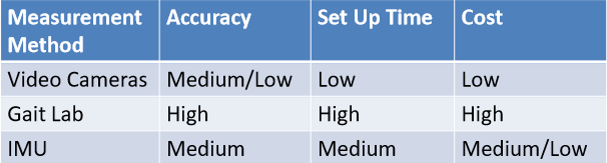
Video Motion Tracking Process
Manual Analysis - approximate body segment angles taken from video footage
Software Analysis - data collected and mapped to a computer model to calculate body loading
Automated real-time motion tracking - mathematical model mapped to video image to approximate joint angles
Gait Laboratory Motion Analysis Process
Infrared sensors used with emitters and detectors to create a capture volume with 8-10 cameras
Force Plates then used - Key measures of ground reaction force in vertical, anterior-posterior and medial-lateral directions
A body segment model is then made with local coordinate frames. Inverse dynamics, anthropometrics and GRF combine together
Inertial Movement Units
Mass puts strain on a sensor
Uses accelerometers, gyroscopes and magnetometers
Software fuses data from multiple sensors
Absolute orientation is determined
Wearable IMUs design considerations
Sensor placement - number of sensors, user acceptance (what people are willing to wear or do)
Calibration - considering differing local coordinate frames, standard vs. real time movement
AI and cloud computing - consider issues around sharing and storing data and large amounts of training data
3 layers of skin
epidermis: protective barrier against environments and regulates water loss
dermis: contains papillary and reticular layers that has blood vessels, nerve endings and dense connective tissue. Provides strength and elasticity
hypodermis: deepest layer containing fat cells and connective tissue. acts as an insulator and provides shock absorption
Skin Function
physical protection
Water and electrolyte balance
temperature regulation
Vitamin D synthesis
Immune Function
Sensation
Energy Storage
Cell actions in skin healing
1) Inflammatory Phase: platelets form clots, neutrophilis clear debris, macrophages remove dead skin cells
2) Proliferative Phase: Keratinocytes re-epithelialize, fibroblasts produce collagen, endothelial cells form new capillaries
3) remodelling phase: fibroblasts strengthen ECM, myofibroblasts contract wound edges, keratinocytes reinforce epidermal barrier.
Cycle of excessive skin loading
1) Primary, direct deformation damage
2) secondary inflammatory edema-related damage
3) Tertiary ischemic damage
People more susceptible to skin injuries
Elderly with aged, underdeveloped tissue
Medical conditions (diabetes)
People on long term coagulant
Design requirements to prevent skin ulcerations from bed pressure
Can adjust pressure distribution during surgery
Will not change whole body position
Can meet safety regulations for medical devices ISO13485
Can be integrated with current surgical table
Prevention and treatment of a diabetic foot ulcer
to prevent: shock absorbing insoles and foot sensors to sense hot spots or areas of high pressure
How does muscle contraction and activation happen?
structure of a muscle fibre
Made up of many myofibrils bundled together. They can split into a number or contractile elements, sarcomeres
Muscle Contraction
Muscles move on commands from the brain
motor neurons fires action potentials down the axon
A chemical is released at the ending
Change in electric field causes contraction in the muscle
Conditions that affect neuromuscular control
Parkinson’s
Post-stroke disability
Multiple Sclerosis
Muscular Dystrophy
Methods to understand muscle activation
Physiological cross-sectional area and muscle structure
Electromyography (EMG, or ECG for heart)
Mathematical modelling
Measuring PCSA
Measure the pennation angle:
PCSA = M cos(theta) / rho*l
Purpose of electromyography
It measures the electrical activity produced from muscle activation. Can be done using surface electrode measurement or intermuscular electrode measurement.
Set up for EMG
one signal electrode, one ground electrode. Signals then amplified using an amplifier. Data acquisition device used.
What are the considerations for good EMG measurement?
Skin cleaning/Abrasion
Electrode placement
Electrode design
Signal cable length/amplification
Post-Processing
What is functional electrical stimulation and how does it work?
electrical stimulation can be used in wound and tissue healing. a stimulator, leads and electrodes are used to stimulate nerves by applying an electrical potential between the electrodes, creating local depolarisation, resulting in action potential. It is both and assistive and rehabilitative technology