Imaging exam
 Call Kai
 Learn
Learn Practice Test
 Spaced Repetition
 Match
Match Flashcards
 Knowt Play
1/83

Earn XP
Description and Tags
- describe the radiological image using appropriate terminology • describe the most important features of pathological abnormalities • describe briefly the working principles of the imaging modality • advantages, disadvantages and contraindications • briefly describe the used contrast agent (where appropriate) • duties of the referring physician related to the imaging study • name the anatomical structures 15 exam topics, 6 images per topic each student gets 1 topic (6 images)
Name | Mastery | Learn | Test | Matching | Spaced | Call with Kai | Chat |
|---|
No analytics yet
Send a link to your students to track their progress
84 Terms
1.1
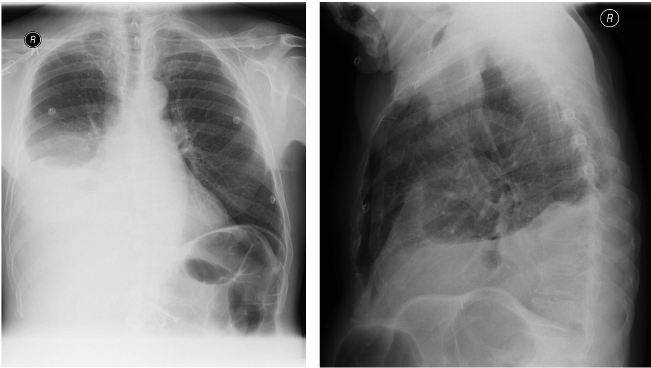
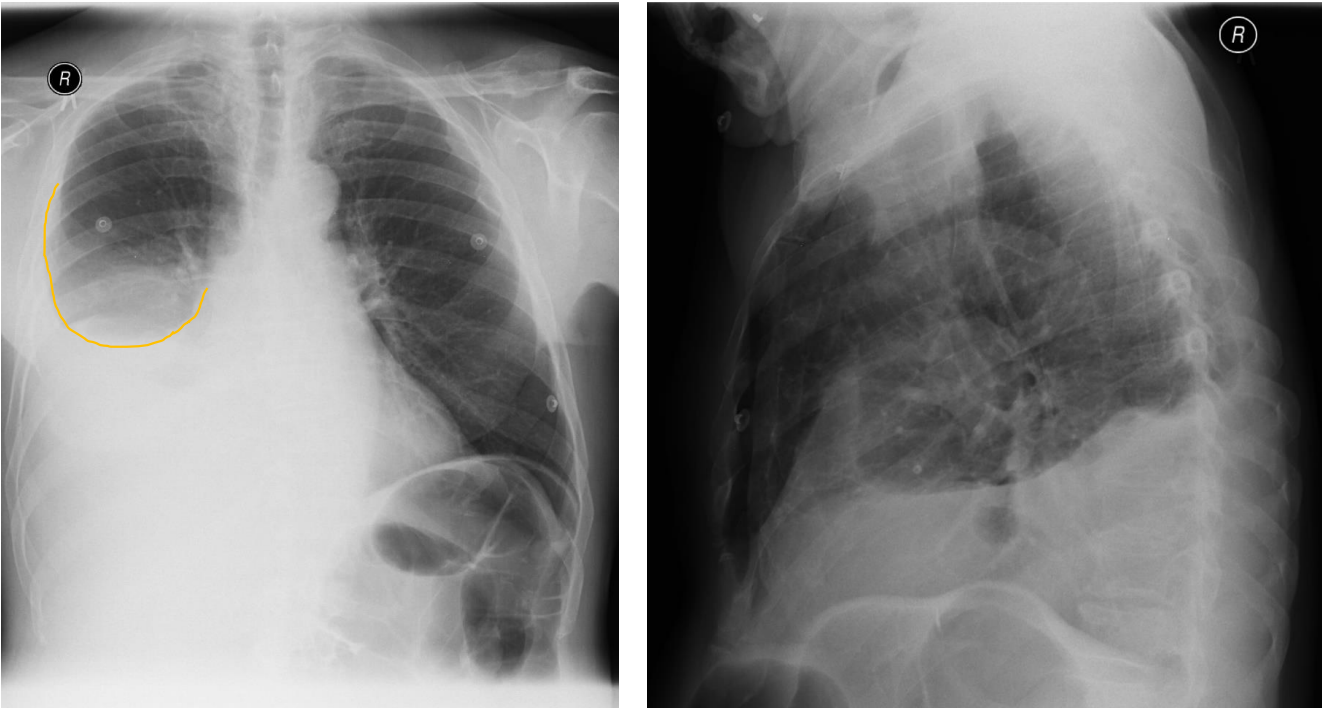
Patient history: abdominal postoperative condition
Modality: PA (left) and lateral (right) chest radiograph; hard-beam technique (120–140 kV)
Region: chest, pleura
Radiologic sign: Decreased transparency at the right lung base showing a meniscus-shaped contour on the right side (yellow line).
Diaphragm and lateral sinuses cannot be differentiated on the right. Left side is normal.
Diagnosis: right-sided pleural effusion
1.2
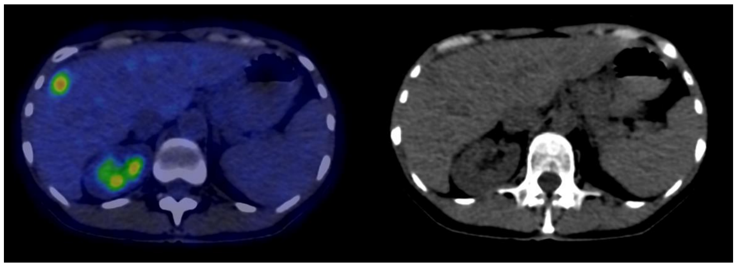
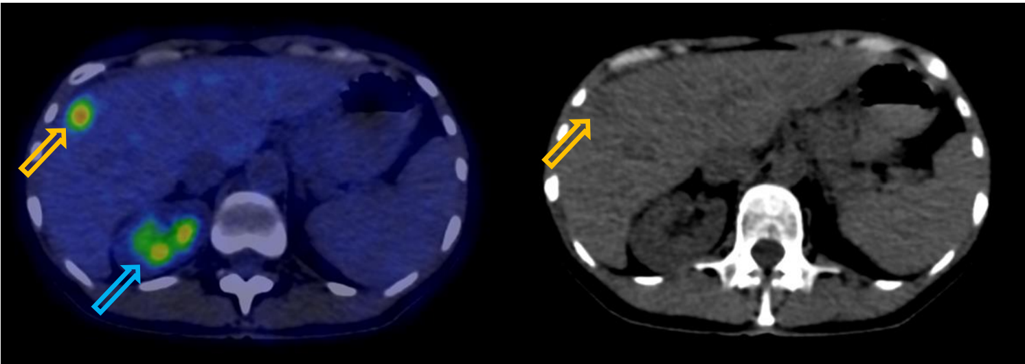
Patient history: colorectal tumor, elevated CEA-level, staging
Modality: FDG PET-CT (right panel: noncontrast CT); 18(F)-fluorodeoxyglucose
Region: upper abdomen, axial view
Sign: increased focal uptake in the liver; physiological high uptake in the right kidney
Diagnosis: malignant tumor (colorectal adenocarcinoma metastasis) in the liver
1.3
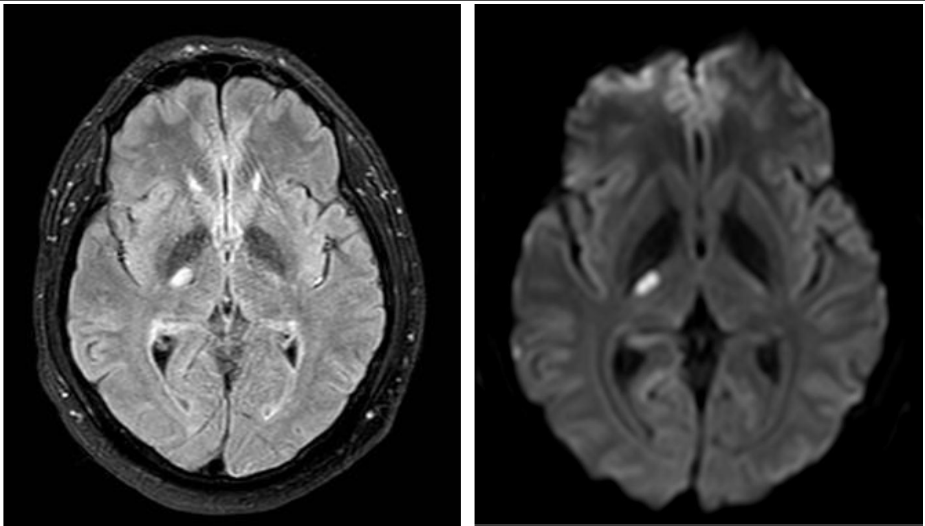
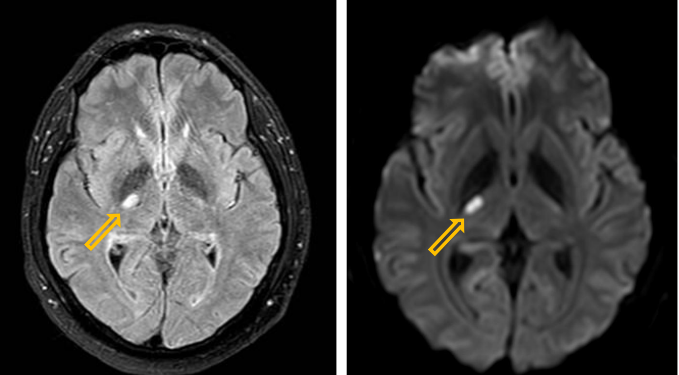
Patient history: at present left-sided hemiparesis (for some hours), with hypertension and diabetes mellitus in patient’s history
Modality: FLAIR (left) and DWI (right) MRI (noncontrast sequences) Region: brain, axial view
Radiologic sign: hyperintense lesion representing edema and restricted diffusion in the posterior genu of the right internal capsule
Diagnosis: small-area acute infarction
1.4
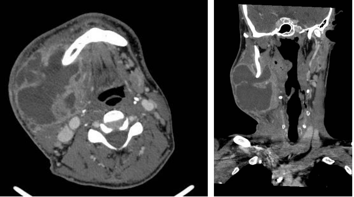
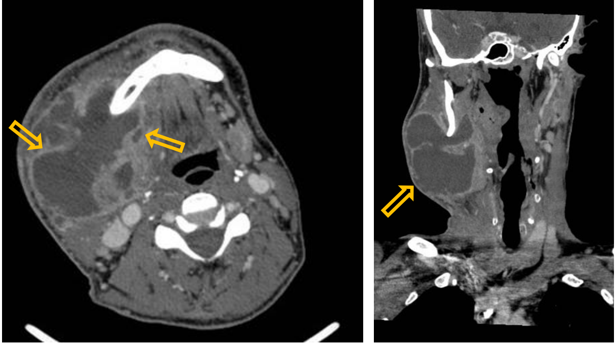
Patient history: right lower wisdom-tooth extraction 6 days ago, painful swollen mass on the right side of the masticator region
Modality: Contrast-enhanced CT, left image – axial view; right image – coronal reconstruction, iv. iodine-based contrast agent
Region: neck, axial (left) and coronal (right) views
Radiologic sign: Right-sided, hypodense submandibular fluid collection with thick, enhancing wall (yellow arrow). Fluid inside the abscess has a density of 0-10 HU and shows no enhancement. Patient had prior wisdom tooth extraction.
Diagnosis: neck abscess
1.5
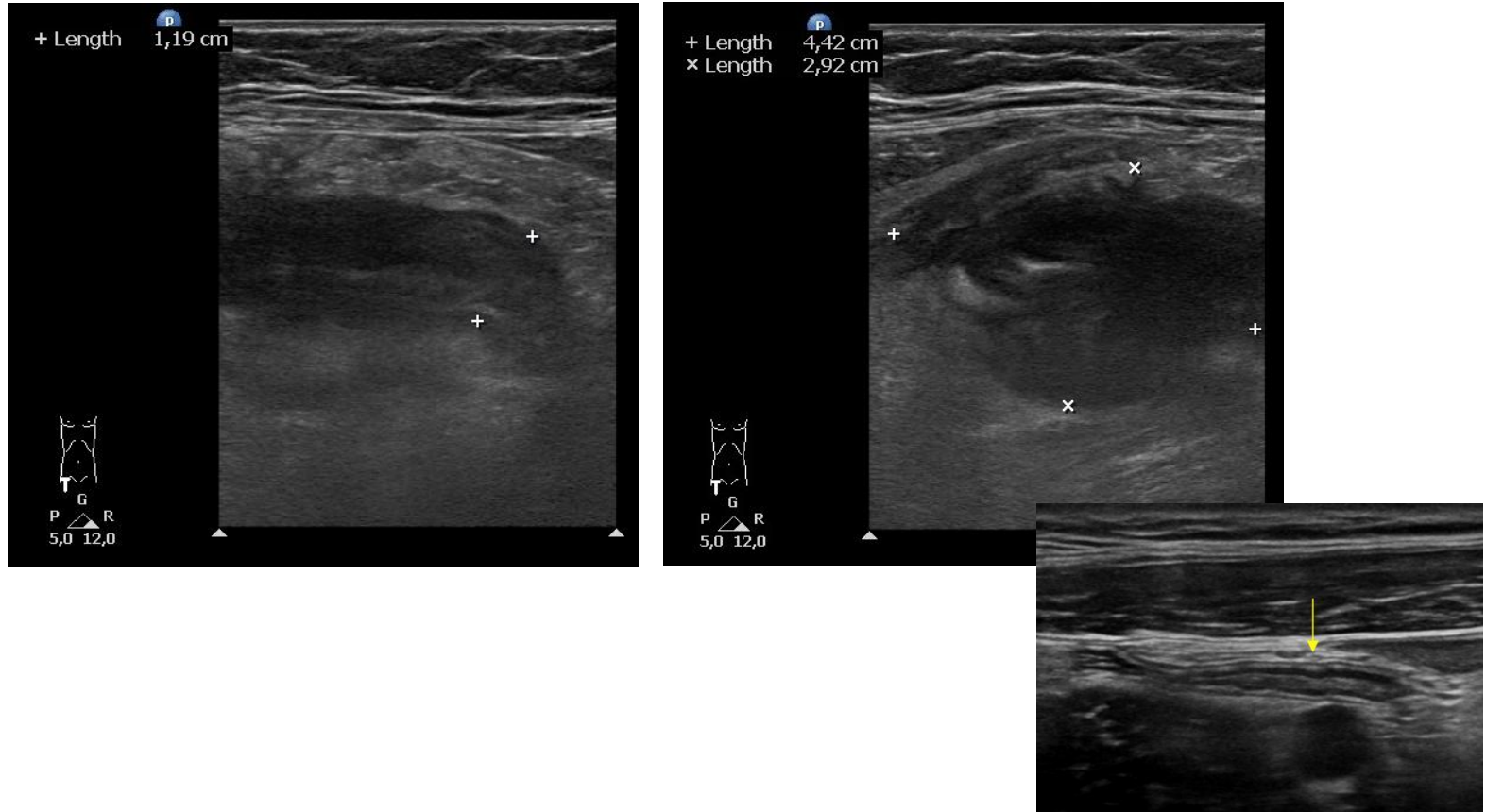
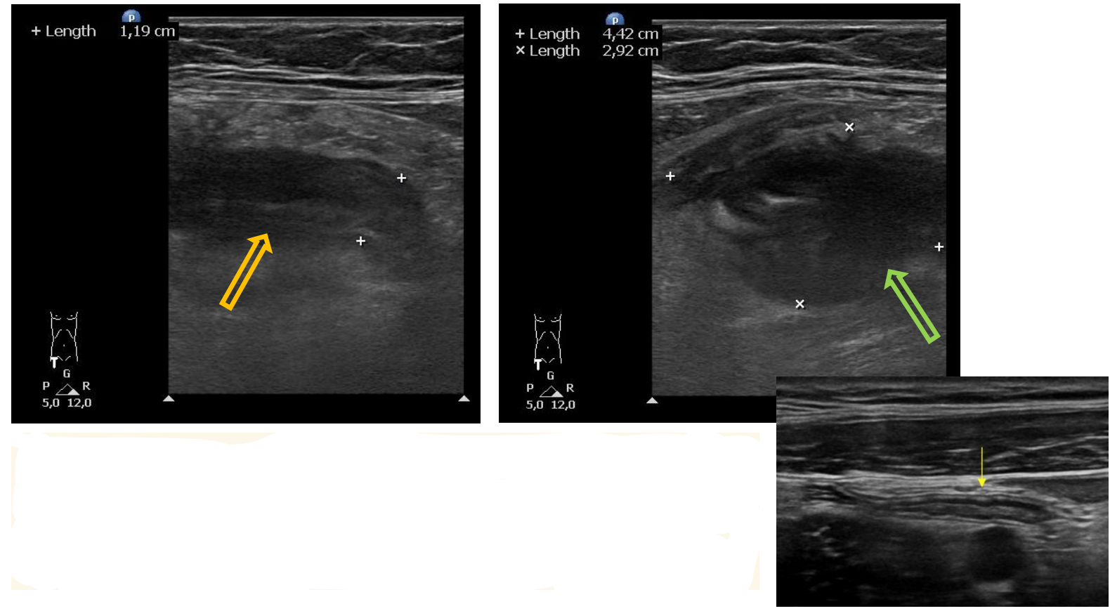
Patient history: acute right lower-quadrant abdominal pain in a young woman with normal body weight
Modality: sonogram of the appendix (linear 7,5–10 MHz probe), B-mode
Region: appendix, longitudinal and cross-sectional views
Radiologic sign: left image – thickened (12 mm), non-compressible hypoechogenic tubular structure
(yellow arrow); right image – hypoechogenic periappendiceal fluid collection around the tip of the
appendix (green arrow); small inlet – normal appendix ultrasonography
Diagnosis: acute appendicitis with periappendiceal abscess
2.1
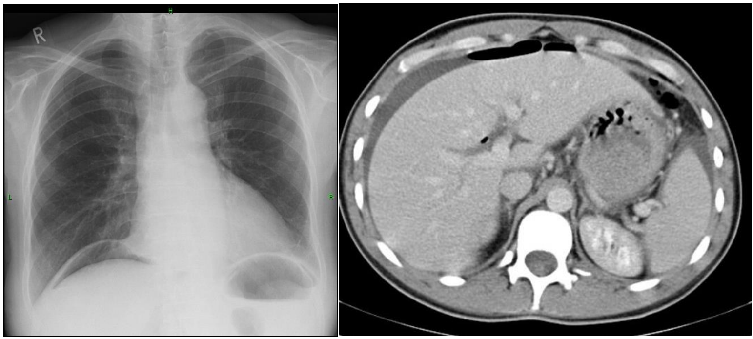
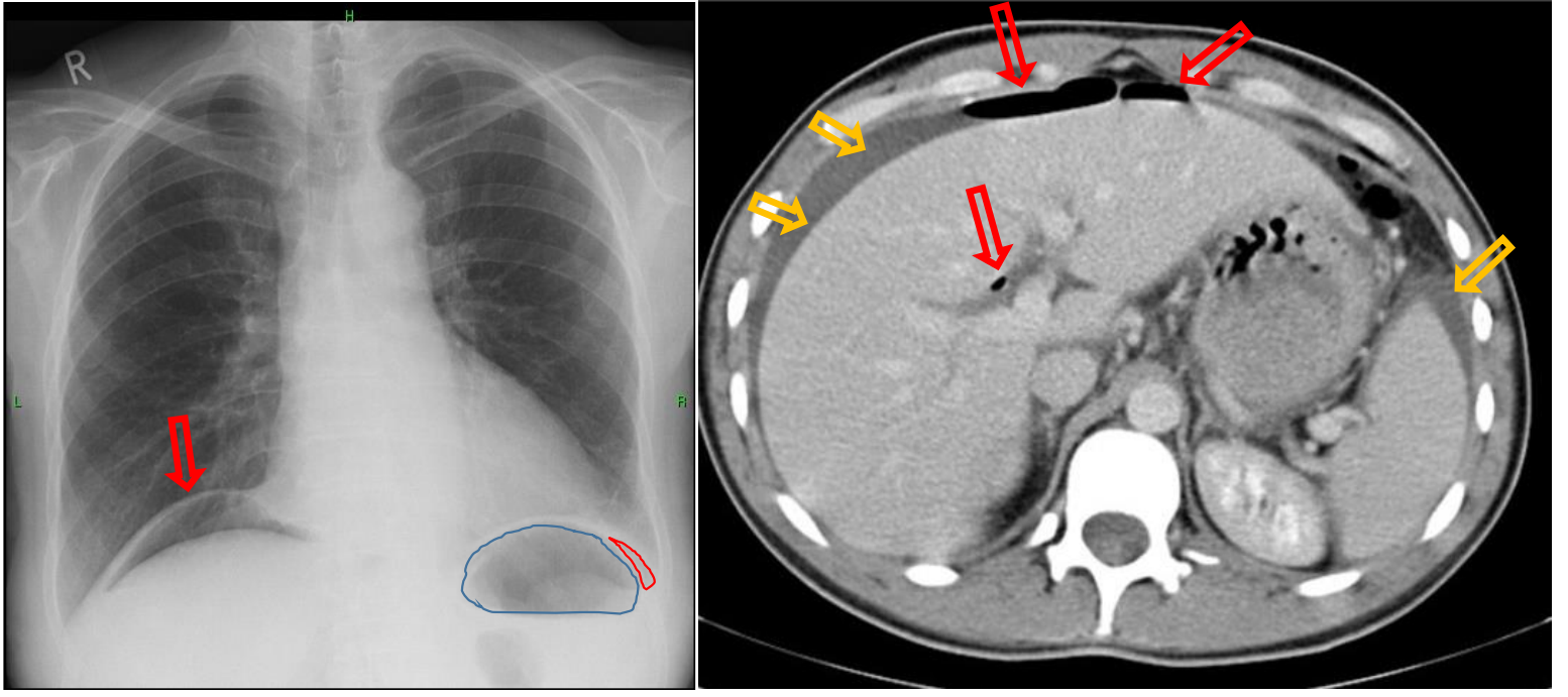
Patient history: sudden and severe upper abdominal pain, vomiting
Modality: left – PA chest radiograph, frontal projection (upright position); right – contrast-enhanced CT, axial view
Region: chest, upper abdomen
Radiologic sign: Crescents of radiolucency under the hemidiaphragms (red arrows and outline) on the radiograph, free air accumulation at the convexity of the liver and at the liver hilum with density of -1000 HU (red arrows), perihepatic and perisplenic free fluid with density of 0 HU (yellow arrows) on CT.
Diagnosis: free abdominal air after ulcer perforation
2.2
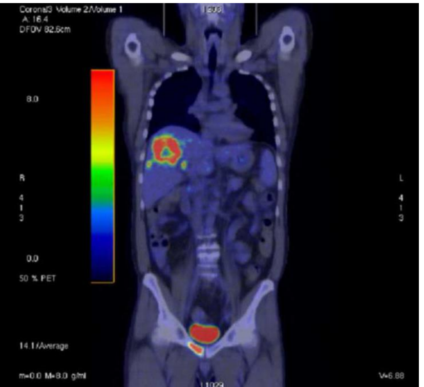
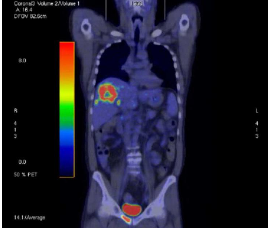
Patient history: breast carcinoma, staging
Modality: PET-CT, 18-fluorodeoxyglucose (18F-FDG) radiotracer; Hybrid imaging method: PET – metabolic activity, CT – morphology
Region: whole-body, coronal view
Radiologic sign: Increased fluorodeoxyglucose (18F-FDG) uptake in liver and pubic bone, high physiologic activity is shown in the bladder (normal finding). Liver lesion shows no central uptake, which is a sign of central necrosis.
Diagnosis: multiple metastases (liver, pubic bone) of breast carcinoma
2.3
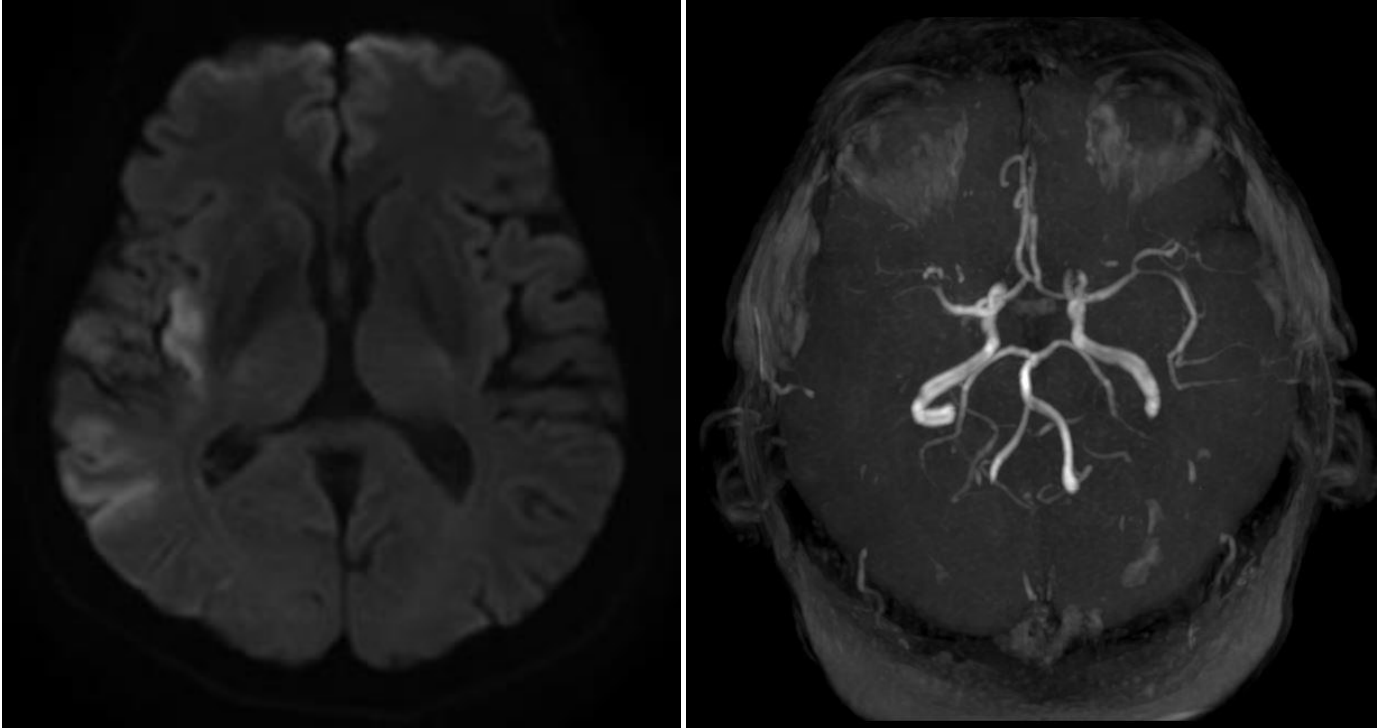
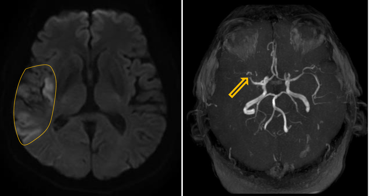
Patient history: leftsidedhemihypesthesia, visualneglect(ignores things on the left side) for a couple of hours
Modality: DWI MRI (left) and 3D TOF (time-of-flight) angiography (right); noncontrast sequence
Region: brain, axial view
Radiologic sign: significant hyperintensity (representing restricted diffusion) in the right parietal lobe with concomitant abrupt filling defect at the right middle cerebral artery
Diagnosis: large acute infarction due to acute thrombosis of the right middle cerebral artery
2.4
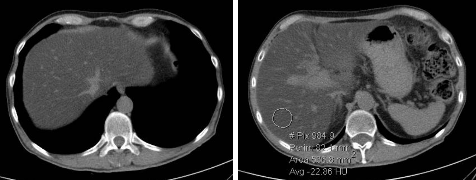
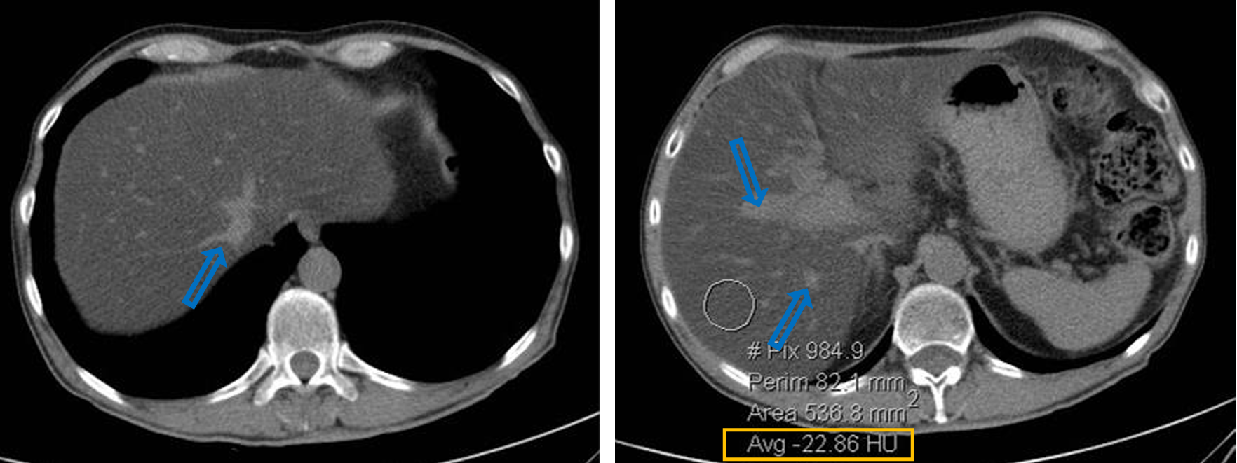
Patient history : elevated ALT (alanine transaminase) in a patient with hyperlipidemia
Modality: noncontrast CT
Region: upper abdomen, axial views
Radiologic sign: Diffusely, homogenously decreased density (cca. - 20 HU) of the liver (hepatic density normally is around 50-60 HU). The vessels (blue arrow) are denser in comparison to the liver parenchyma.
Diagnosis: steatosis hepatis (fatty liver)
2.5
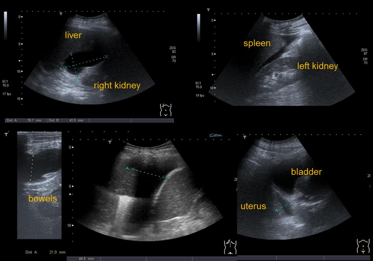
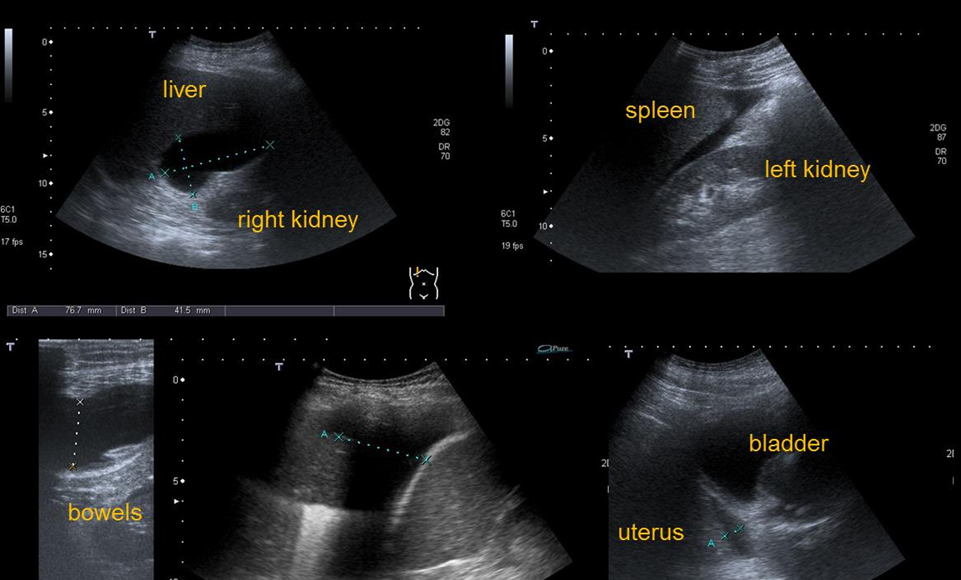
Patient history: blunt abdominal trauma
Modality: FAST (Focused Assessment with Sonography for Trauma) scan
Region: Abdomen, pleural sinuses, pericardium, aorta
Radiologic sign: Free abdominal fluid in the hepatorenal fossa, around the spleen, among the bowel loops and in Douglas pouch.
Diagnosis: Free abdominal fluid after blunt trauma, suspicious for internal hemorrhage.
3.1
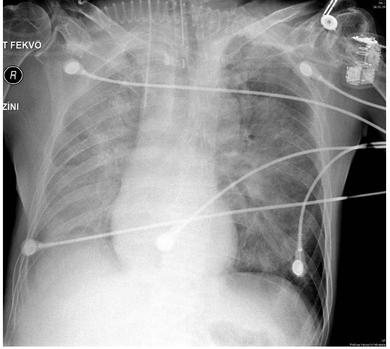
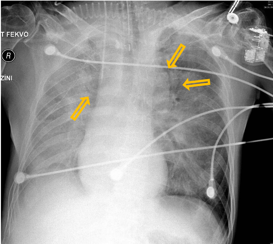
Patient history: postoperative condition, cardiac failure
Modality: AP chest radiograph
Region: chest, lungs
Radiologic sign: Extensive, bilateral perihilar opacities with air bronchograms (yellow arrow). Right diaphragm cannot be differentiated, on this side homogeneous loss of transparency can be seen due to the extending pleural effusion.
Diagnosis: severe pulmonary edema, right-sided hydrothorax
3.2
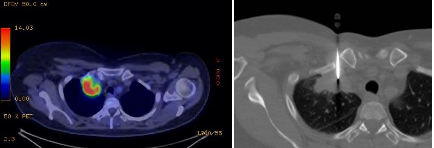
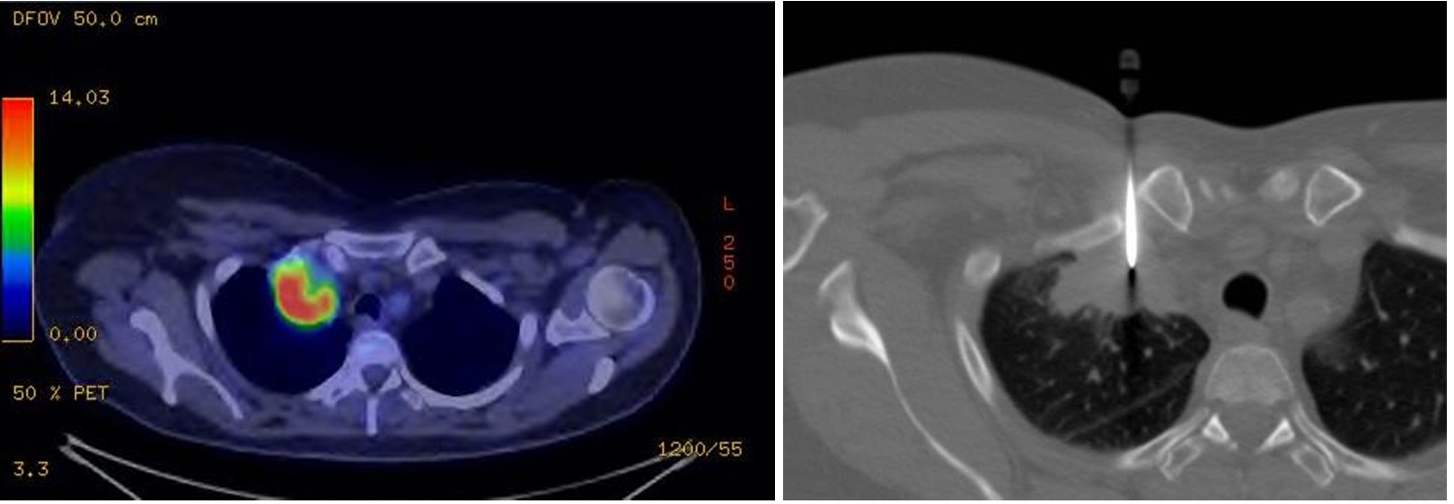
Patient history: heavy smoker for three decades, right shoulder pain at present in a middle-aged patient
Modality: PET-CT; 18-fluorodeoxyglucose (18F-FDG) radiotracer fused image (left); non-enhanced chest CT with bone window, core biopsy (right)
Region: chest, lung
Radiologic sign:Increased 18F-FDG uptake in the right apical mass;
Diagnosis: CT-guided core biopsy from the right apical mass. Pancoast-tumor, lung core biopsy
3.3
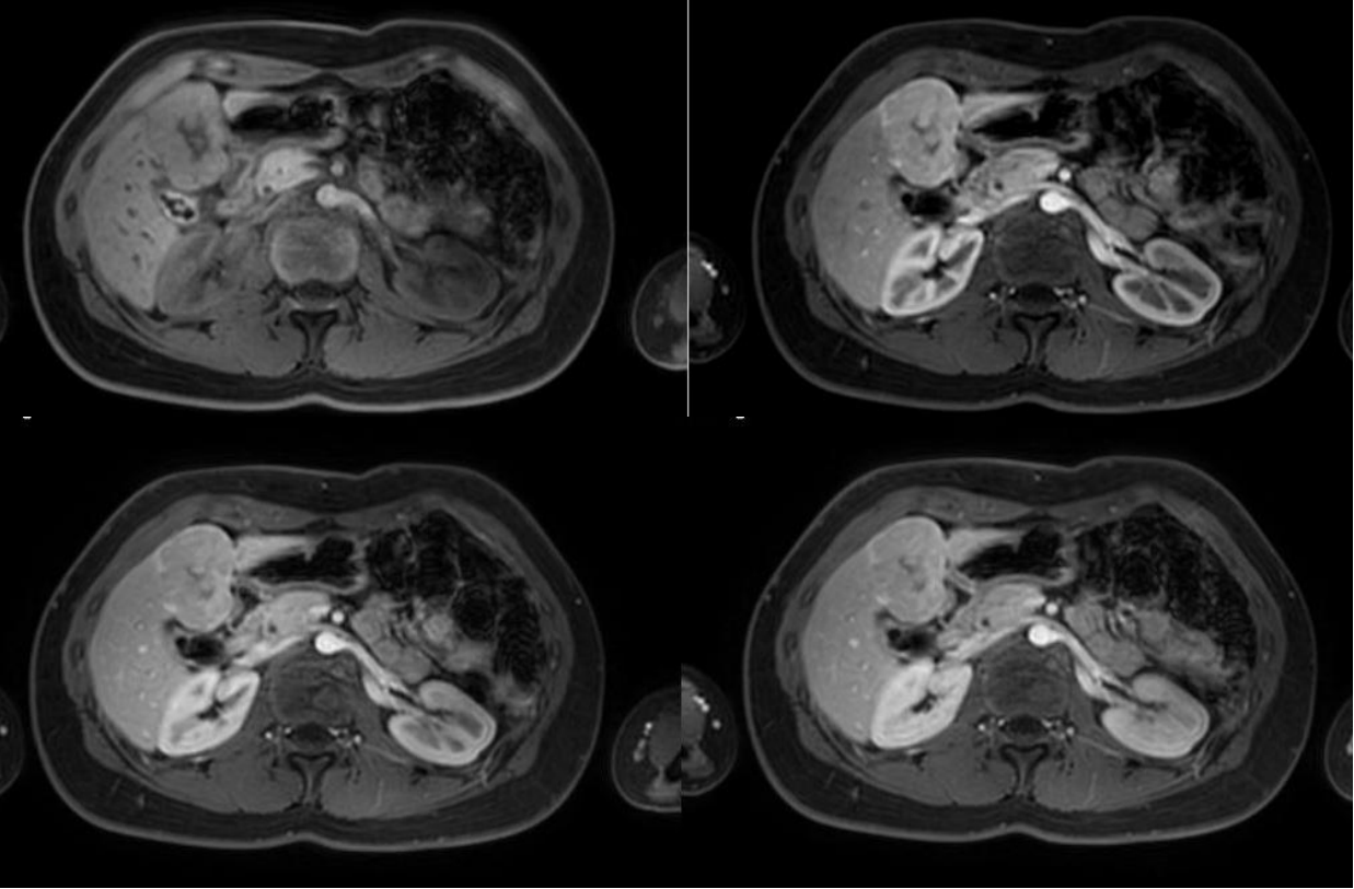
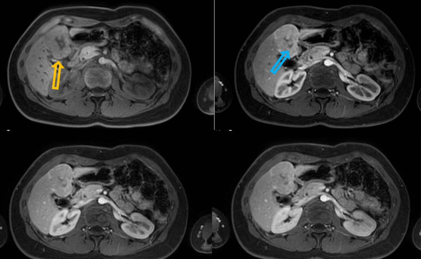
Patient history: liver mass was discovered on a routine abdominal ultrasound, work-up imaging
Modality: dynamic contrast-enhanced MR (T1 with fatsat) after iv. gadolinium (non-contrast, arterial phase, portal phase, venous phase), axial views
Region: upper abdomen
Radiologic sign: large hepatic mass (yellow arrow), intensive arterial enhancement, no „wash-out”, non-enhancing central scar (blue arrow).
Diagnosis: focal nodular hyperplasia (FNH)
3.4
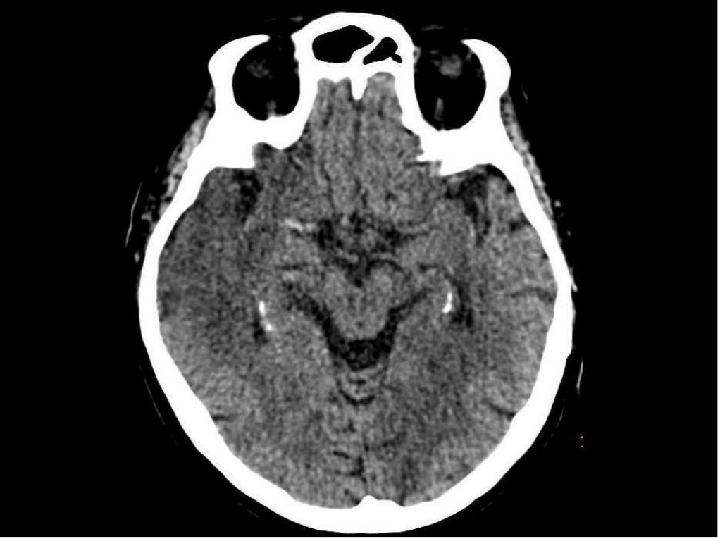
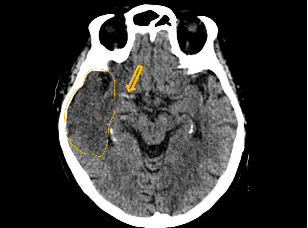
Patient history: epilepticseizure, hallucination, suddenmemoryimpairment
Modality: non-enhanced CT, brain window setting (window level: 40 HU; window width: 80 HU)
Region: brain, axial view
Radiologic sign: Cortical-subcortical border disappeared/blurred due to edema (blue area), “hyperdense media”-sign – thrombosis of the right middle cerebral artery (yellow arrow).
Diagnosis: acute/subacute ischaemia in the territory of the right MCA
3.5
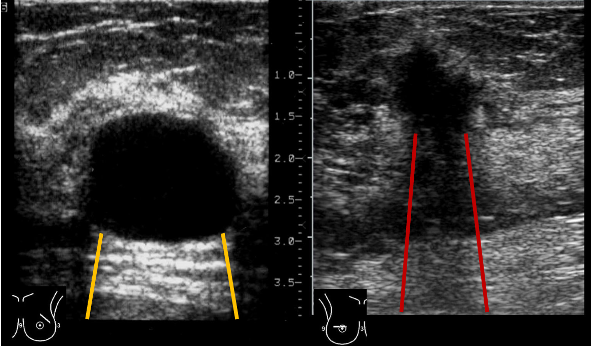
Patient history: asymptomatic patients, complex breast screening studies
Modality: breast sonography with linear probe (7.5–10 MHz), B-mode
Region: Breast, axilla
Radiologic sign: left image – well-defined anechoic lesion with posterior acoustic enhancement (btw yellow lines); right image – ill-defined hypoechogenic lesion with partial acoustic shadowing (btw red lines), “taller than wide” solid mass, suspicious of cancer
Diagnosis: simple cyst / breast carcinoma
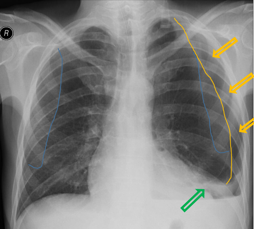
4.1
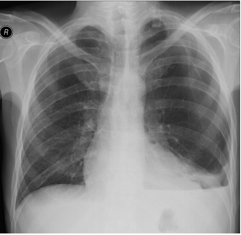
Patient history: blunt chest trauma in a young patient
Modality: PA chest radiograph
Region: chest, pleura
Radiologic sign: Left peripheral space is radiolucent without lung markings (yellow arrow); visible visceral pleural line (yellow line); slight mediastinal shift to the right. Left diaphragm and lateral sinus cannot be differentiated due to pleural fluid (horizontal fluid level is visible), while little above left lung basis partially collapsed (green arrow). Scapula lines are signed with blue lines.
Diagnosis: left-sided hydropneumothorax
4.2
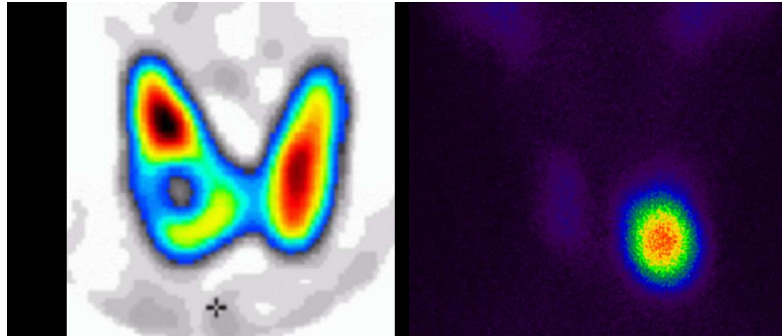
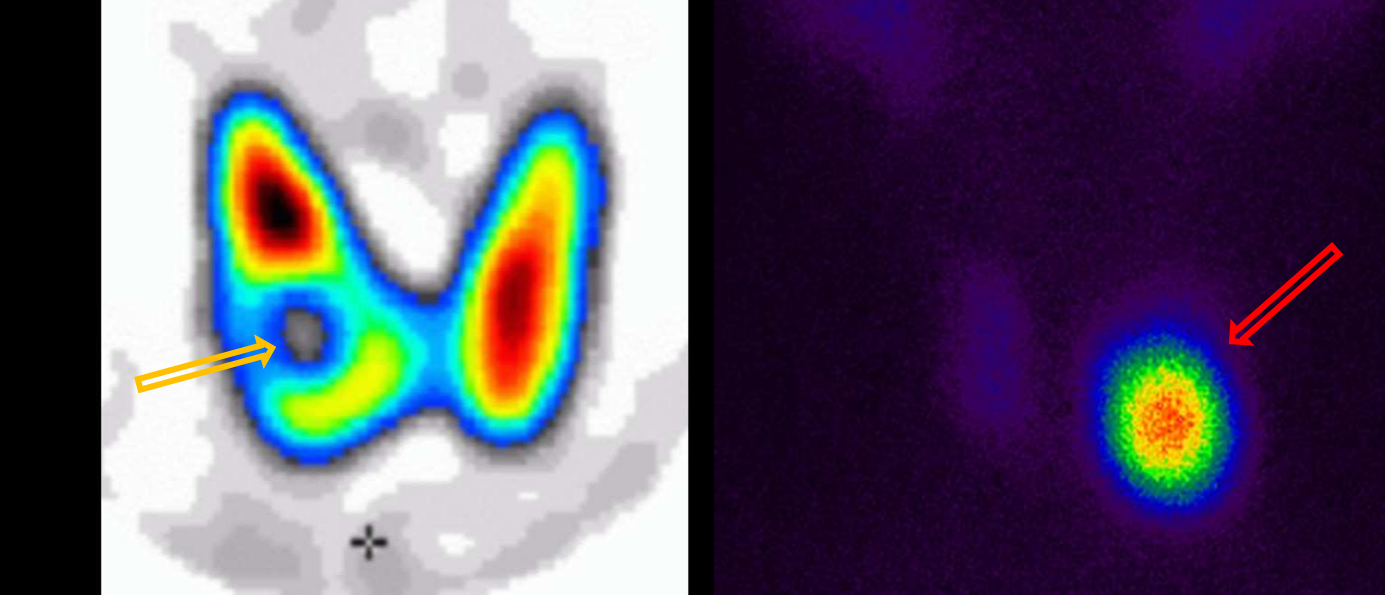
Patient history: thyroid masses found on routine ultrasonography
Modality: thyroid gland scintigram, Technetium-99m (99m Tc)-pertechnetate scintigraphy
Region: thyroid
Radiologic sign: left image - Focal lesion showing decreased tracer activity in the right lobe (yellow arrow); right image - increased uptake in the left lobe of the thyroid, low uptake in other parts of the gland (red arrow).
Diagnosis: left – cold nodule, which arise the suspicion of malignancy; right – hot, most probably benign nodule causing suppression of normal gland function
4.3
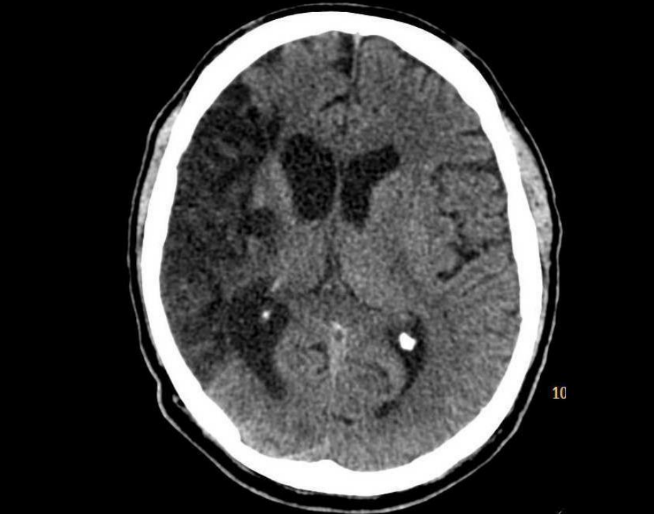
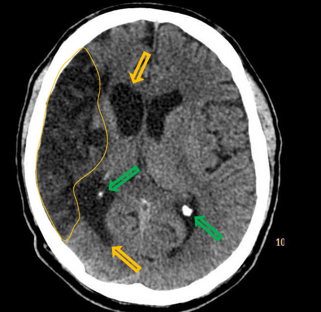
Patient history: elderly patient with chronic symptoms such as severe, left spastic hemiparesis with hemihypesthesia
Modality: Non-enhanced CT, brain window setting (window level: 40 HU; window width: 80 HU)
Region: brain, axial view
Radiologic sign: Extensive hypodense (20-25 HU) brain parenchyma, concomitant dilatation of the right lateral ventricle (yellow arrows) due to brain tissue loss; calcifications of the choroideal plexuses (common finding, green arrows)
Diagnosis: chronic ischemic lesion in the territory of right MCA
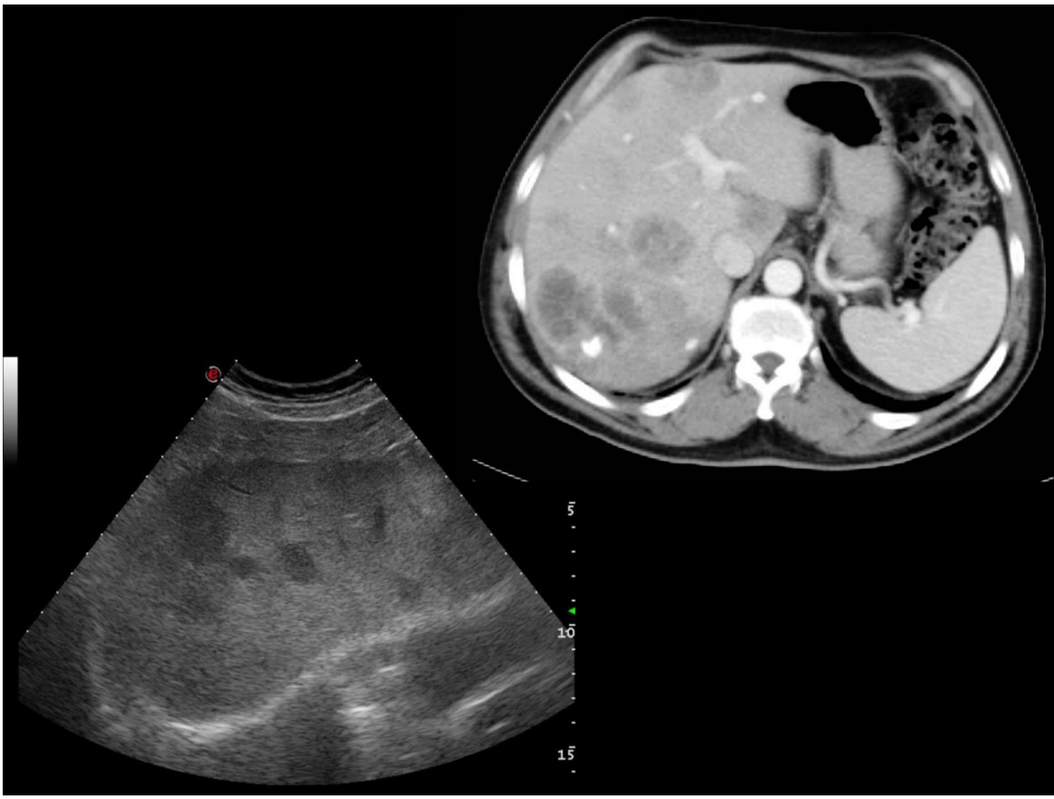
4.4. upper
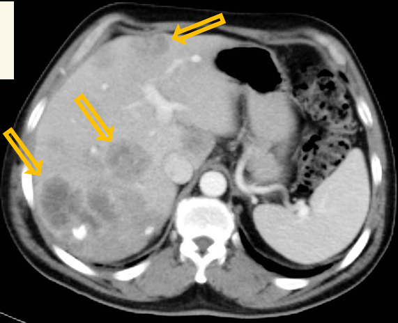
Modality: Region: contrast-enhanced
CT, portal phase, iv. iodine-based contrast agent upper abdomen, axial view
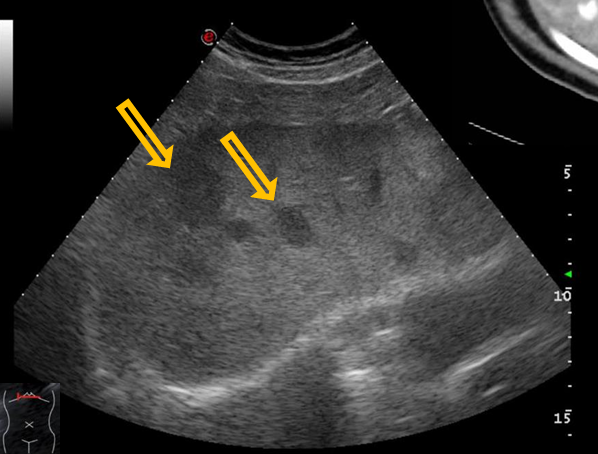
Radiologic sign: multiple hypoenhancing lesions of the liver, normal spleen
Diagnosis: multiple liver metastases (most commonly from colon adenocc.)
4.4 lower
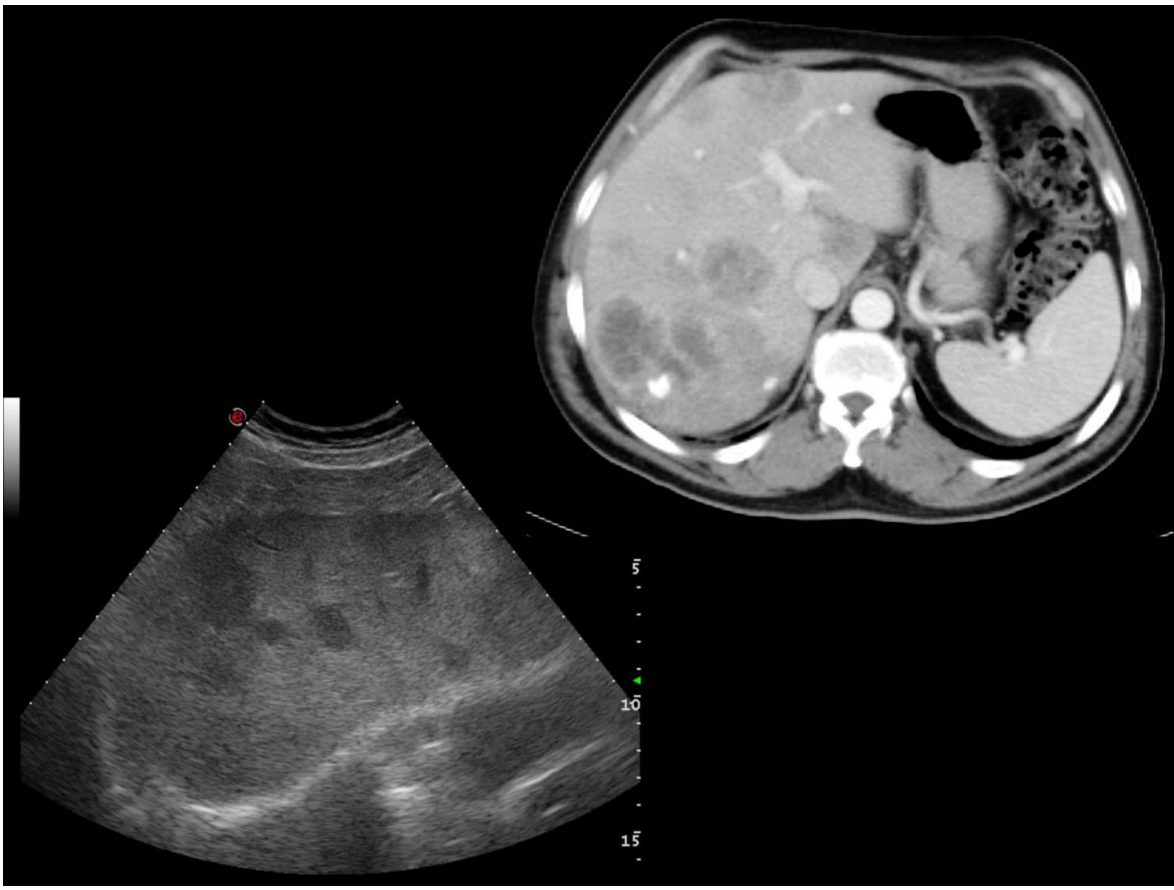
Patient history: colorectal tumor, increased CEA level at present
Modality: Liver ultrasound with convex probe (3.5-5 MHz), B-mode
Region: liver, right subcostal view
Radiologic sign: Diagnosis: hypoechoic lesions of the liver multiple liver metastases
4.5
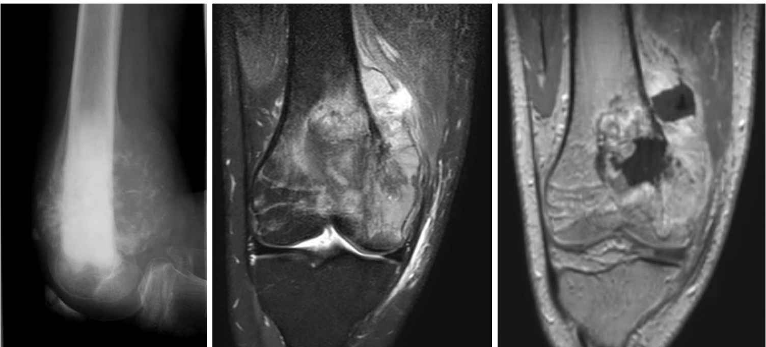
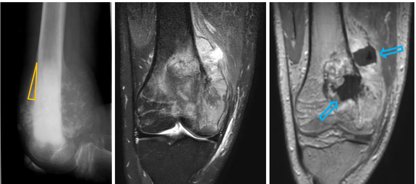
Patient history: soft tissue swelling and pain of the distal femur in a 22-year-old patient
Modality: X-ray of the left knee (left), PD FS (proton density fatsat) and T1W postcontrast MRI (middle and right, respectively)
Region: left knee, lateral view (radiograph), coronal views (MRI)
Radiologic sign: Inhomogeneous destructive metaphyseal lesion of the left femur. Periosteal reaction, Codman triangle (yellow triangle) and invasion of the surrounding soft tissue is also present (radiograph). Inhomogenously enhancing metaphyseal mass infliltrating the surrounding soft tissue (blue arrows).
Diagnosis: osteosarcoma of the femur
4.6
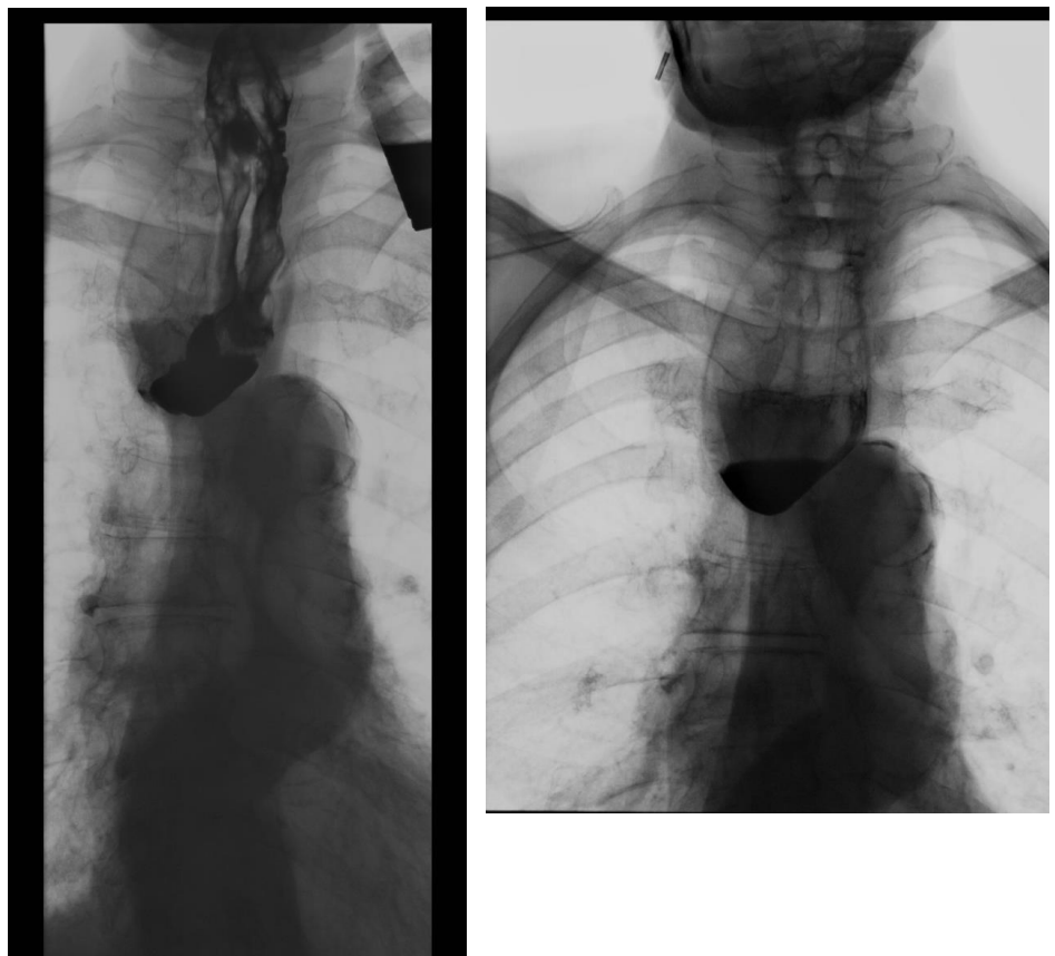
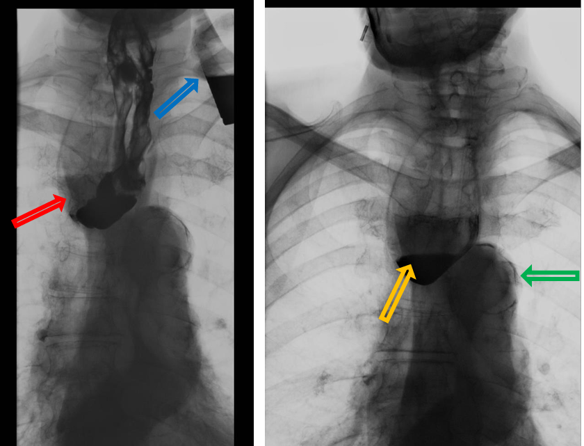
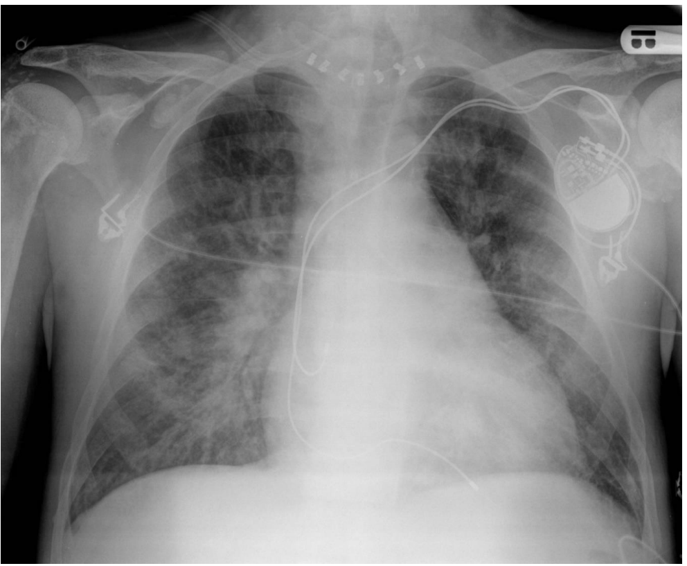
Patient history: dysphagia and sensation of food stuck
Modality: barium swallow test, fluoroscopy
Region: esophagus Radiologic sign: outpouching (red arrow) filled with contrast material near the pharyngo-esophageal junction; air-fluid level (yellow arrow) in the outpouching; dilated, sclerotic aorta (green arrow); glass filled with contrast material (blue arrow)
Diagnosis: Zenker’s diverticulum, aortosclerosis
5.1
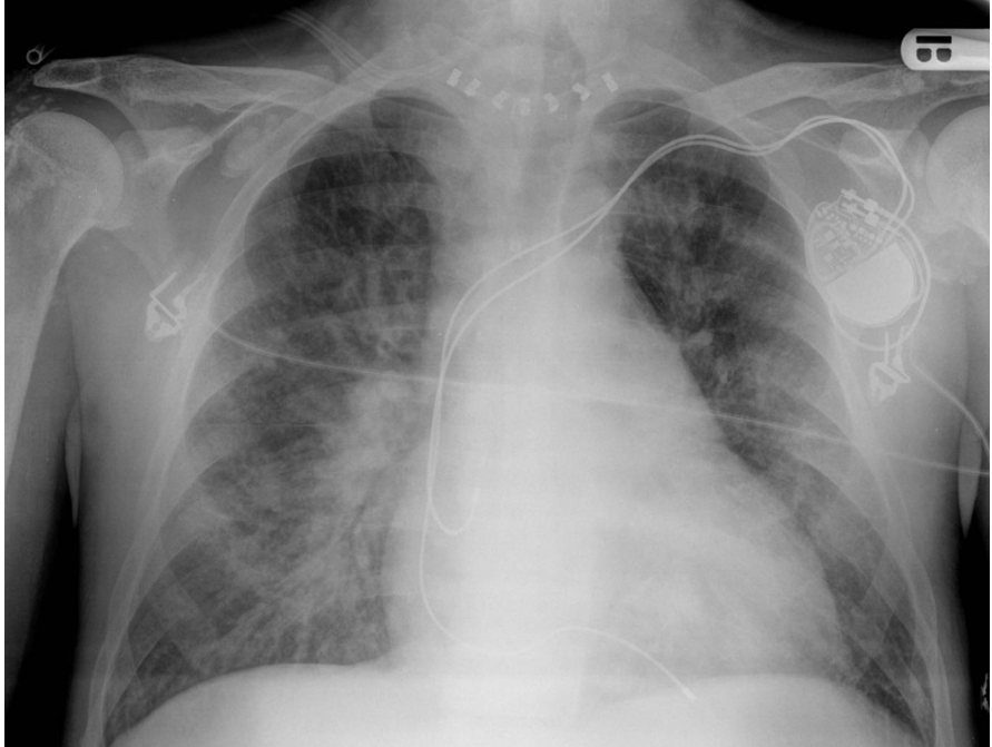
Patient history: postoperative condition (thyreoidectomy)
Modality: AP (prone) chest radiograph
Region: chest, lungs Radiologic sign: Cephalisation, bilateral perihilar consolidation and cardiomegaly (CTI > 0.5). Foreign bodies: pacemaker device and electrodes, surgical clips following thyroid surgery, ECG
Diagnosis: pulmonary edema
5.2
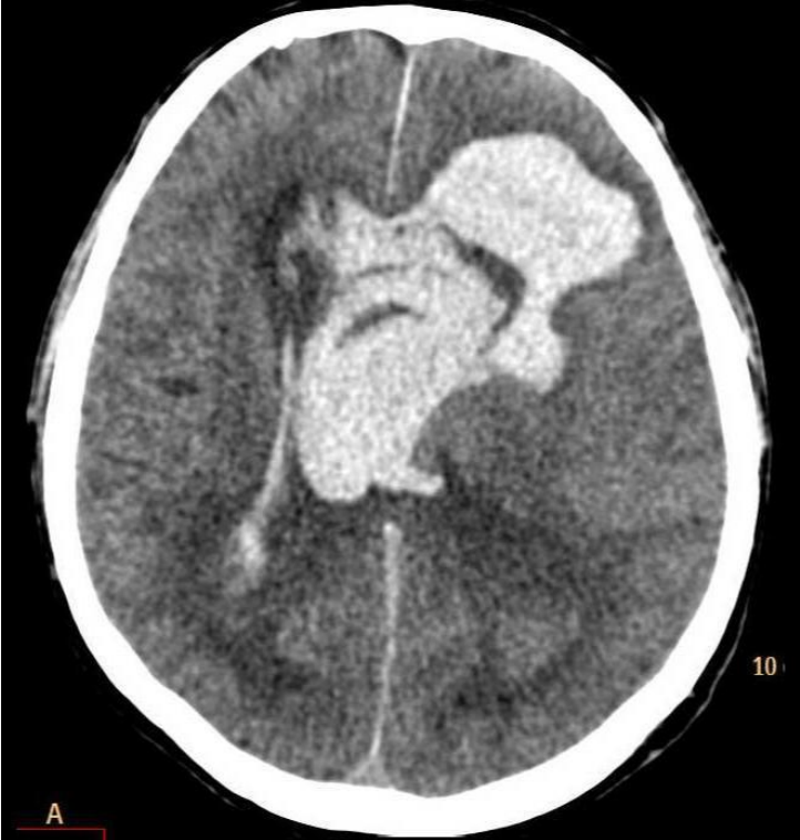
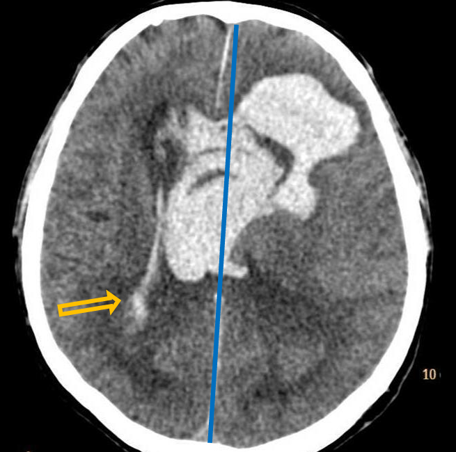
Patient history: sudden onset of altered mental status, loss of consciousness in an elderly patient with untreated hypertension
Modality: non-enhanced CT, brain window setting (window level: 40 HU; window width: 80 HU)
Region: brain, axial view Radiologic sign: Large hyperdense area (density: 60-70 HU) extending into the ventricles, slight midline shift to the right and compressed right lateral ventricle due to mass effect (yellow arrow).
Diagnosis: acute cerebral apoplexy
5.3
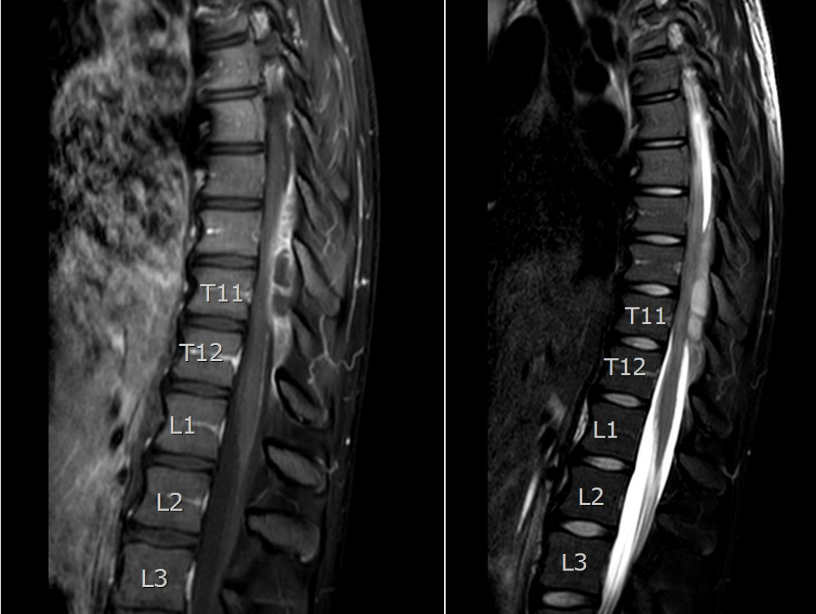
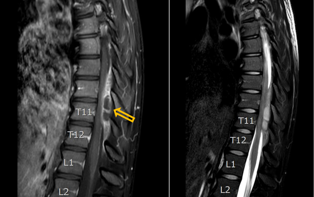
Patient history: days with progrediatingsevere lower back pain, fever, paraparesis, urinary retention disturbance in an immunocompromised patient
Modality: left – T1W fatsat postcontrast MRI after iv. gadolinium administration; right – T2W fatsat MRI (sagittal view)
Region: thoracic spine
Radiologic sign: epidural enhancing mass at the level of Th 9-12th vertebras (yellow arrow), no signal loss on fatsat image; non-enhancing fluid signal intensity inside of the mass
Diagnosis: epidural abscess
5.4
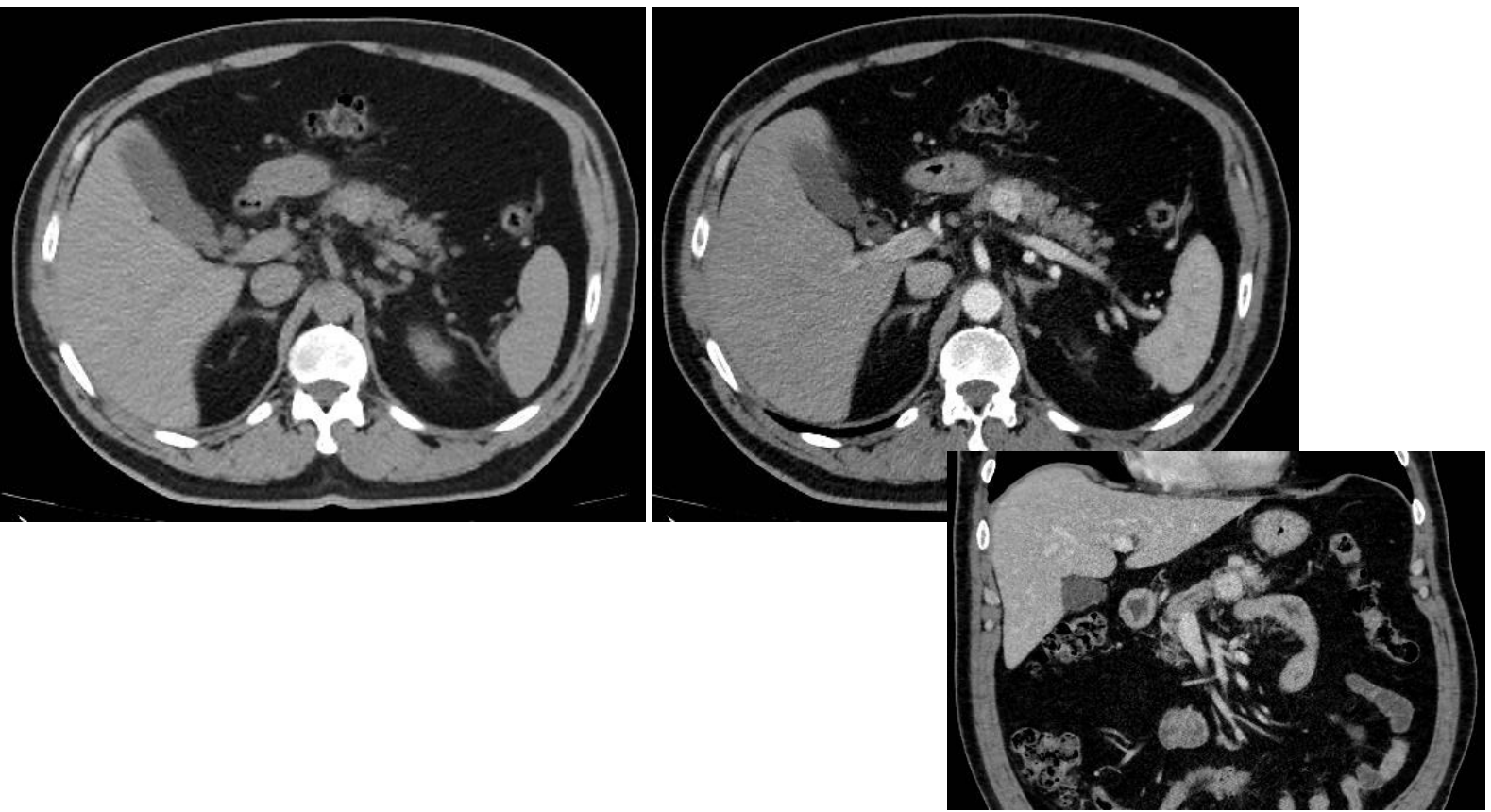
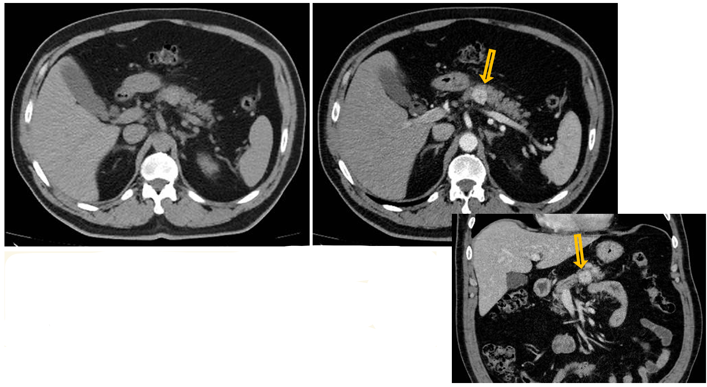
Patient history: symptoms of hypoglycemia, sweating and weight gain in a 52-year-old patient
Modality: left – non-enhanced CT, axial plane; right – early arterial phase of dynamic CT (iv. iodinebased contrast agent), axial plane, and (bottom right) coronal reconstruction of the same study
Region: upper abdomen
Radiologic sign: solid mass in the pancreas body showing intensive early arterial contrast enhancement (yellow arrow)
Diagnosis: pancreas insulinoma
5.5
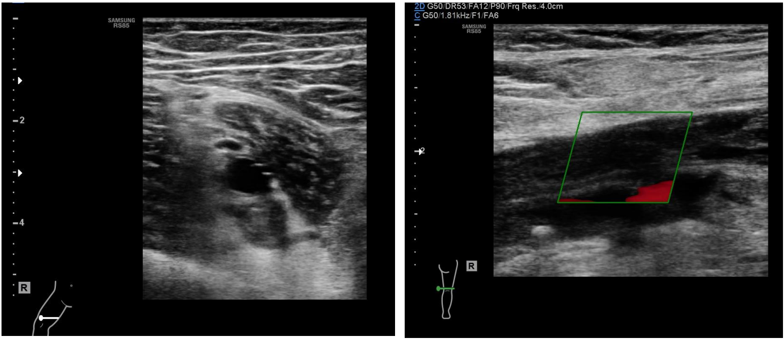
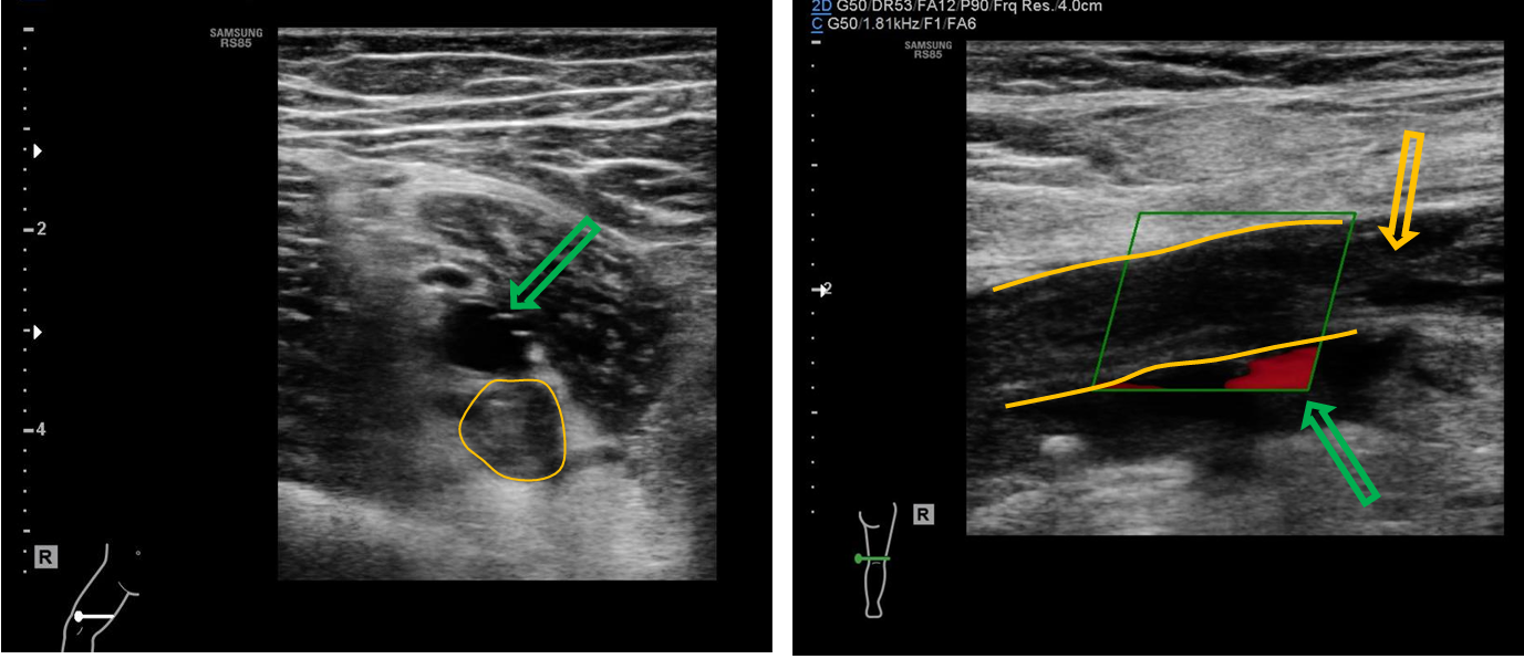
Patient history: calf pain and swelling of the right leg, plaster cast due to trauma in recent weeks
Modality: Lower extremity venous ultrasound, linear probe (7,5-10 MHz), B-mode and Color-Doppler mode (right panel)
Region: femoral superficial and popliteal veins; left image – transverse section, right image – sagittal section
Radiologic sign: Veins are filled with hypoechoic thrombus (yellow lines and arrow); the lumen is non-compressible demonstrating no flow (lack of Doppler-signal); beneath the popliteal artery has normal flow signal (green arrow).
Diagnosis: deep vein thrombosis (DVT)
6.1
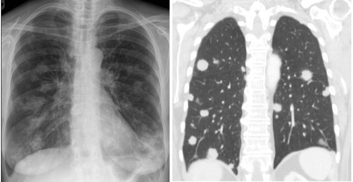
Patient history: melanoma malignum, staging
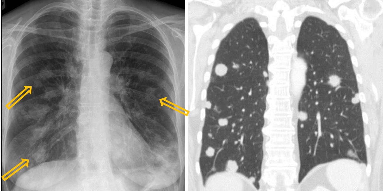
Modality: left image – PA chest radiograph (upright); right image – postcontrast chest CT; lung window setting, iv. iodine-based contrast agent
Region: chest, lung
Radiologic sign: Multiple soft tissue lesions showing different size and rounded shape (yellow arrows), number of nodules is higher at the base than the apex due to its higher perfusion.
Diagnosis: multiple lung metastases
6.2
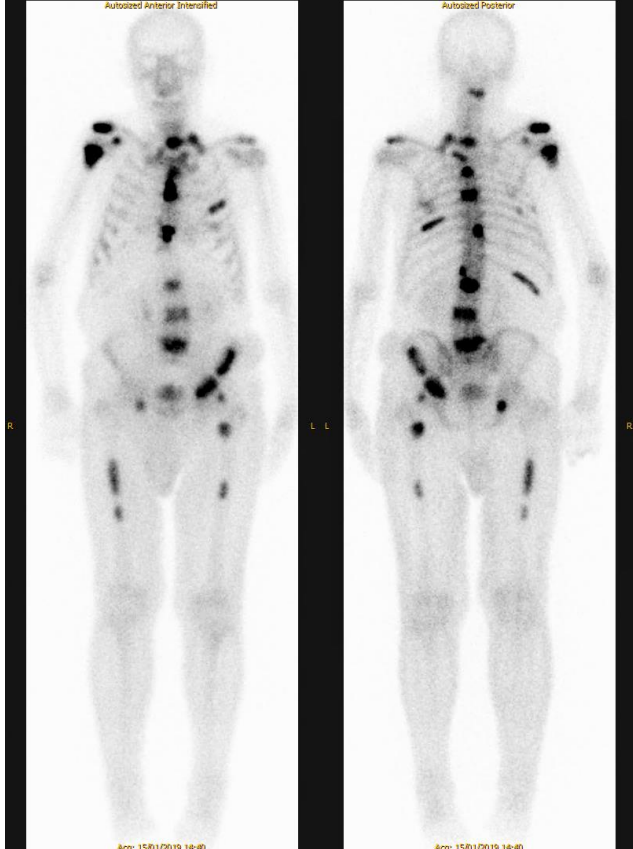
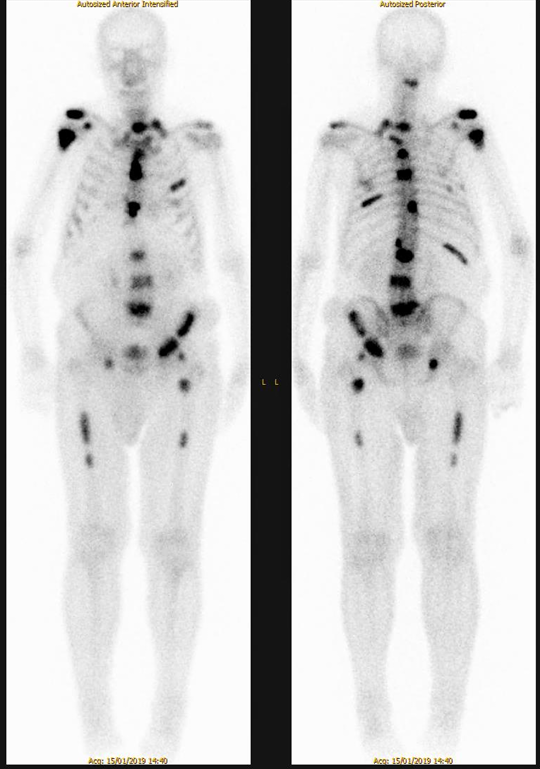
Patient history: history of prostate cancer, elevated PSA level at present
Modality: bone scintigraphy with 99mTc withmethylene diphosphonate(MDP) radiotracer
Region: whole body, AP and PA views
Radiologic sign: multiple foci of increased radiopharmaceutical uptake in the bones
Diagnosis: multiplex osseal metastases, scoliosis
6.3
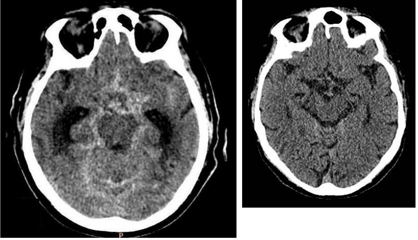
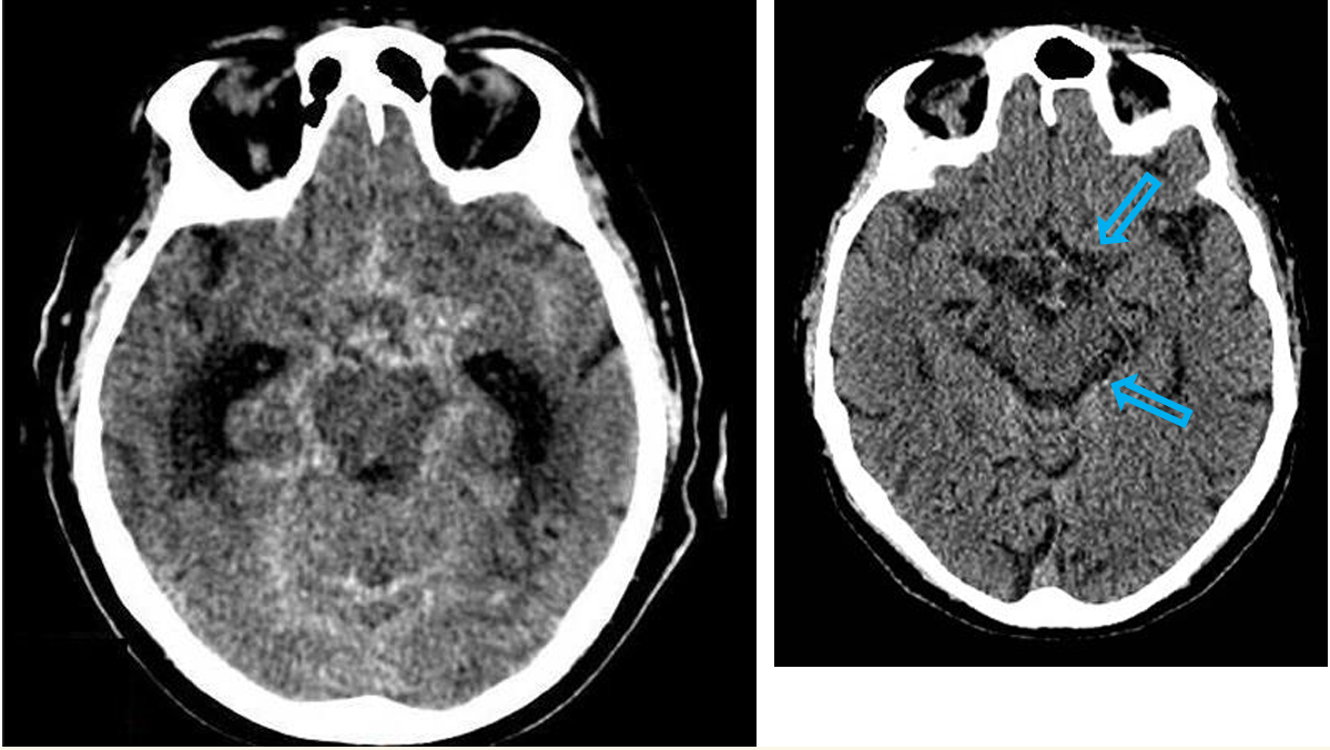
Patient history: “worst headache” in life with acute onset and nausea in a 42-year-old patient
Modality: non-enhanced CT
Region: brain, axial view
Radiologic sign: cast-like hyperdensity filling the basal cisterns and sulci (normal hypodens, liquor-filled cysterns are shown on the right image, blue arrow)
Diagnosis: acute subarachnoid hemorrhage, most commonly due to berry aneurysm rupture
6.4
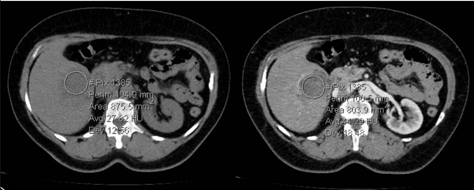
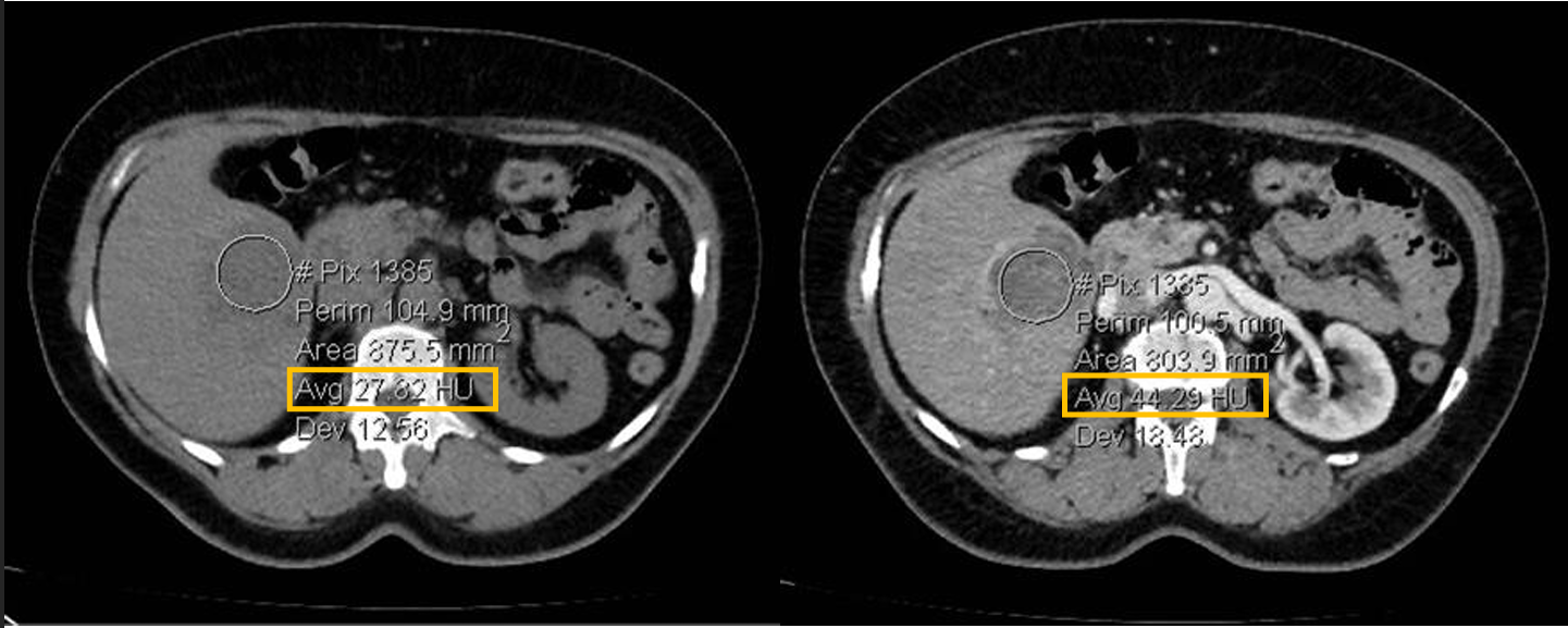
Patient history: tumor search in a middle-aged patient with fatigue and weight loss
Modality: left image – non-enhanced CT; right image – arterial phase enhanced CT (iodine-based contrast agent)
Region: upper abdomen, axial views
Radiologic sign: solid, contrast-enhancing (from 27 HU to 44 HU), hypodense mass in the 5-6th segment of the liver
Diagnosis: cholangiocarcinoma
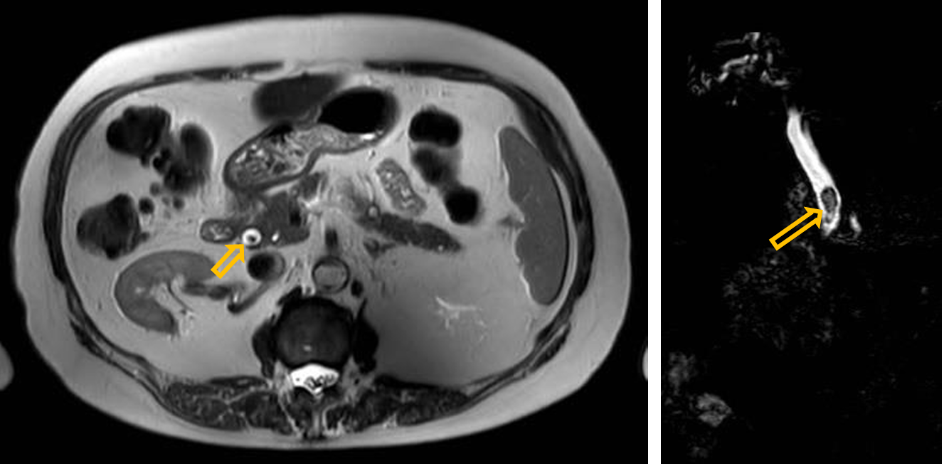
6.5
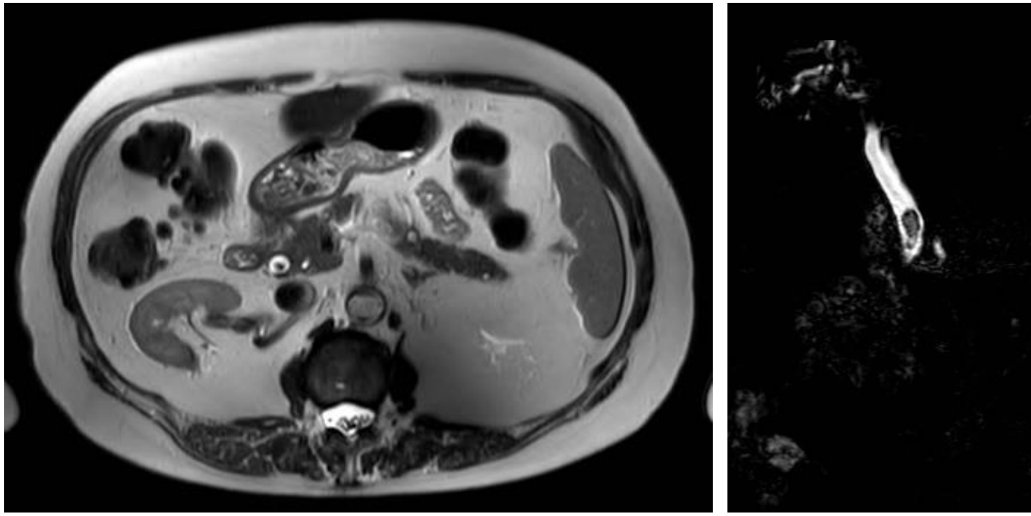
Patient history: chronic right upper abdominal pain
Modality: T2W MRI (left); 3D MRCP (a heavily T2-weighted sequence, right), noncontrast studies
Region: upper abdomen, axial (left) and coronal (right) views Radiologic sign: filling defect in the fluid-filled, hyperintense common bile duct
Diagnosis: choledocholithiasis
7.1
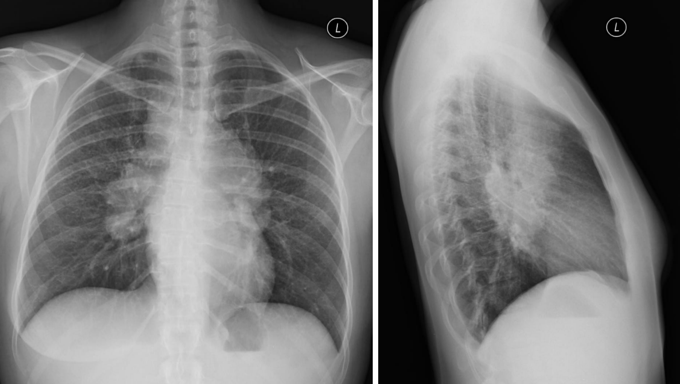
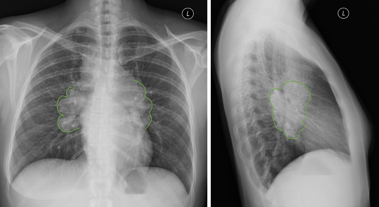
Patient history: chronic fatigue and weight loss in a 39-year-old patient
Modality: left – PA chest radiograph right – left lateral chest radiograph
Region: chest, lungs Radiologic sign: bilateral hilar enlargement (green area), middle mediastinal mass, no alteration in lung parenchyma
Diagnosis: bilateral hilar lymphadenopathy in sarcoidosis (stage 1)
7.2
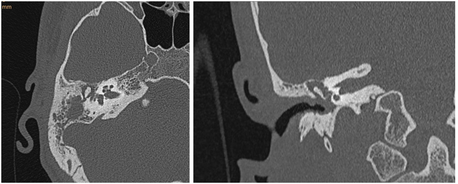
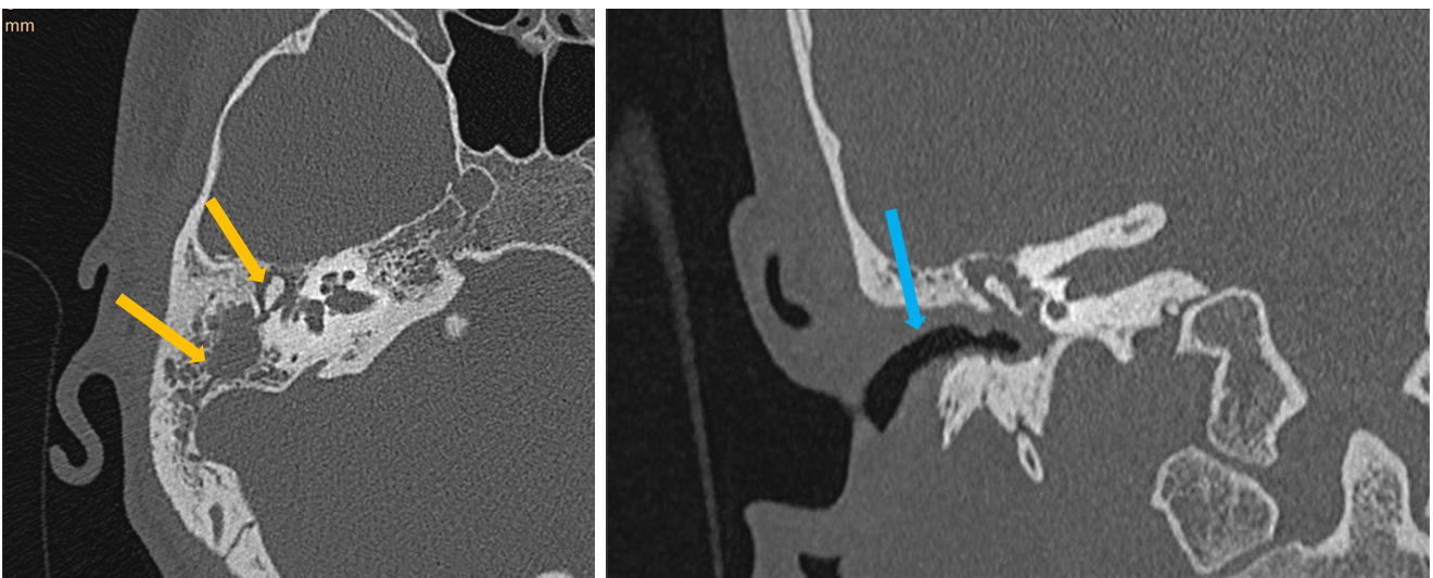
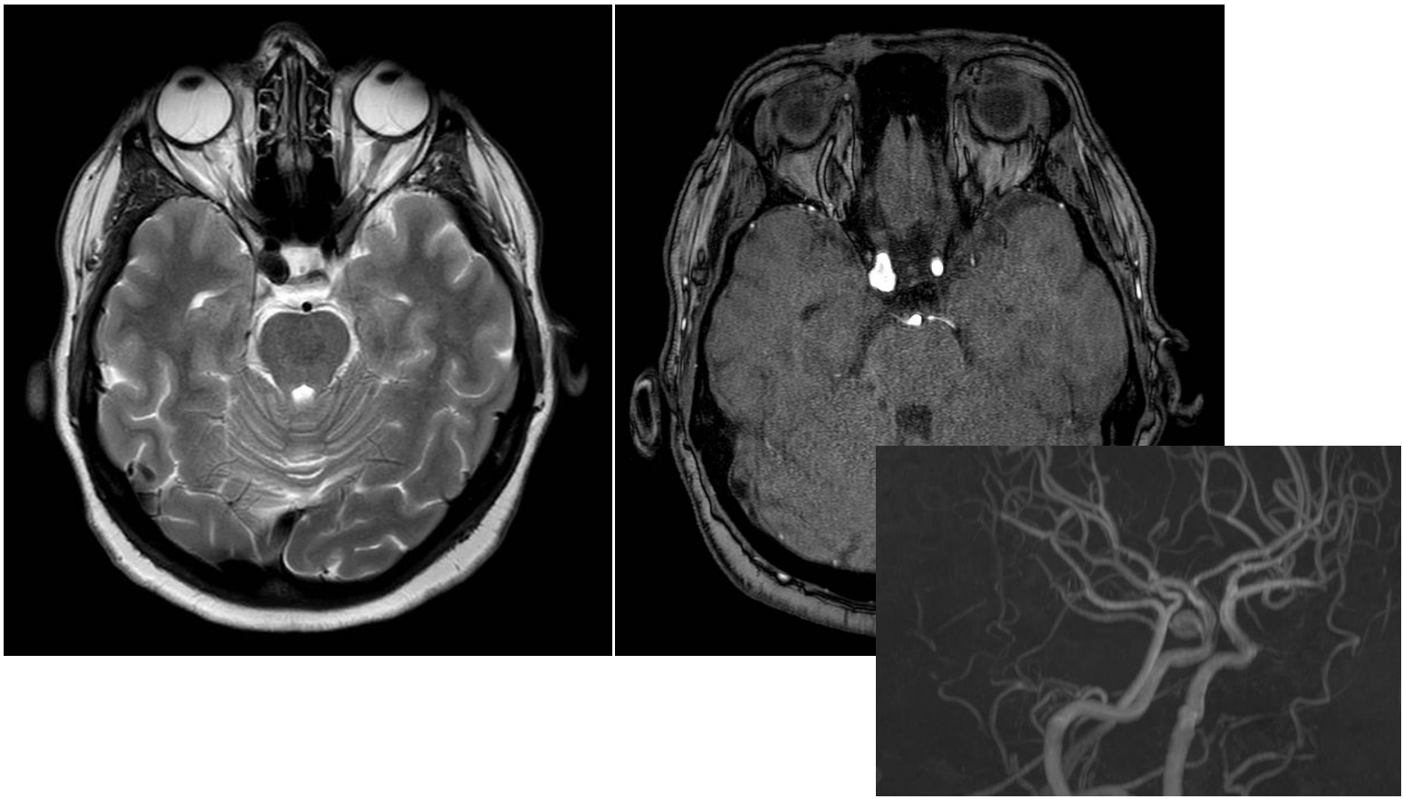
Patient history: Modality: ear pain and discomfort, poor hearing non-enhanced HRCT of the temporal bone
Region : temporal region, axial and coronal views
Radiologic sign: fluid-filled middle ear and mastoid cells (yellow arrows); bone erosion of the latter; thickened outer ear wall (blue arrow)
Diagnosis : recurrent otitis media
7.3
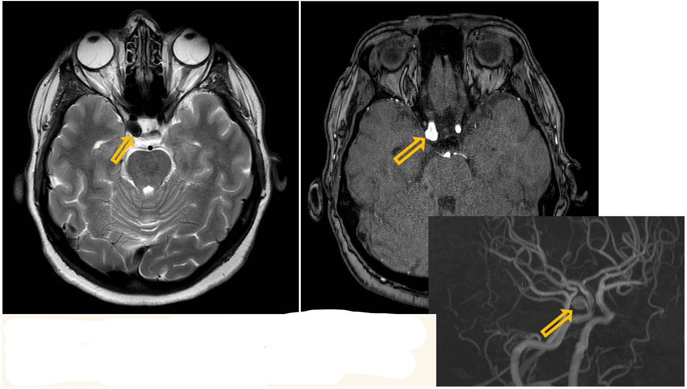
Patient history: young asymptomatic patient with positive family history of cerebral aneurysm
Modality: T2W MRI and 3D TOF angiography (noncontrast)
Region: brain, axial view
Radiologic sign: circumscribed, larger “flow-void” on T2W MRI and dilatation of the right internal carotid artery on TOF (yellow arrow)
Diagnosis: aneurysm of the right internal carotid artery (cavernous part)
7.4
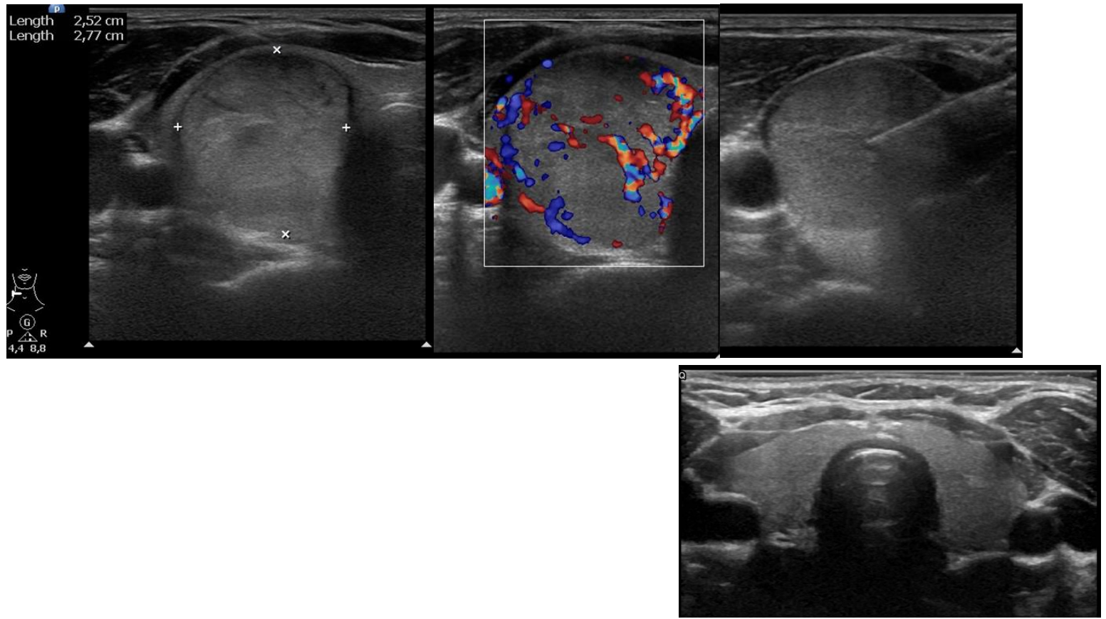
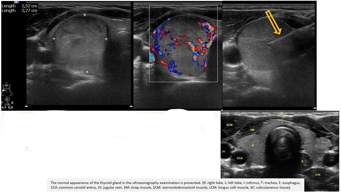
Patient history: palpable, unpainful mass in the right side of the neck
Modality: linear probe (7,5–10 MHz), left – B-mode US, middle – Color Doppler US, right – B-mode US
Region: neck, thyroid gland
Radiologic sign: well-circumscribed, isoechoic, vascularized nodule; FNAB (fine-needle aspiration biopsy), echodense needle (yellow arrow)
Diagnosis: suspicious thyroid nodule, cytology proved benignity
7.5
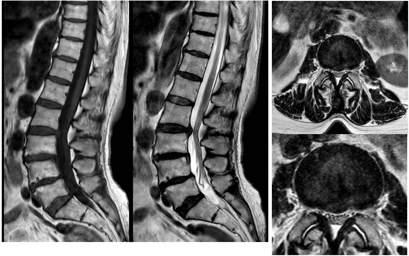
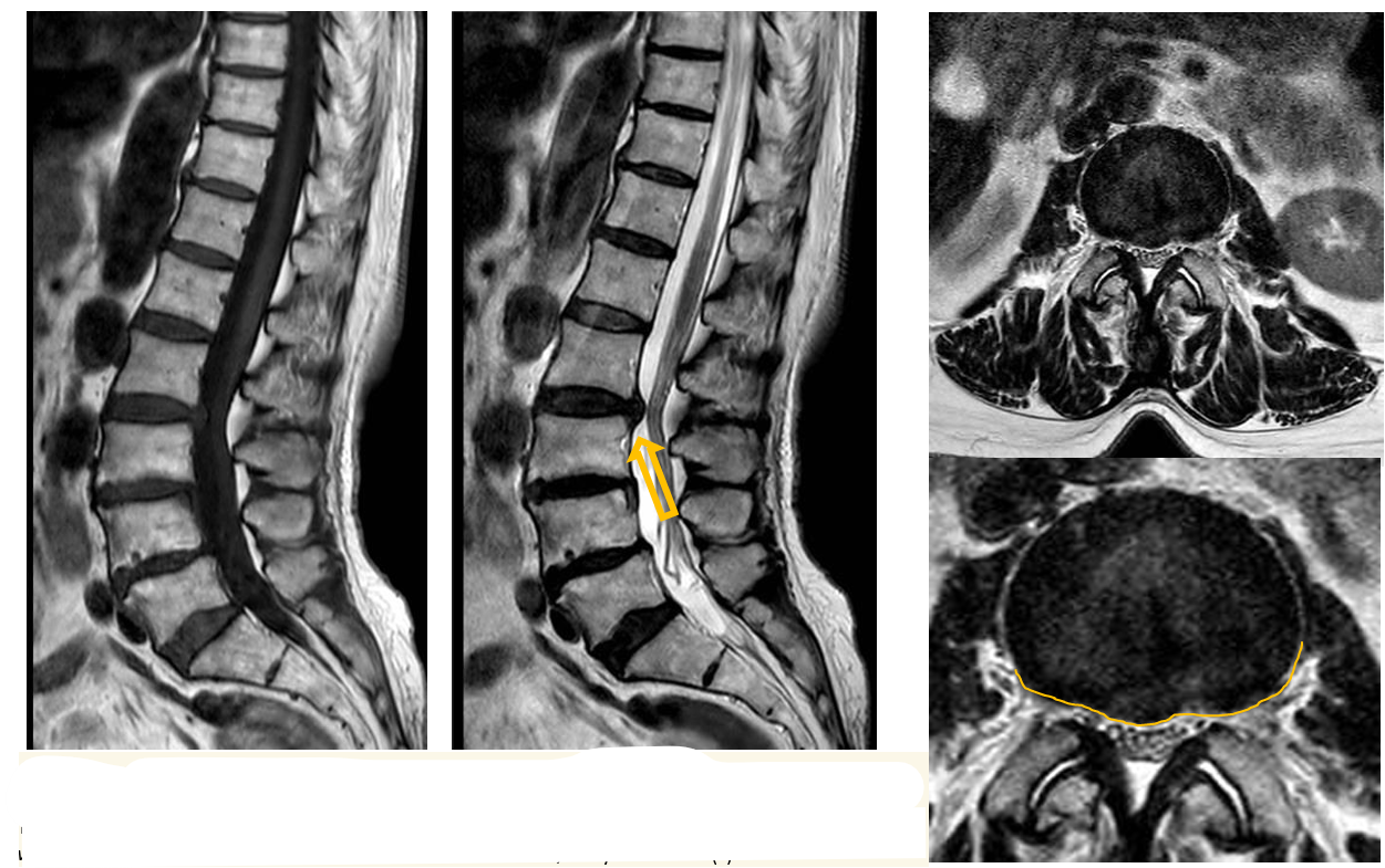
Patient history: pain radiating to the right L2 dermatome, without paresis or urinary disturbance.
Modality: Non-enhanced MRI (left: T1WI sagittal, center: T2WI sagittal, right: T2W axial)
Region: lumbar spine
Radiologic sign: Btw L2/3 hypointense (signaling low water content) disc protrudes into the spinal canal (yellow arrow)
Diagnosis: disc herniation between L2 and L3 level; dehydrated disc(s)
8.1
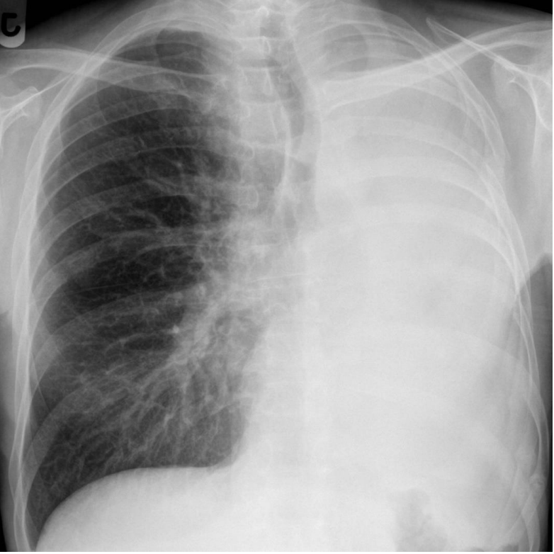
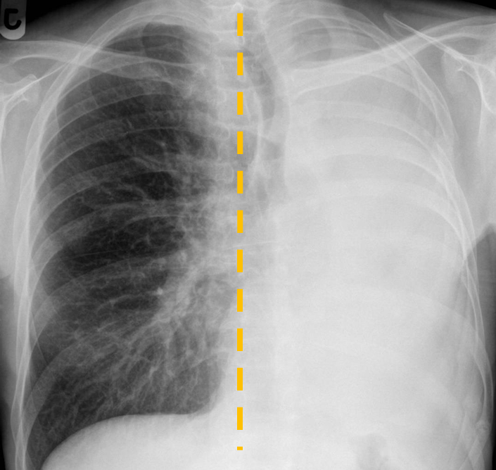
Patient history: left-sided pneumonectomy, control Modality: PA chest radiograph
Region: chest, lungs
Radiologic sign: In the projection of the left lung, air-filled lung parenchyma does not present; the trachea and the heart are deviated toward the pathological side (yellow dashed line)
Diagnosis: “white lung”, state of postpneumonectomy on the left side
8.2
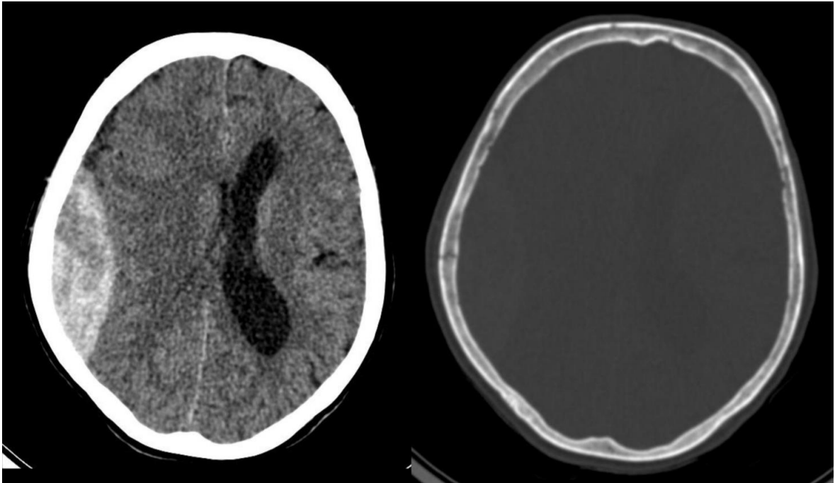
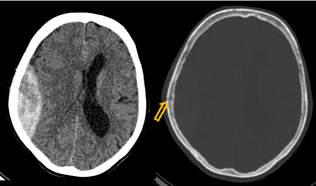
Patient history: loss of consciousness following a bike accident
Modality: Non-enhanced CT; left – brain window setting (window level: 40 HU; window width: 80 HU), right – bone window setting (window level: 600 HU; window width: 2800 HU)
Region : Brain, axial views
Radiologic: Lens-shaped hyperdense mass and a radiolucent line at identical position (yellow arrow); compressed right lateral ventricle and midline sign shift due to mass effect.
Diagnosis : right-sided epidural hematoma with skull vault fracture
8.3
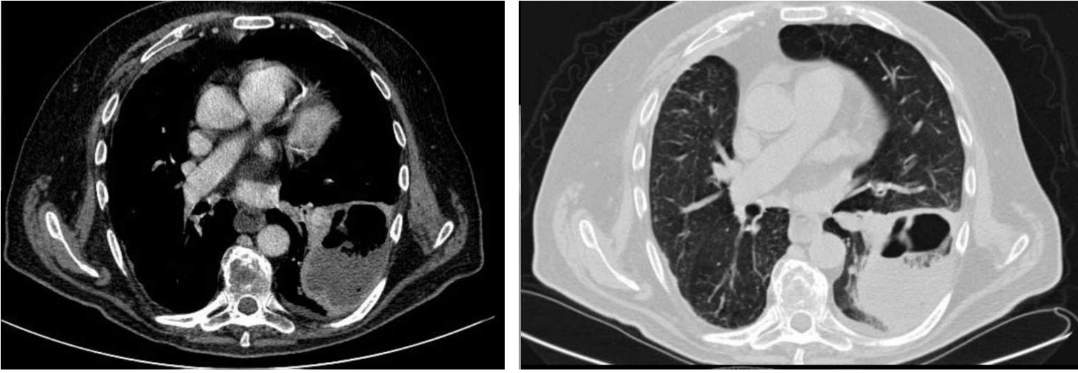
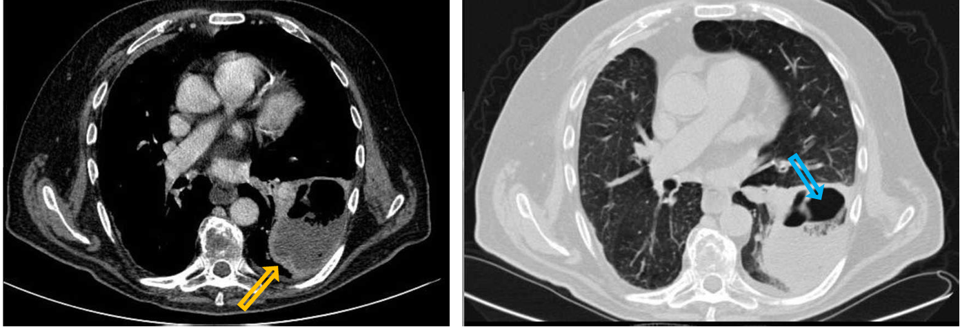
Patient history: chest pain, cough and fatigue in an immunocompromised patient
Modality: Contrast-enhanced chest CT, lung window setting (right panel); mediastinal window setting (left panel), iv. iodine-based contrast agent
Region: chest, axial views
Radiologic sign: air-containing (blue arrow) fluid collection with thick enhancing rim (yellow arrow)
Diagnosis: left-sided lung abscess
8.4
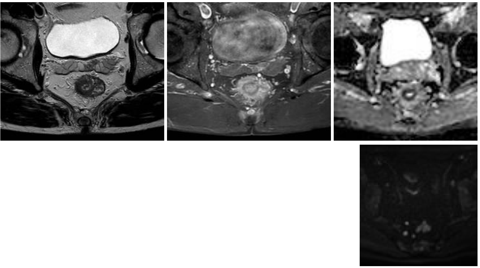
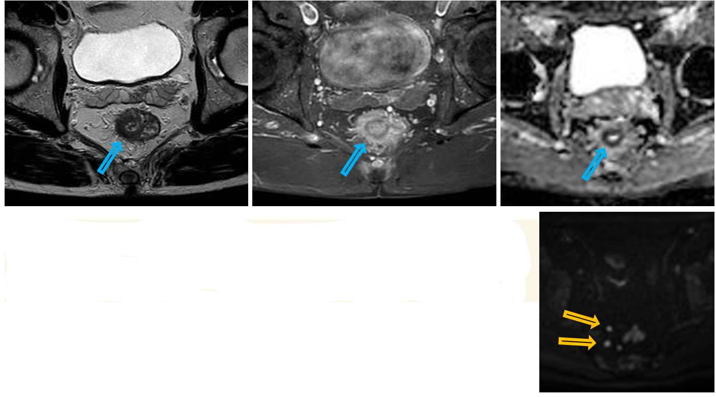
Patient history: bloody stool, colonoscopy showed high-grade rectal adenocarcinoma, staging MRI
Modality: pelvis, axial views
Region: T2W MRI (left), T1 fatsat contrast-enhanced MR (middle) after iv. gadolinium, ADC map MRI (right); DWI (lower)
Radiologic sign: semicircular thickened rectal wall with moderate contrast enhancement and concomitant restricted diffusion (blue arrows), enlarged locoregional lymph nodes with restricted diffusion (yellow arrows)
Diagnosis: rectal adenocarcinoma
8.5
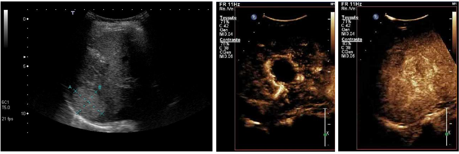
Patient history:
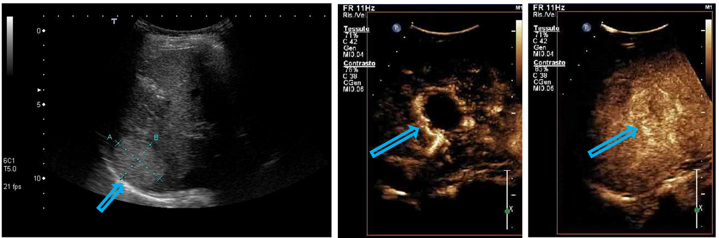
Modality: asymptomatic young patient, routine abdominal ultrasonography followed by CEUS work-up left – convex probe, B mode US; middle – contrast-enhanced ultrasonography (CEUS) after iv. microbubble administration, arterial phase; right – CEUS, late phase
Region: liver, right subcostal view
Radiologic sign: Hyperechoic, well-circumscribed hepatic mass showing peripheral nodular enhancement in the arterial phase and complete filling in the late phase.
Diagnosis: hepatic hemangioma
9.1
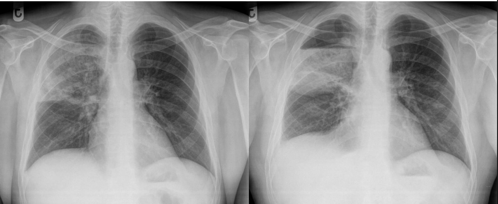
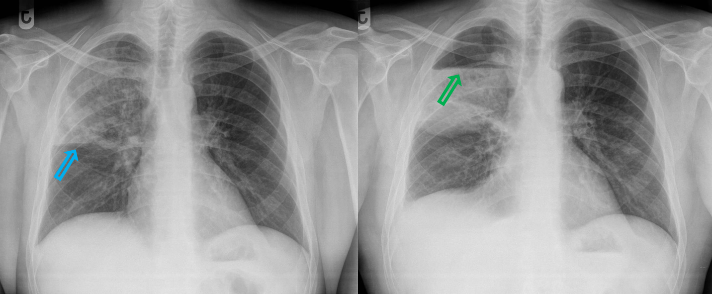
Patient history: chills, chest pain and cough with purulent sputum in an immunocompromised patient
Modality : PA chest radiograph
Region : chest, lungs
Radiologic sign : left - right-sided upper lobar consolidation right - abscess with air-fluid level two weeks later (blue arrow – horizontal fissure; green arrow – air-fluid level)
Diagnosis: lobar pneumonia in the right upper lobe, which later develops an abscess
9.2
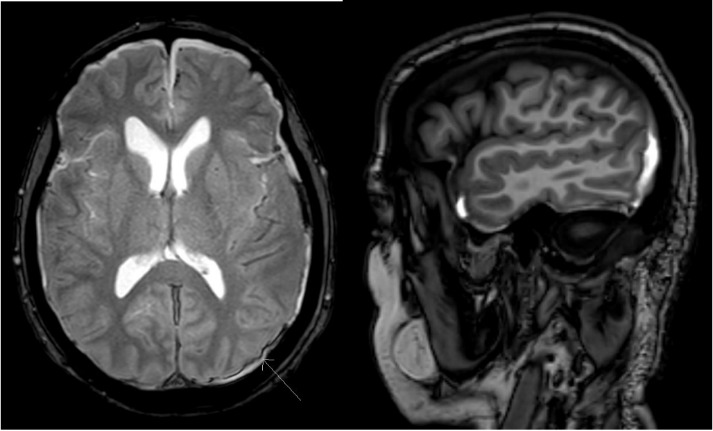
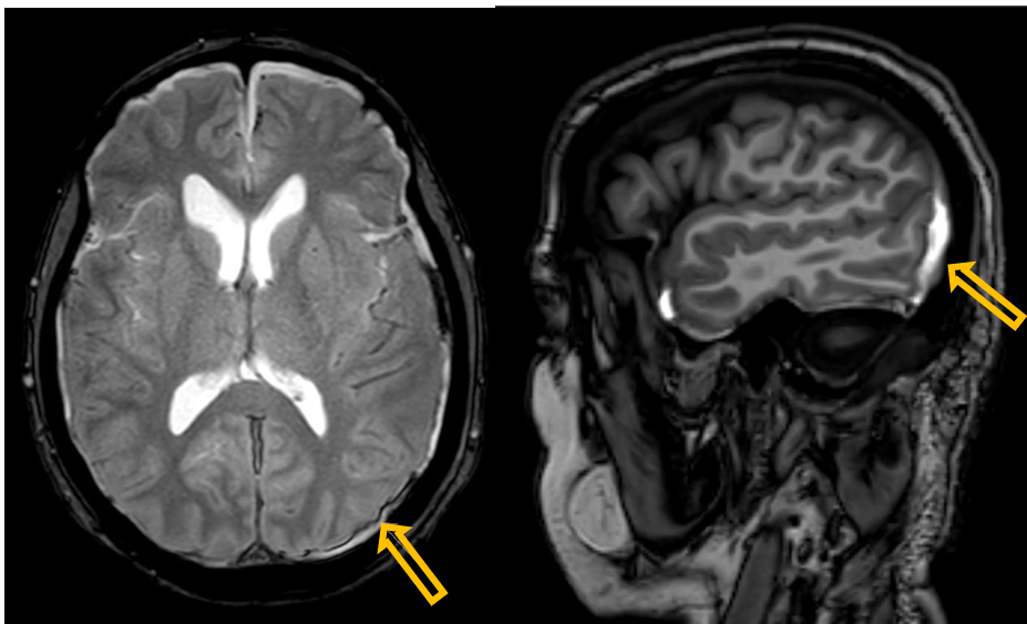
Patient history: headache after head trauma
Modality: left – T2W axial MRI; right – T1W sagittal MRI (without iv. contrast agent)
Region: brain, axial (left and middle) and sagittal (right) planes
Radiologic sign: crescent-shaped, hyperintense area in the subdural space (yellow arrow)
Diagnosis: left-sided acute parieto-occipital subdural hematoma
9.3
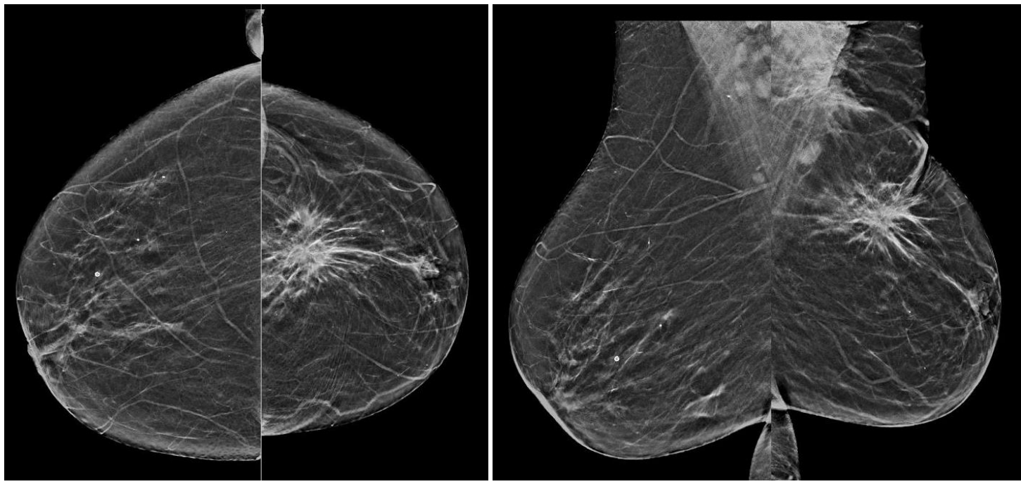
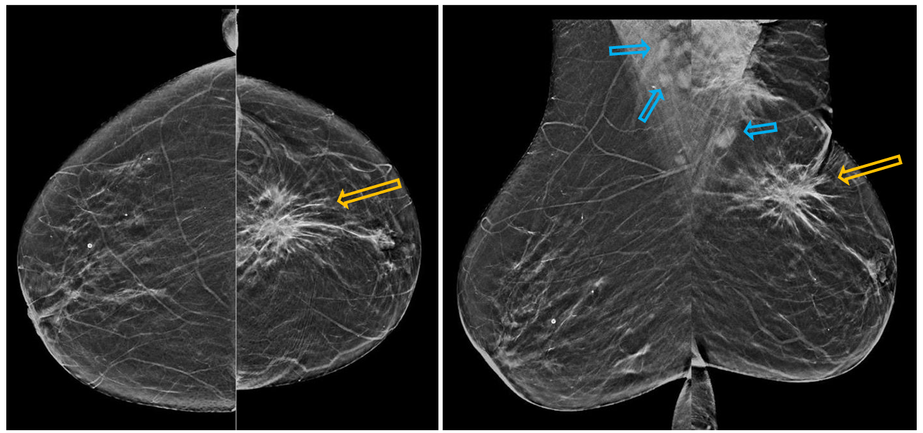
Patient history: palpable mass in the left breast in a 41-year-old female patient
Modality: mammograms of the right and left breast, low-energy beam technique (30 kV)
Region: left and right breasts, craniocaudal (CC, left images) and mediolateral oblique (MLO, right images) views
Radiologic sign: spiculated asymmetric lesion of the left breast (upper quadrants, on the border of medial and lateral quadrants). It is easier to detect lesions in adipose (fatty) breast than in dense breast. Axillary metastatic lymph nodes in the projection of the pectoral muscle (blue arrows).
Diagnosis: invasive carcinoma of the left breast, normal right breast
9.4
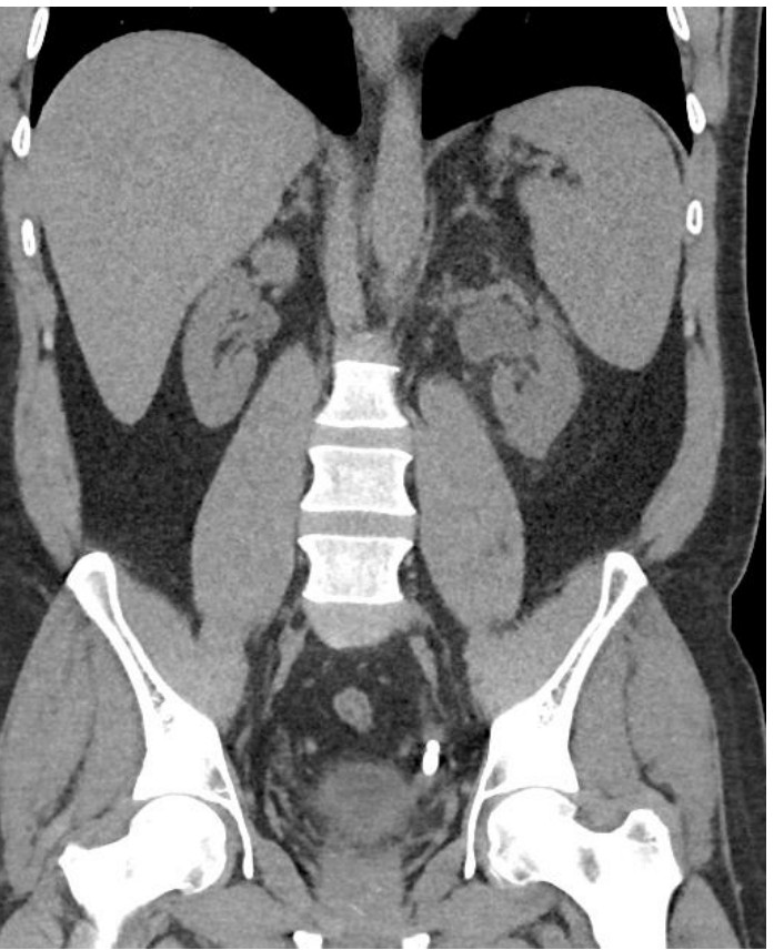
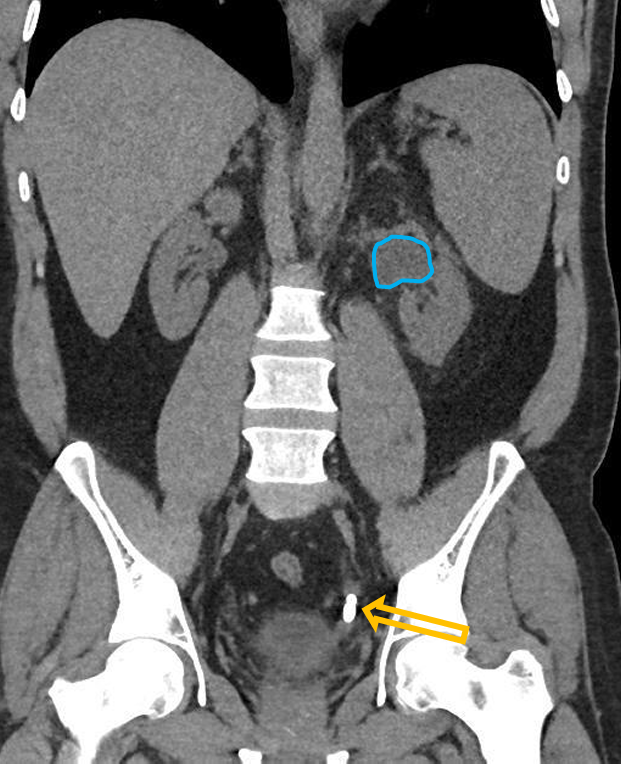
Patient history: acute severe left-sided flank pain, hematuria in a young patient
Modality: low-dose non-enhanced CT scan
Region: abdomen, coronal view
Radiologic sign: juxtavesical hyperdensity (yellow arrow, 450-500 HU) in the left distal ureter with ipsilateral hydronephrosis (blue area – dilated pyelon)
Diagnosis: obliterative juxtavesical ureter stone with hydronephrosis
9.5
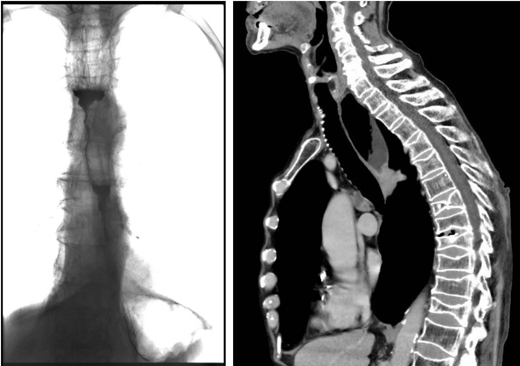
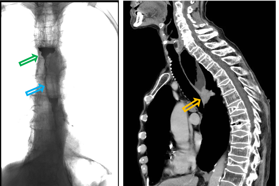
Patient history: dysphagia and weight loss in a middle-aged male patient with alcoholism
Modality: left – Barium swallow test, fluoroscopy, coronal view; right – contrast enhanced CT (iv. contrast agent), soft tissue window, sagittal view
Region: chest
Radiologic sign: contrast enhancing mass (yellow arrow) causing dilatation of the upper third of the esophagus; cranially to the mass the oral contrast agent is accumulated (green arrow), only a small amount of contrast agent passes through the stenosis (blue arrow).
Diagnosis: squamous cell carcinoma of the esophagus
9.6
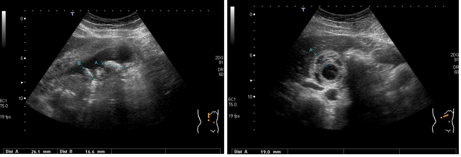
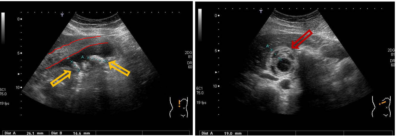
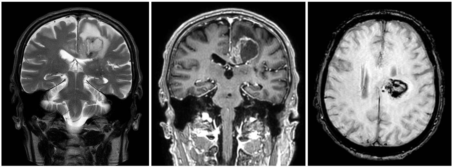
Patient history: fever, nausea, strong right upper abdominal pain Modality: B-mode gallbladder ultrasonography, convex probe (3.5–5 MHz)
Region: gallbladder, right subcostal view, longitudinal (left) and transverse (right) planes
Radiologic sign: echodense structure in the gallbladder lumen (yellow arrow) with acoustic shadowing; thick, stratified, oedematous wall (red arrow and red lines)
Diagnosis: cholecystitis with gallstone
10.1
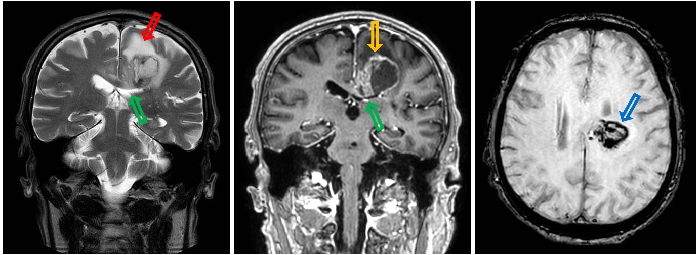
Patient history: mutism (inability to speak) and right lower extremity hemiparesis with spastic muscle tone in a 79-year-old male patient
Modality: left panel – coronal view T2W MRI; middle panel – coronal view CE T1W MRI; right panel – axial view SWI MRI
Region: brain, axial and coronal views
Radiologic sign: Intraaxial left-sided parenchymal mass in the corona radiata and corpus callosum. The lesion has large perifocal edema (hyperintensity on T2W, red arrow), rim-enhancement (T1W hyperintensity, yellow arrow), and compresses the left lateral ventricle (green arrow). Intralesional hemorrhage is observable on SWI (low signal intensity, blue arrow).
Diagnosis: glioblastoma multiforme
10.2
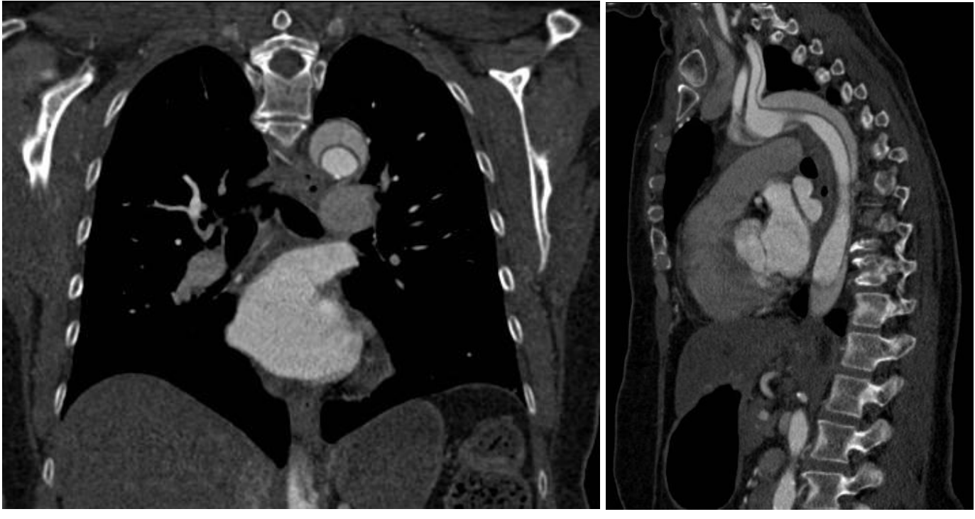
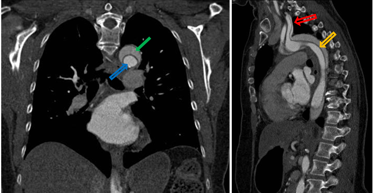
Patient history: sudden severe chest / upper back pain, shortness of breath in a middle-aged patient
Modality: aortic CT angiography;
Region: chest; left – coronal view, right – sagittal view, iv. iodine-based contrast agent
Radiologic sign: Hypodense intima flap (yellow arrow), crescent-shaped false lumen (green arrow), oval true lumen (blue arrow) in the aortic arch and descending aorta. True lumen is denser due to its higher contrast agent concentration.
Diagnosis: Stanford A aortic dissection, affecting the left subclavian artery (red arrow)
10.3
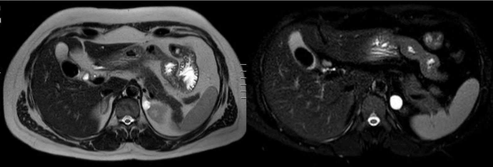
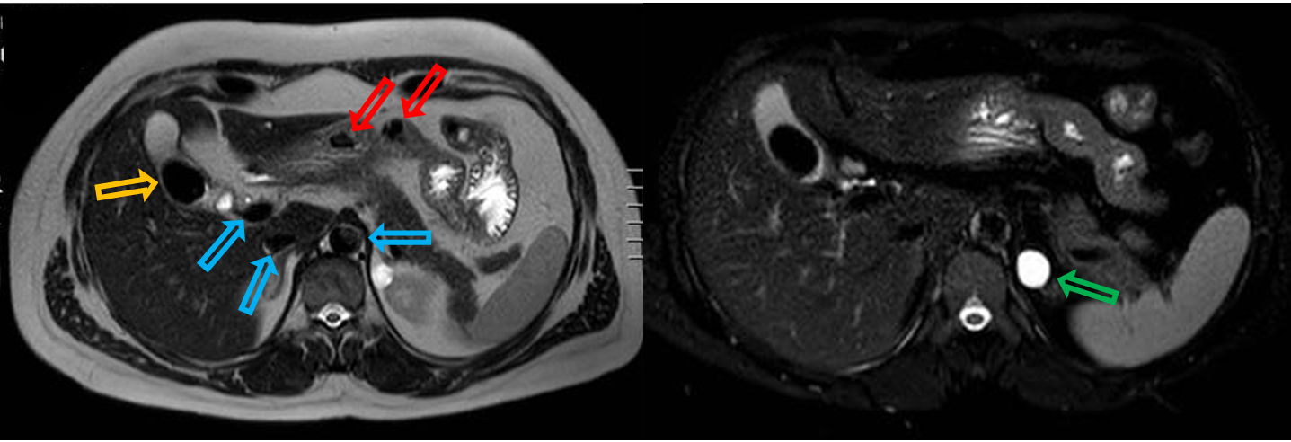
Patient history: upper abdominal MRI scan in a middle-aged patient (scanned for pelvic symptoms)
Modality: left – T2W axial MRI; right – T2W fat suppressed axial MRI (without iv. contrast administration)
Region: upper abdomoen
Radiologic sign: circumscribed signal loss in the gallbladder (yellow arrow); hyperintense well circumscribed lesion in the upper pole of the upper abdomen left kidney (green arrow). Signal loss detected in several additional places: flow-void (in the aorta, inferior vena cava, portal vein, blue arrows); and gas bubbles in the intestines (red arrows)
Diagnosis: cholecystolithiasis, kidney cyst
10.4
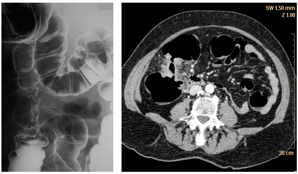
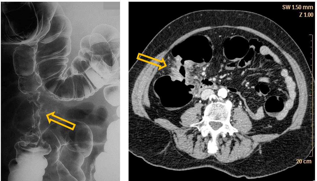
Patient history: change in bowel habits and weight loss
Modality: left - Barium enema study, double-contrast method (barium and air); right – contrast-enhanced CT, venous phase, iv. iodine-based contrast agent
Region: Abdomen, axial view
Radiologic sign: Annular irregular stenosis and bowel wall thickening (apple-core sign), tumorous infiltration of the surrounding fat (increased density)
Diagnosis: colon adenocarcinoma
10.5
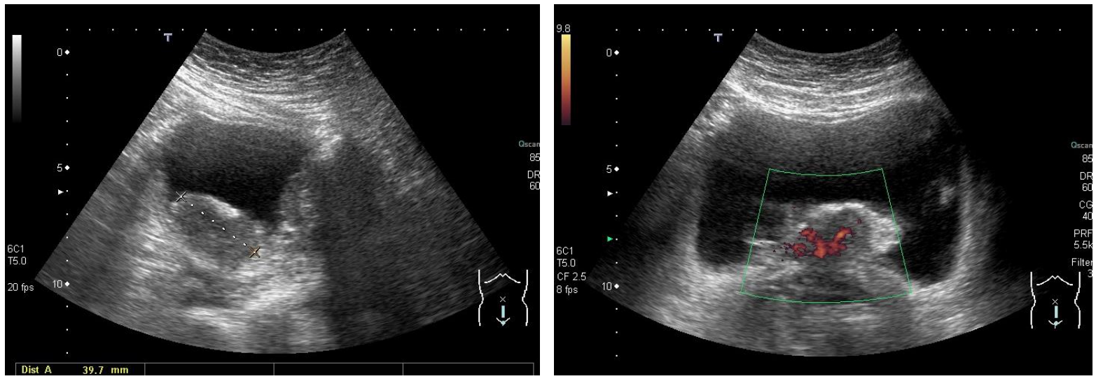
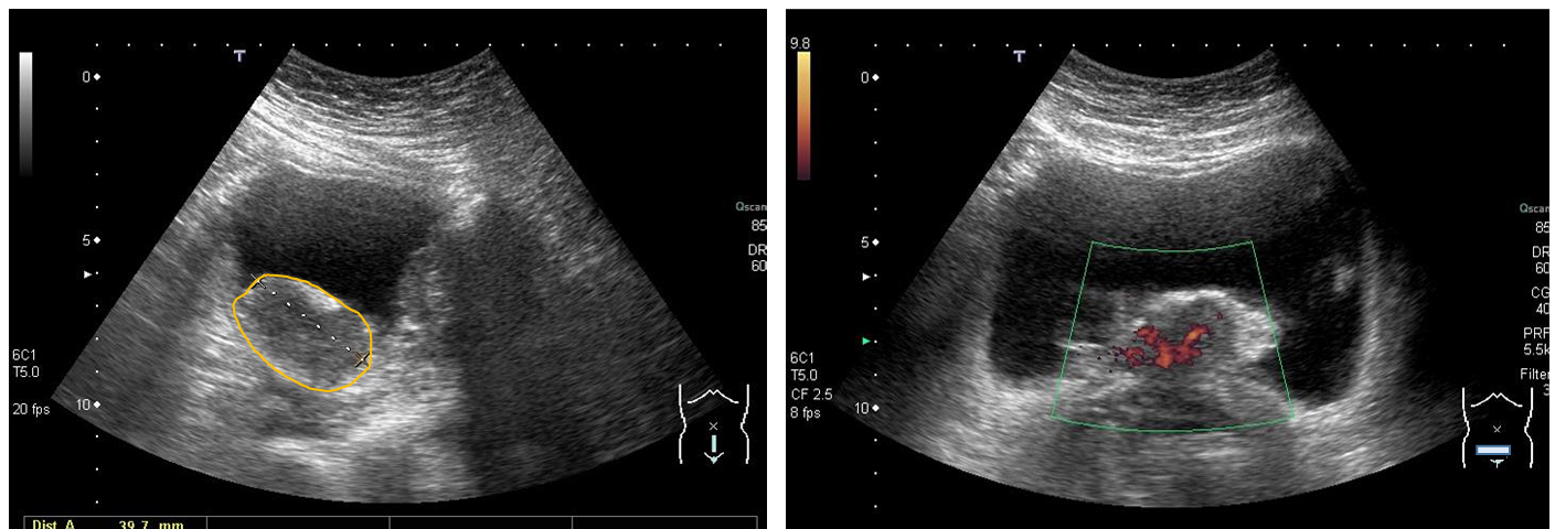
Patient history:elderly patient with macroscopic hematuria
Modality: Ultrasonography of the urinary bladder with convex probe (3.5–5 MHz), B mode (left image) and Power Doppler mode (right image) Region: pelvis, sagittal and transverse views
Radiological sign: Lobulated, hypoechoic, vascularized (Doppler-sign detectable) solid mass, which protrudes into the lumen of the urinary bladder. The bladder contains anechoic urine.
Diagnosis:urinary bladder transitional cell carcinoma
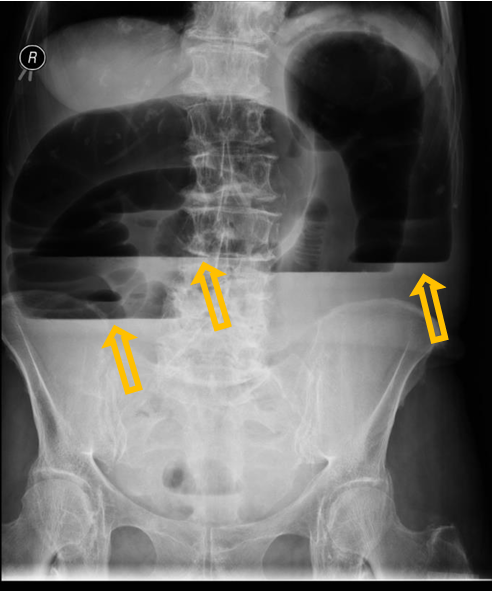
10.6
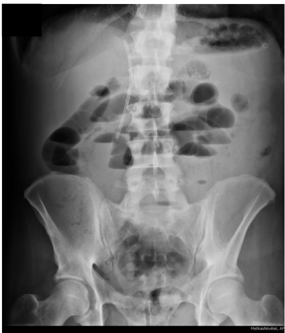
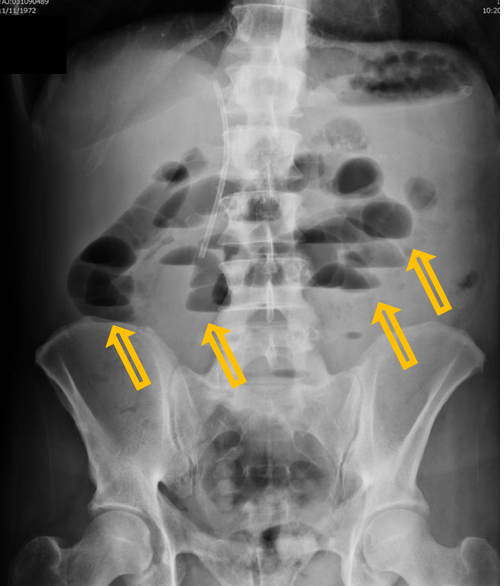
Patient history: vomiting and acute abdominal pain, swelling in the groin region
Modality: abdominal radiograph, erect position
Region: abdomen and pelvis
Radiologic sign: Several, distended (2-3 cm in diameter), meteoristic small bowel loops in the upper abdomen containing air-fluid levels (yellow arrows). The colon contains less gas than normal. A stent is visible in the extrahepatic bile ducts.
Diagnosis: mechanical small bowel ileus caused by incarcerated inguinal hernia
11.1
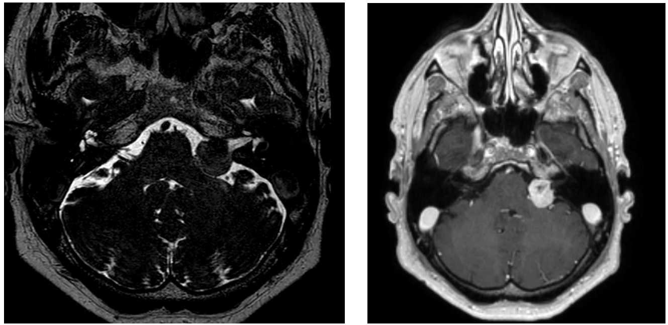
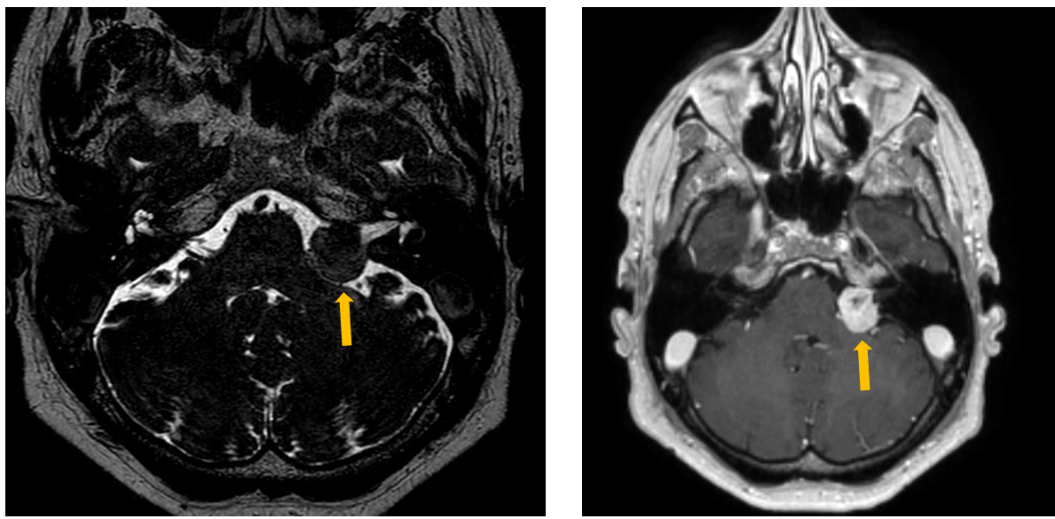
Patient history: neural hearing loss, that developed over months
Modality: left – T2 Drive MRI (non-contrast), right – post-contrast T1W MRI (after iv. gadolinium administration)
Region: brain, axial view
Radiologic sign: well-circumscribed, contrast-enhancing mass in the left cerebellopontine angle (yellow arrows)
Diagnosis: vestibular schwannoma (acoustic neuroma)
11.2
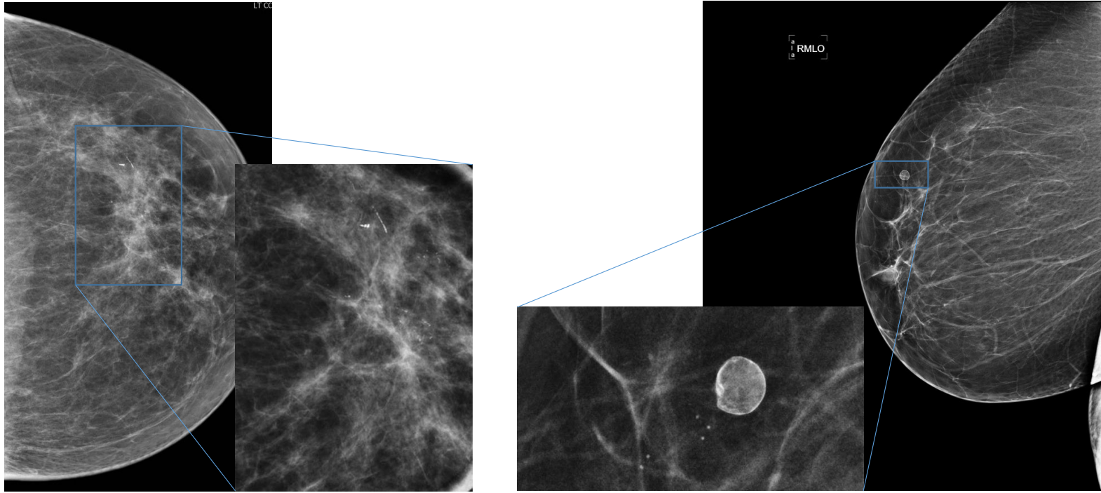
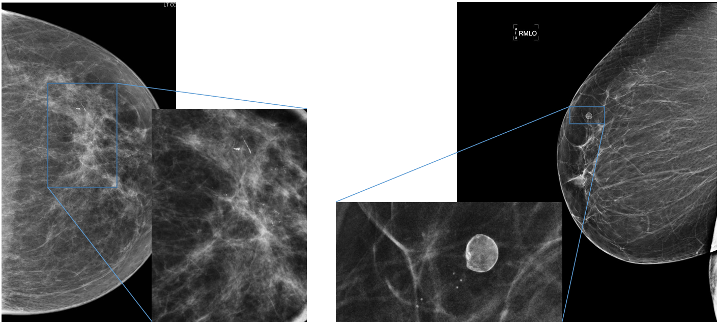
Patient history: screening mammograms in asymptomatic patients
Modality: mammograms of the left breast in craniocaudal (CC) view (left panels) and of the right breast in mediolateral oblique (MLO) view (right panels), low-energy beam technique (30 kV) + magnified compression images
Region: breasts
Radiologic sign: left panels – polymorph microcalcification; right panels – well circumscribed, eggshell calcification
Diagnosis: invasive ductal carcinoma / benign oil cyst
11.3
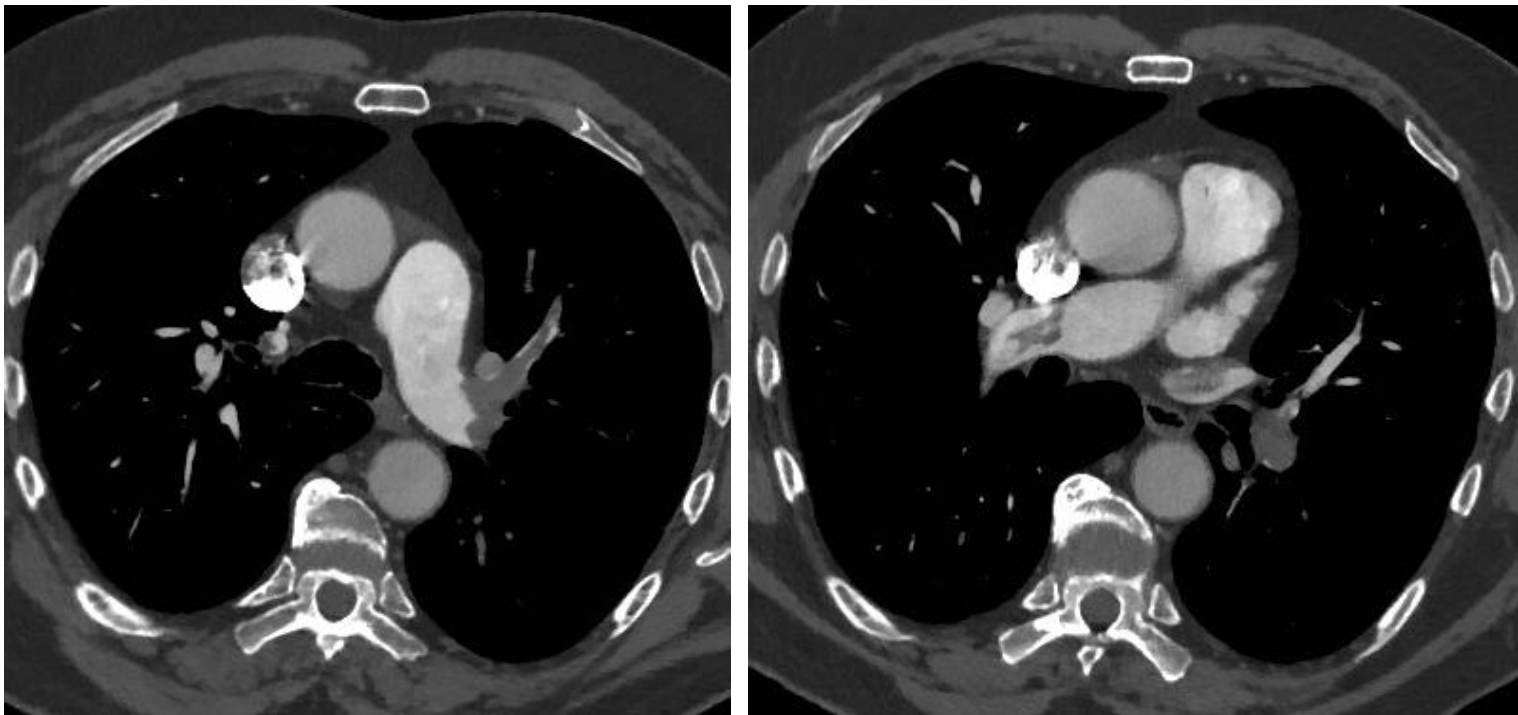
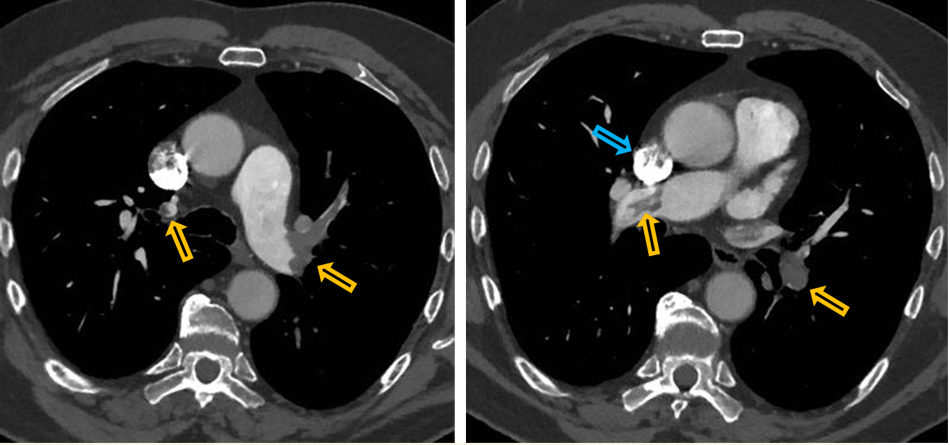
Patient history: elevated D-dimer, dyspnoe and heart palpitations in a female patient after cesarean section
Modality: Pulmonary CT-angiography, iv. iodine-based contrast agent
Region: chest, axial views at the level of the pulmonary bifurcation
Radiologic sign: Large hypodense filling defects in the pulmonary arteries on both sides (yellow arrows). Highest density can be observed in the SVC due to high contrast agent concentration, where it causes artefact (blue arrow).
Diagnosis: bilateral pulmonary embolism
11.4
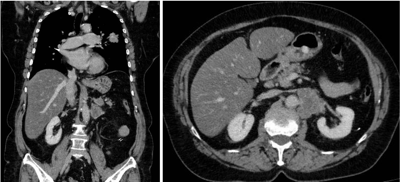
Patient history: staging CT after positive chest x-ray screening
Modality: contrast-enhanced CT scan, venous phase; left – coronal reconstruction; right – axial view
Region: chest and upper abdomen
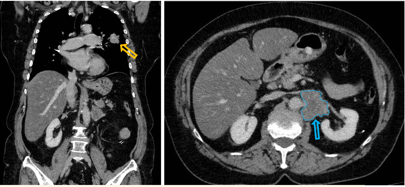
Radiologic sign: lobulated, contrast-enhancing mass in the lower lobe of the left lung (yellow arrow), contrast enhancing solid mass in the left adrenal gland with similar morphology (blue arrow)
Diagnosis: primary non-small cell lung cancer with metastasis in the left adrenal gland
11.5
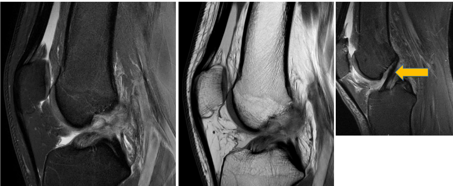
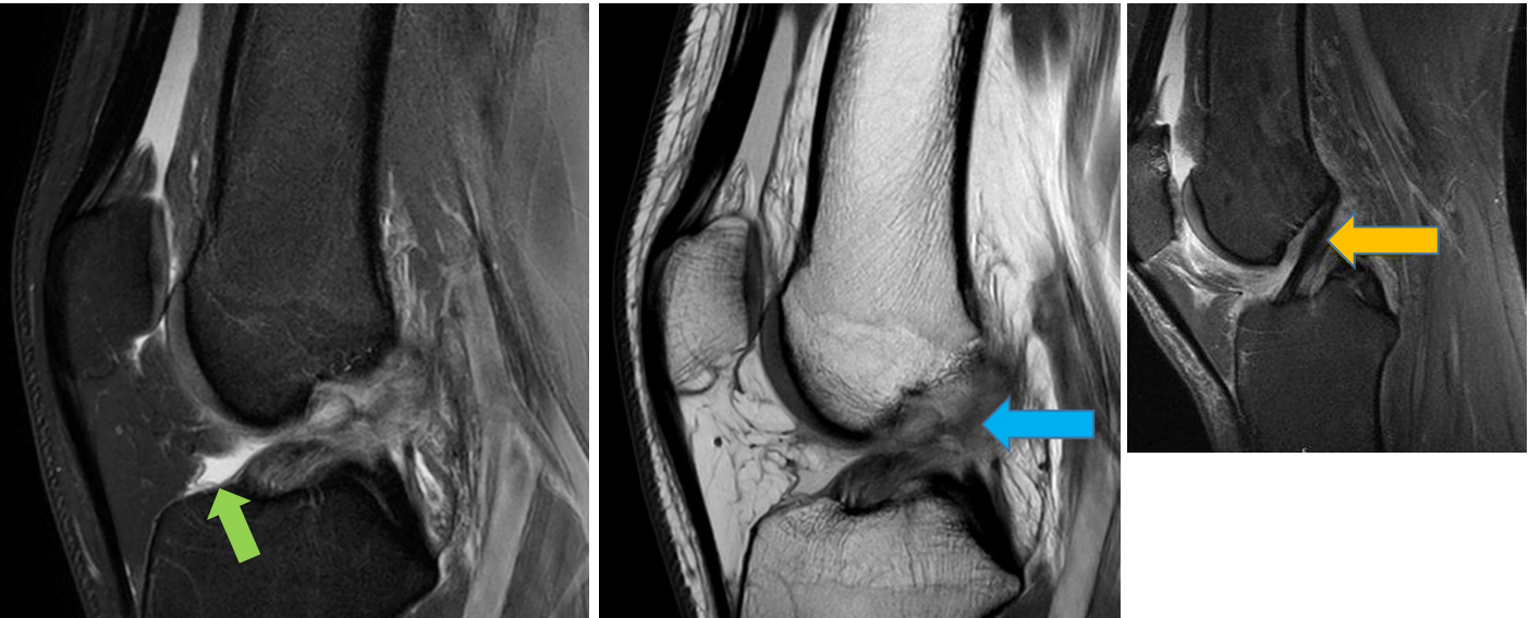
Patient history: young patient with unstable knee after ski accident
Modality: left– fatsat Proton Density (PD) MRI (sagittal view); middle – PD MRI (sagittal view); right (smaller) image – normal anatomy (normal, hypointense anterior crucial ligament marked with yellow arrow, visible through its whole length); without iv. contrast administration
Region: knee
Radiologic sign: normal anterior crucial ligament cannot be detected (blue arrow), small amount of hyperintense synovial fluid (green arrow)
Diagnosis: tear of the anterior crucial ligament caused by sport injury
11.6
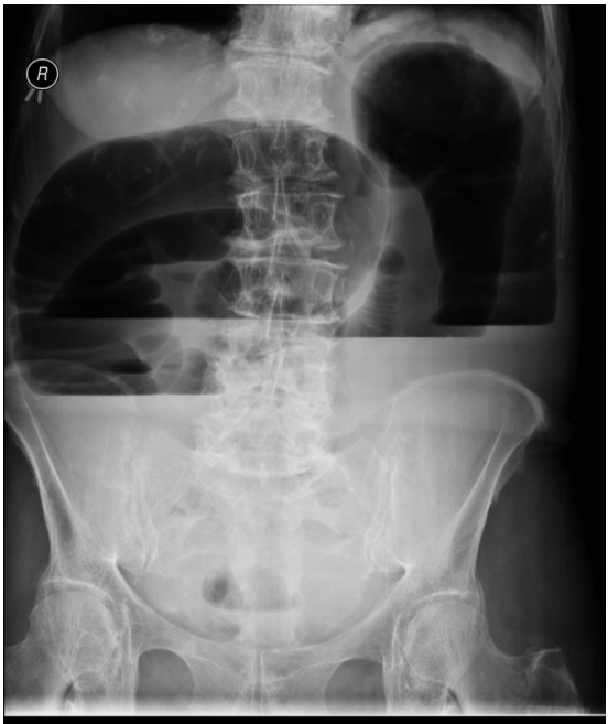
Patient history: pelvic tumor in history, at present severe abdominal pain and cramping in an elderly female patient
Modality: abdominal radiograph, erect position
Region: abdomen and pelvis Radiologic sign: Distended, meteoristic ascending, transverse and descending colon with several large air-fluid levels up to 10-15 cm in diameter (yellow arrows). Small bowels containing less gas than normal, few non distented jejunum loops visible epigastically.
Diagnosis: mechanical large intestine ileus caused by ovarial cancer infiltrating the sigmoid colon
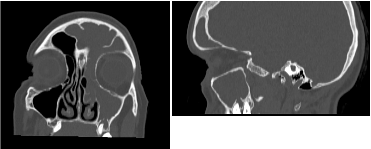
12.1
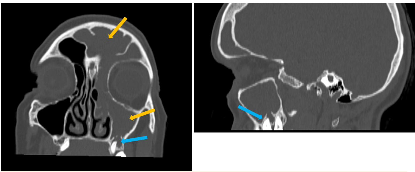
Patient history: chronic headache and cough
Modality: low-dose non-contrast CT scan, coronal and sagittal reconstructions, bone window
Region: maxilla, paranasal sinuses
Radiologic sign: mucosal thickening and fluid collection in the left frontal and maxillary sinus (0-20 HU) (yellow arrows), partially filled ethmoidal cells; periapical, radiolucent area can be observed around the left upper molar tooth (blue arrow) ; normal, air-containing (-1000 HU) right maxillary sinus
Diagnosis: odontogenic sinusitis
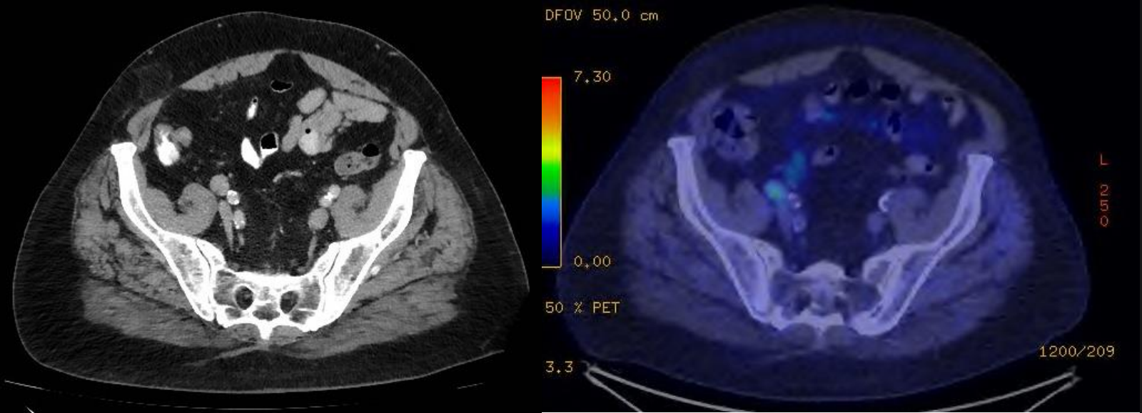
12.2
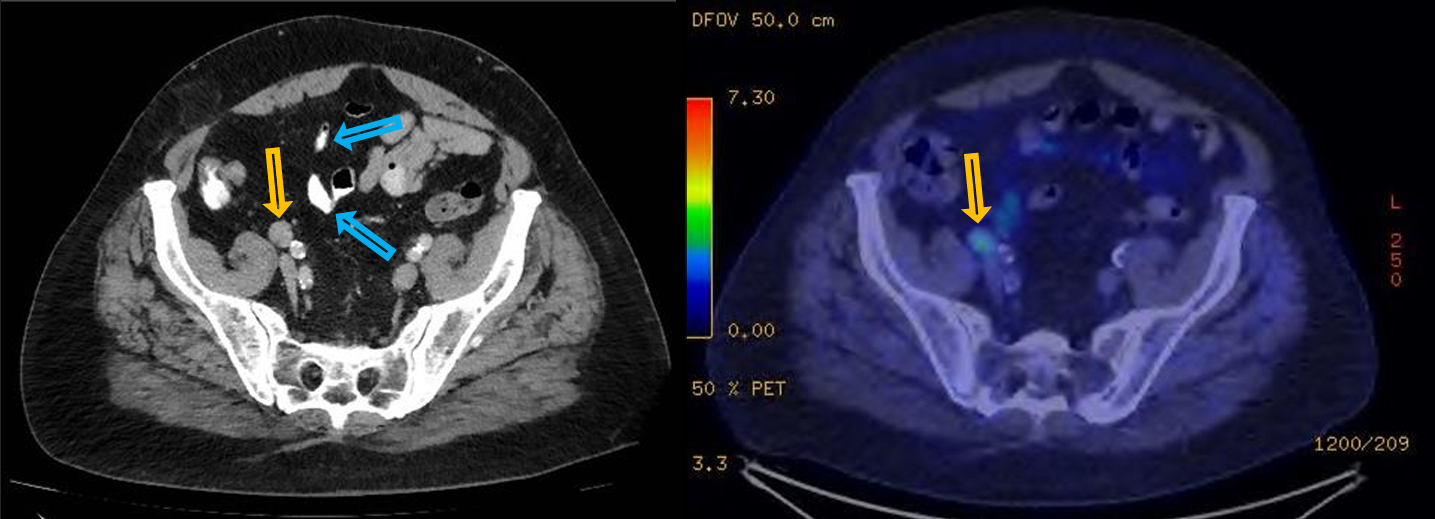
Patient history: follow-up study in a patient with colorectal tumor
Modality: left – contrast-enhanced CT scan, venous phase, soft tissue window (iv. and oral iodine-based contrast agent); right – PET-CT, iv. 18-FDG (fluorodeoxyglucose) radiotracer
Region: pelvis, axial views
Radiologic sign: Pathological (16 mm shorter diameter) lymph node in the right parailiac region (yellow arrow). In the same localization, increased FDGuptake can be observed on PET-CT scan 10 days later (yellow arrow). Oral contrast agent containing bowel loops are marked with blue arrows.
Diagnosis: FDG-avid, pathological lymph node, suspicious for metastasis.
12.3
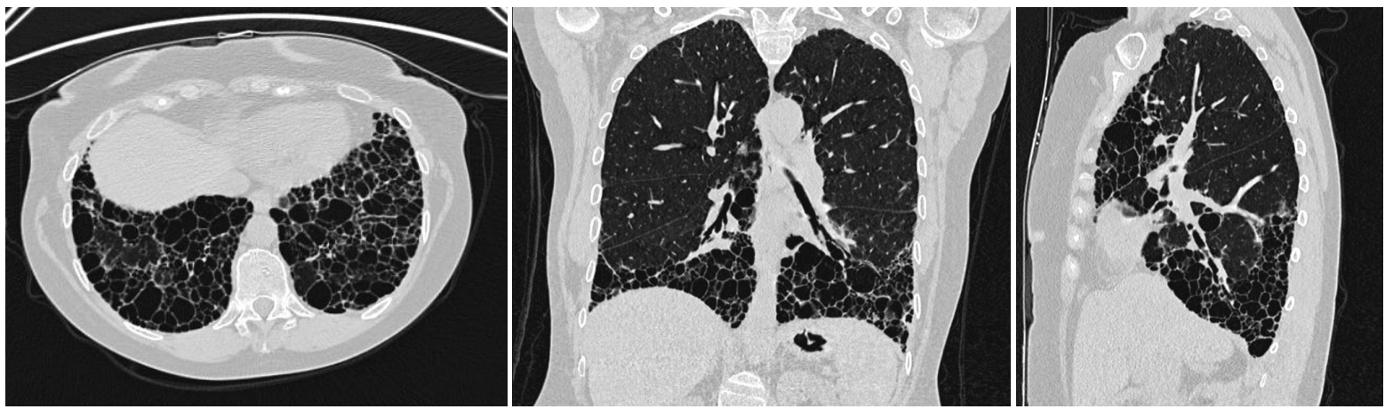
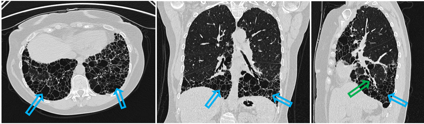
Patient history: 56-year-old female patient with systemic lupus erythematosus with moderate restrictive ventilation problems
Modality: HRCT of the lung (non-contrast study), lung window
Region: chest, lung, axial-coronal-sagittal views
Radiologic sign: view bilateral dorsobasal predominant honeycombing and reticulation (blue arrows), traction bronchiectasis and thickened bronchial wall (green arrow)
Diagnosis: large-area interstitial lung disease, UIP (usual interstitial pneumonia) pattern
12.4 upper
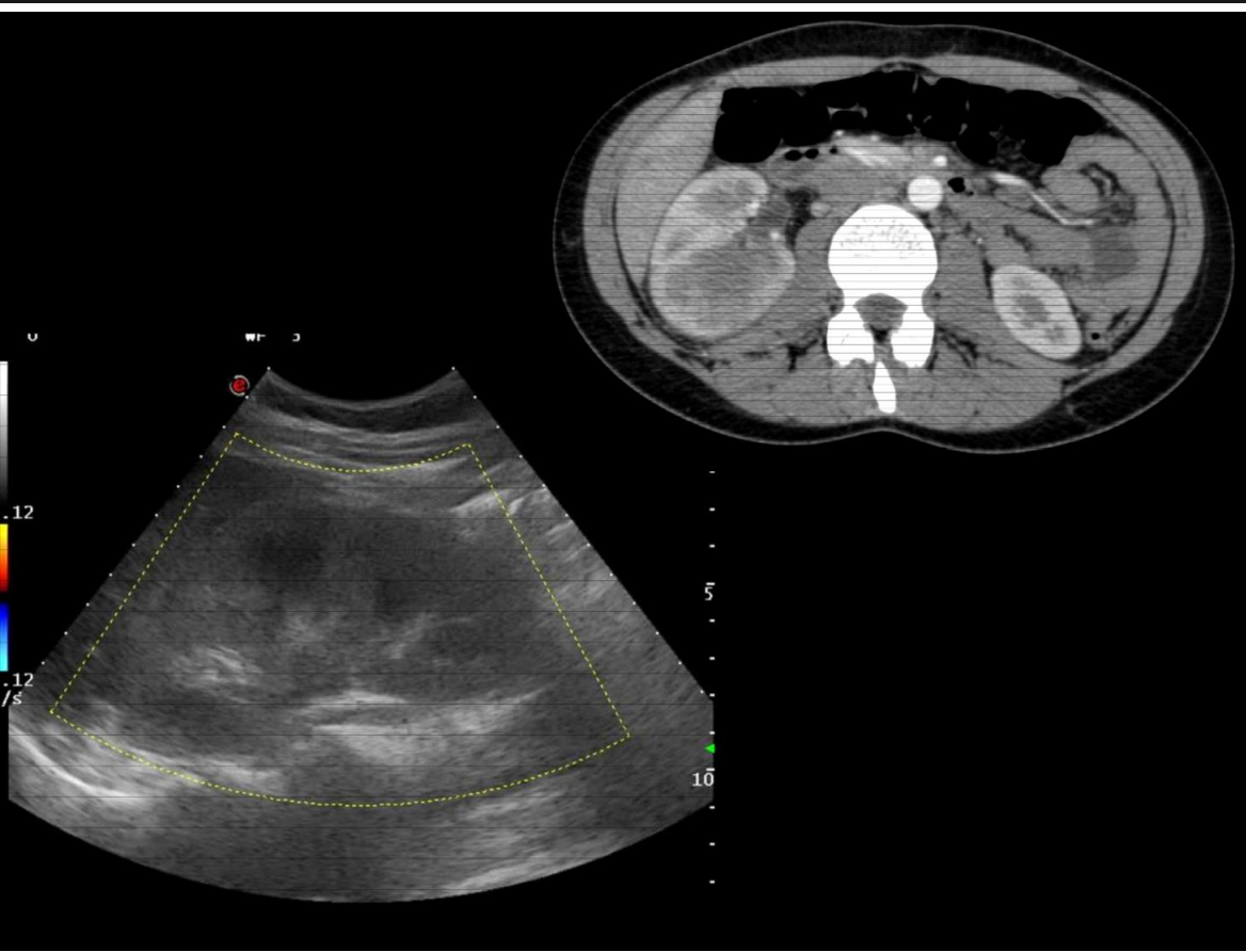
Modality: contrast-enhanced CT; arterial phase
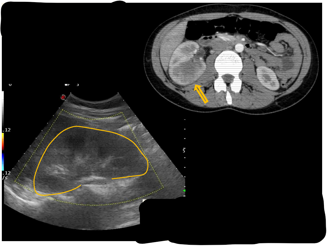
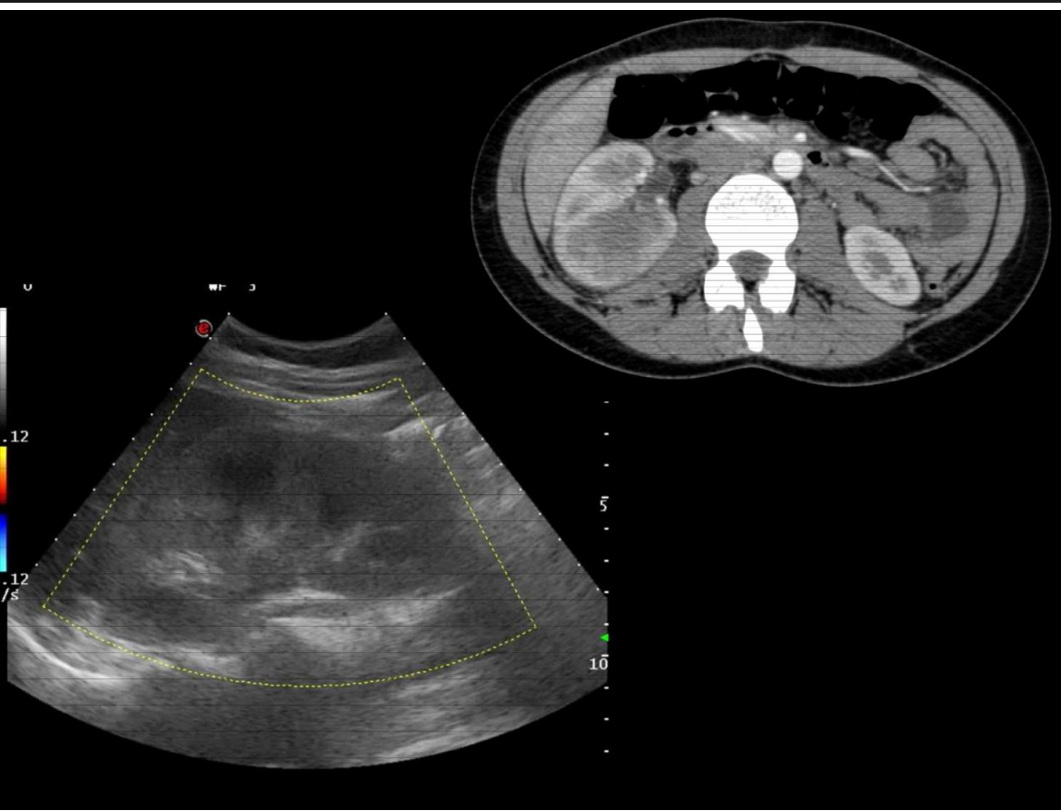
Region: abdomen, axial view
Radiologic sign: inhomogenic, enlarged right kidney with patchy hypoenhancement
Diagnosis: acute pyelonephritis (right kidney)
12.4 lower
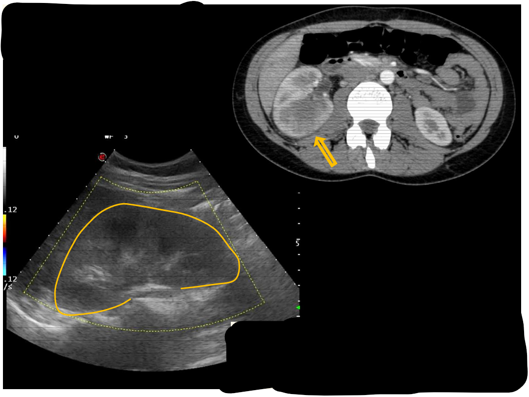
Patient history: fever, flank pain and bacteriuria Modality: kidney ultrasound with convex probe (3.5–5 MHz), B-mode & color Doppler
Region: abdomen, longitudinal view of the right kidney
Radiologic sign: enlarged right kidney shows ill-marginated central echo group and parenchymal hypovascularization.
Diagnosis: acute pyelonephritis
12.5
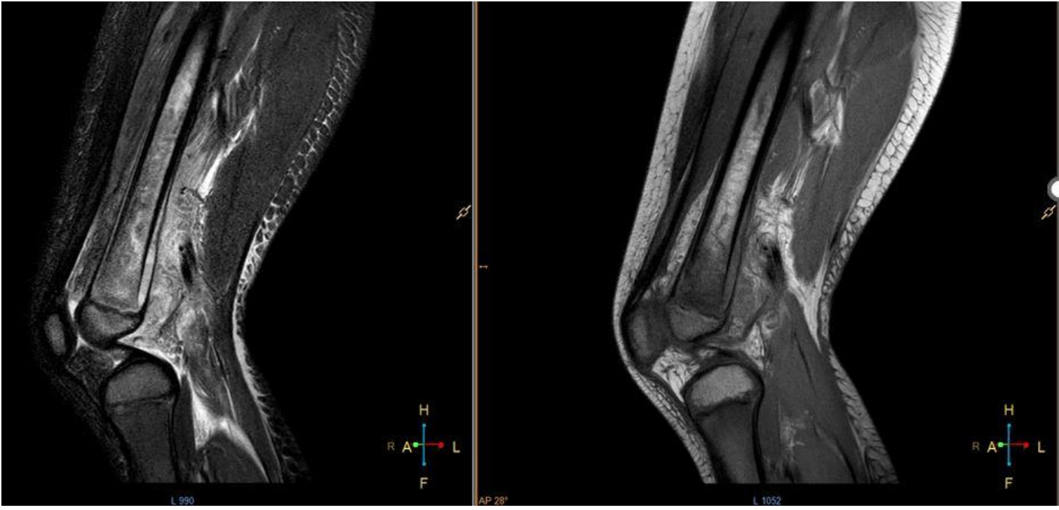
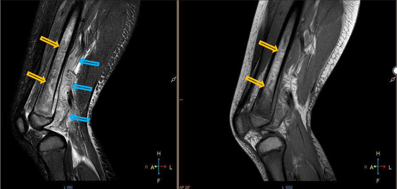
Patient history: swollen and painful area above the knee in a young adult
Modality: left – fatsat Proton Density (PD) MRI; right – PD MRI
Region: knee, sagittal views
Radiologic sign: Bone marrow hyperintensity detected in the middle and distal diaphysis and in the distal metaphysis of the femur due to edema (yellow arrows), significant periosteal reaction and edema in the sorrounding soft tissue (blue arrows – extensive hyperintensity in the sorrounding soft tissue)
Diagnosis: acute osteomyelitis of the femur
13.1
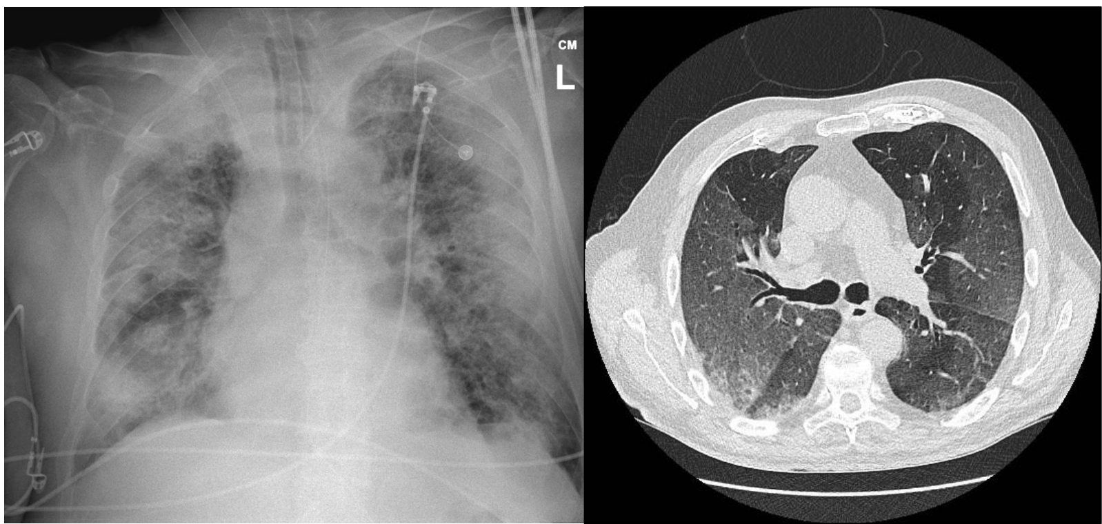
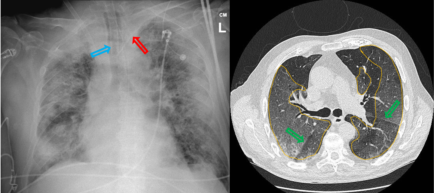
Patient history: acutely developed fever, shortness of breath, cough and fatigue in a 72-year-old patient
Modality: left– AP prone chest radiograph right–non-enhanced chest CT,axial plane
Region: chest
Radiologic sign: peripheral ground-glass opacity in all lung lobes (“ground-glass opacity” – yellow area) and consolidations; oblique fissures (green arrow); foreign bodies: endotracheal tube (blue arrow), ECG electrodes, left v. subclavia catheter (red arrow)
Diagnosis : Covid 19 pneumonia involving more than 80% of the lung parenchyma, CO-RADS 5
13.2
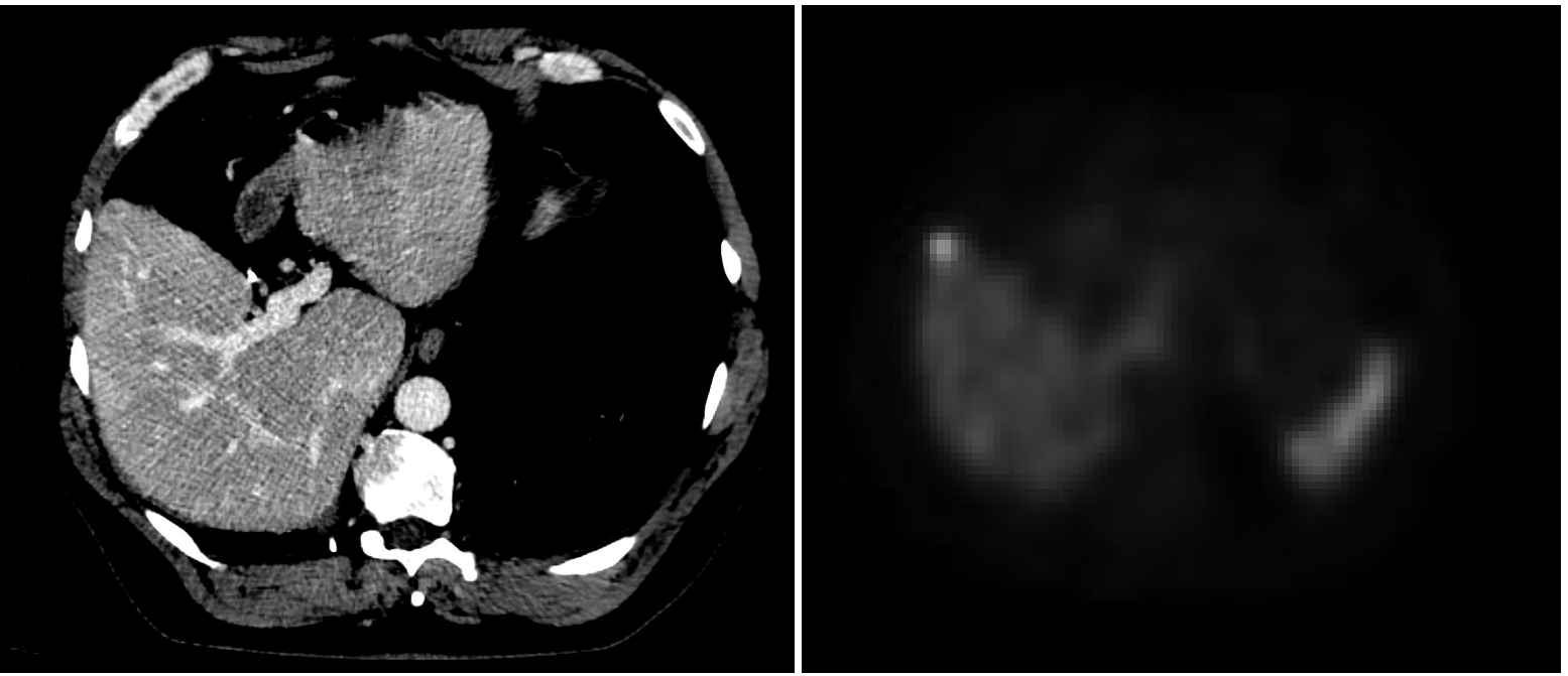
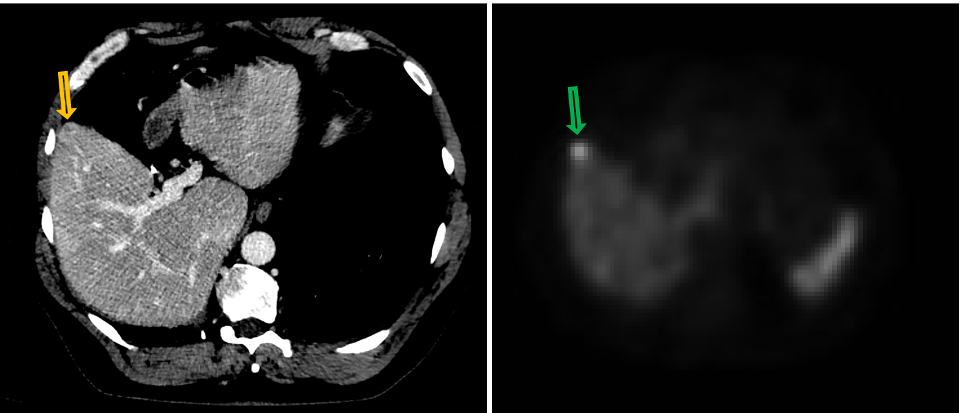
Patient history: follow-up studies of a patient under treatment of small bowel neuroendocrine tumor
Modality: left–contrast enhanced venous phase, soft tissue window (iv. iodine-based contrast agent); right– SPECT (applied radiopharmacon: Tektrotyd Tc-99m)
Region: upper abdomen, axial views
Radiologic sign: Small-sized, intensively enhancing ring-like, circumscribed lesion that can be hardly suspected in the right lobe of the liver (yellow arrow). At the same location SPECT shows increased radiopharmacon uptake (green arrow).
Diagnosis: liver metastasis of small bowel neuroendocrine tumor
13.3
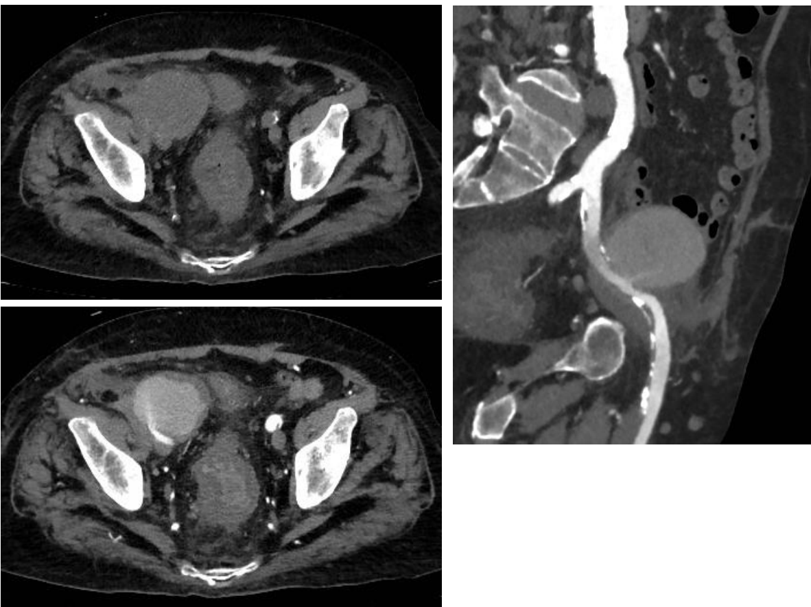
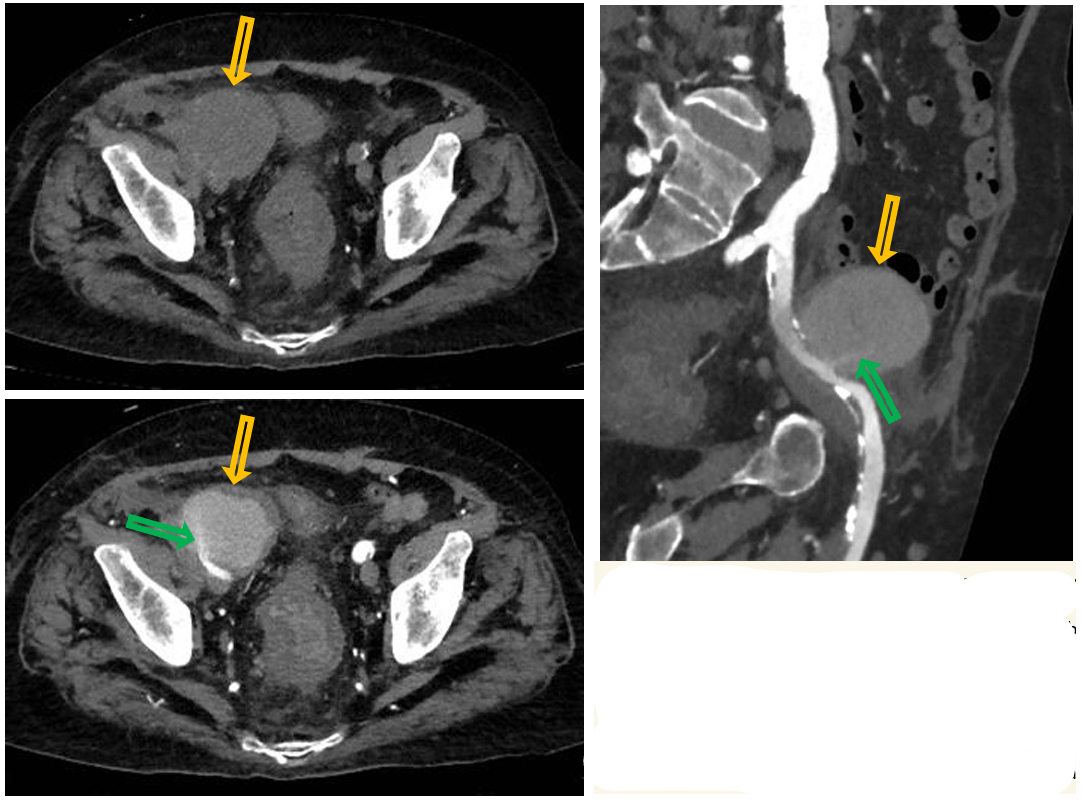
Patient history: elderly patient with end-stage renal disease, transplant kidney removal surgery a few hours before, large hematoma on US
Modality: left upper and lower images – non-enhanced abdomen and pelvis CT and CTA, axial plane, soft tissue window (iv. iodine-based contrast agent); right – CTA MPR (multiplanar reconstruction)
Region: pelvis
Radiologic sign: Mass along the right external iliac artery with an approximate noncontrast density of 25 HU, the density measured on CTA is around 100 HU. As seen on CTA, the contrast agent is flowing into the lesion in a narrow lane (green arrow).
Diagnosis: post-surgical right external iliac artery pseudoaneurysm
13.4
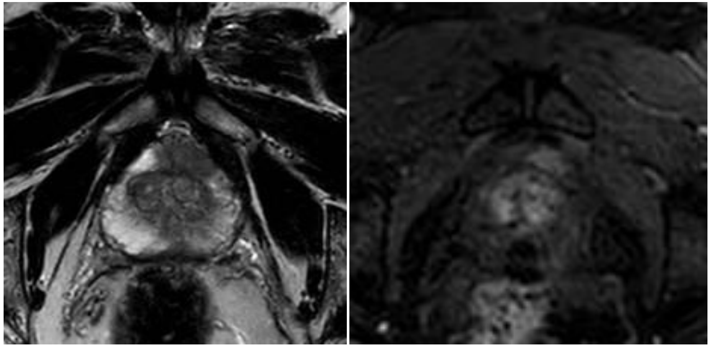
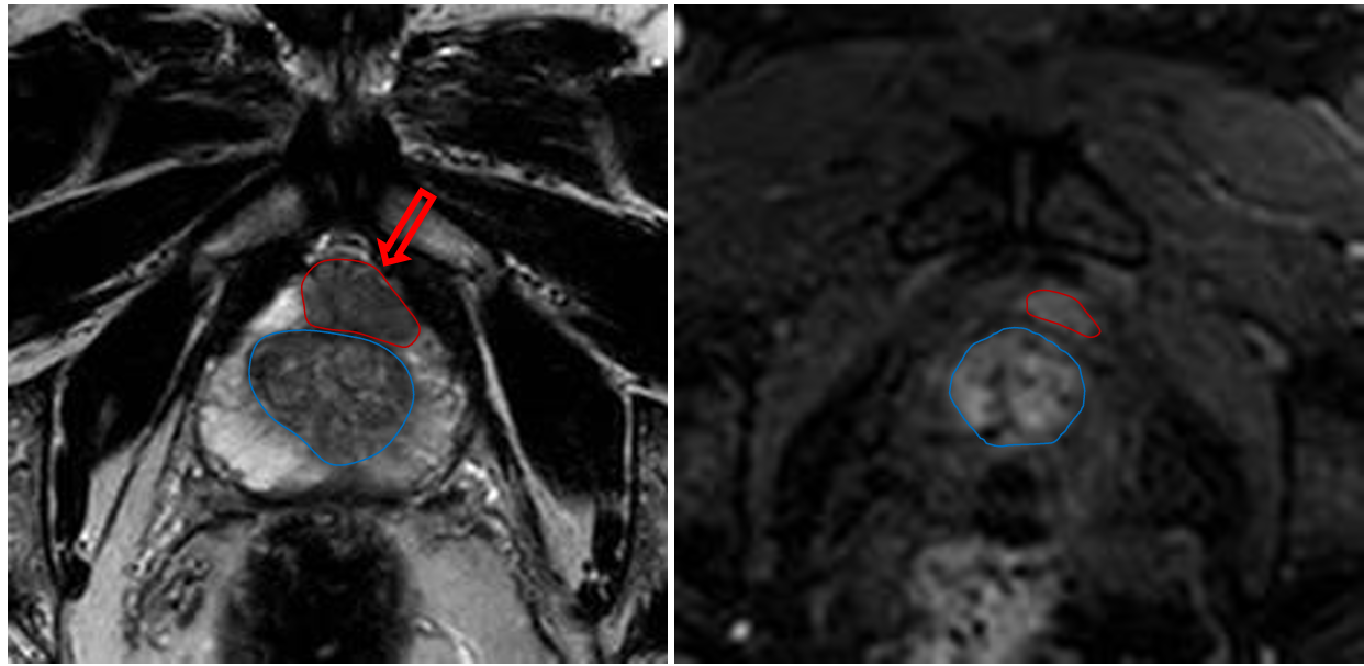
Patient history: elevated PSA level and positive transrectal ultrasonography study in an elderly patient
Modality : T2W MRI and dynamic contrast-enhanced MRI of the prostate after iv. gadolinium contrast agent administration
Region : pelvis, axial views
Radiologic sign: T2 hypointensity in the peripheral zone (left panel); intensive contrast-enhancing mass in the same area (signed with red); normal transitional zone (signed with blue)
Diagnosis: prostate adenocarcinoma
13.5
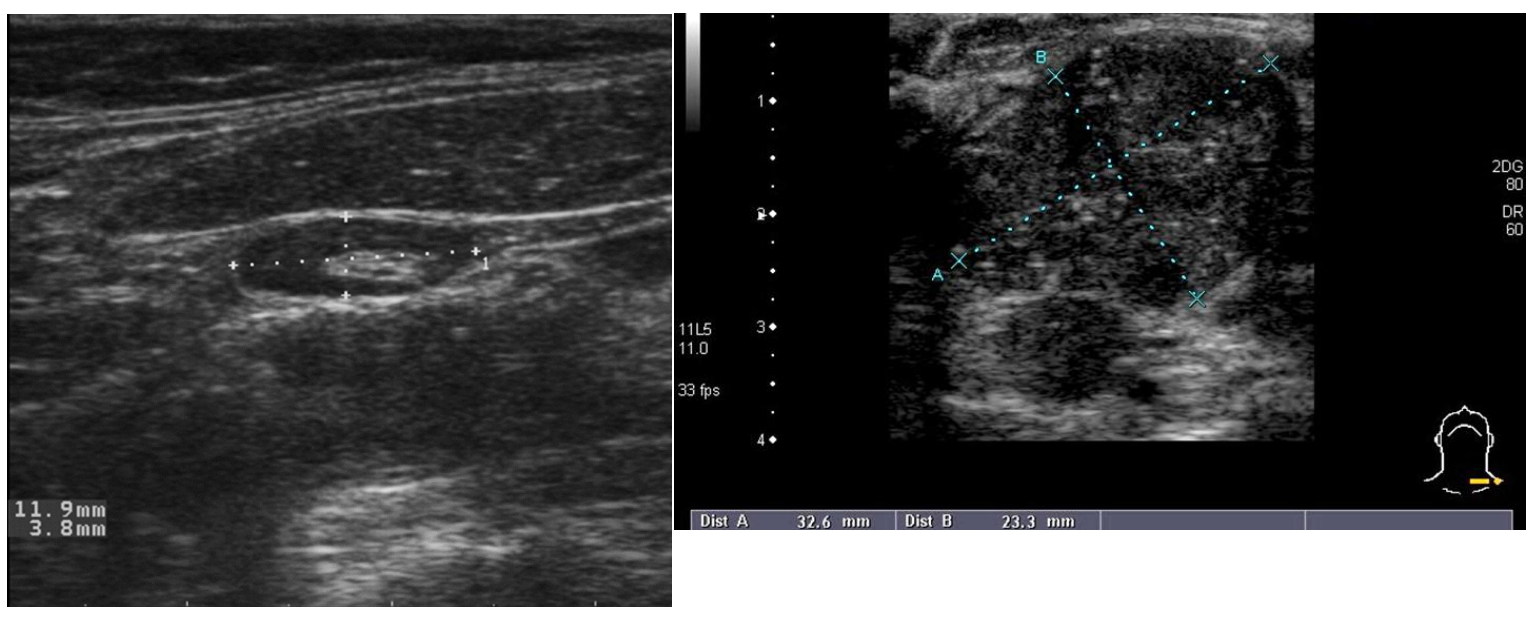
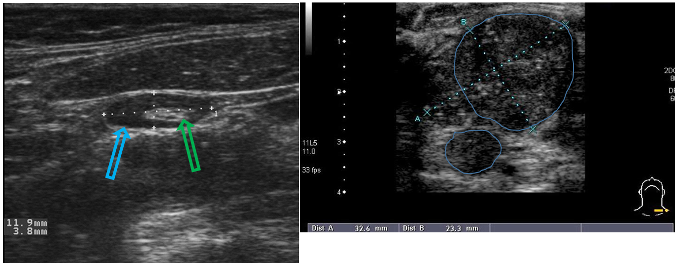
Patient history: right image – palpable left supraclavicular mass in a middle-aged patient with pancreatic adenocarcinoma (left panel – asymptomatic)
Modality: ultrasound, linear high frequency transducer (7,5-12 MHz)
Region: superficial neck soft tissues
Radiologic sign: structure), larger than 3 cm left image – lymph node with normal size and morphology (hyperechoic, fatty hilum and - green arrow; hypoechoic cortex - blue arrow), short diameter is less than 10 mm; right image – enlarged lymph node conglomerate with pathologic appearance (hypoechoic, round, loss of hilar
Diagnosis: normal (left) and metastatic (right) lymph nodes (later proven to be a metastasis from pancreatic adenocarcinoma)
13.6
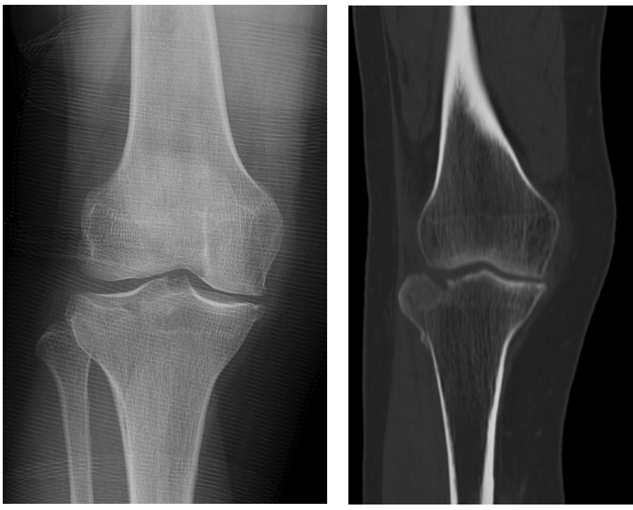
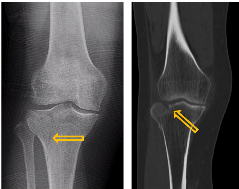
Patient history: knee trauma
Modality: left image – AP knee radiograph; right image – noncontrast CT, coronal plane
Region: right knee
Radiologic sign: radiolucent fracture lines, interruption of the anatomic continuity of bone cortex
Diagnosis: acute fracture of tibial plateau
14.1
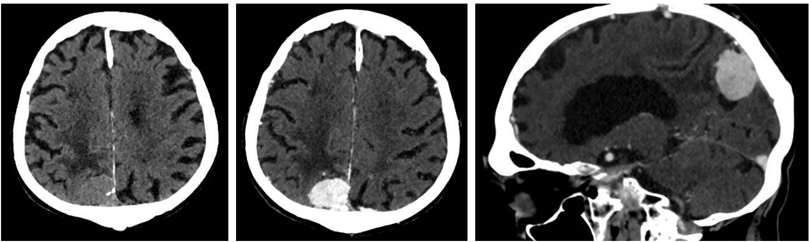
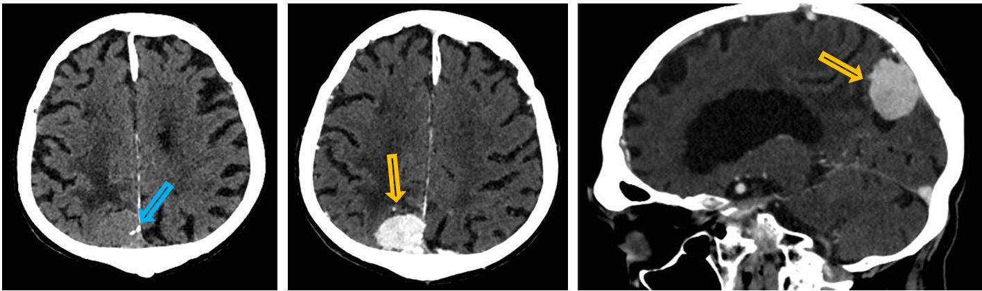
Patient history: epileptic seizure in a middle-aged patient with non-MR-compatible pacemaker Modality: left image – noncontrast CT, axial plane; middle and right images – contrast enhanced CT, axial and sagittal plane (iv. iodine-based contrast agent)
Region: brain
Radiologic sign: Right parietal extra-axial mass with small hyperdense calcifications (blue arrow) displaying intensive contrast-enhancement (yellow arrow). The mass exerts external compression on the brain parenchyma.
Diagnosis: meningioma (most common extra-axial solid mass)
14.2
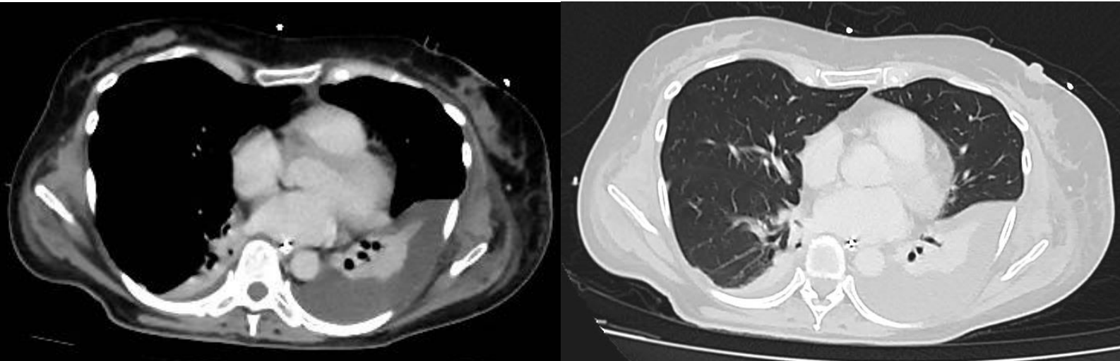
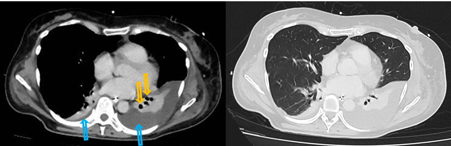
Patient history: postoperative CT scan after abdominal surgery
Modality : left – contrast-enhanced CT, soft tissue window; right – contrast-enhanced CT, lung window
Region: chest, axial planes
Radiologic sign: Minimal pleural effusion on the right side with an approximate density of 0 HU (blue arrow). 3 cm-wide pleural effusion on the left side (blue arrow), left lower lobe is devoid of air, air bronchograms can be seen (yellow arrows).
Diagnosis: bilateral hydrothorax with concomitant left lower lobe atelectasis
14.3
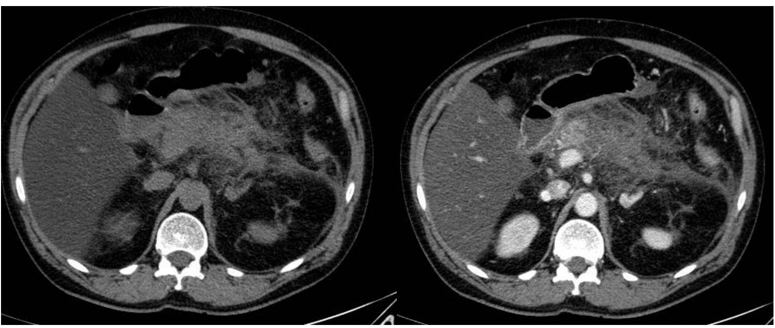
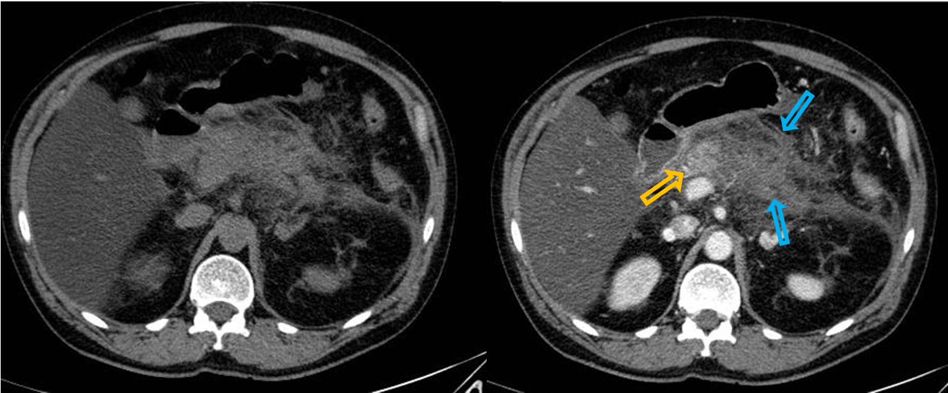
Patient history: fever, intense upper abdominal pain radiating to the back, massively elevated amylase and lipase levels
Modality: left – Noncontrast CT, right – Contast-enhanced CT, arterial phase, iv. iodine-based contrast agent
Region: upper abdomen
Radiologic sign: Enlarged pancreas with indistinct margins (blue arrow); enhancing pancreatic head (yellow arow) reflects living tissue, while hypodens, non-enhancing body and tail suggests necrosis. Increased density of the surrounding mesenterial fat due to fat necrosis; decreased liver density on non-contrast series (see Image 14).
Diagnosis: acute necrotizing pancreatitis, fatty liver
14.4
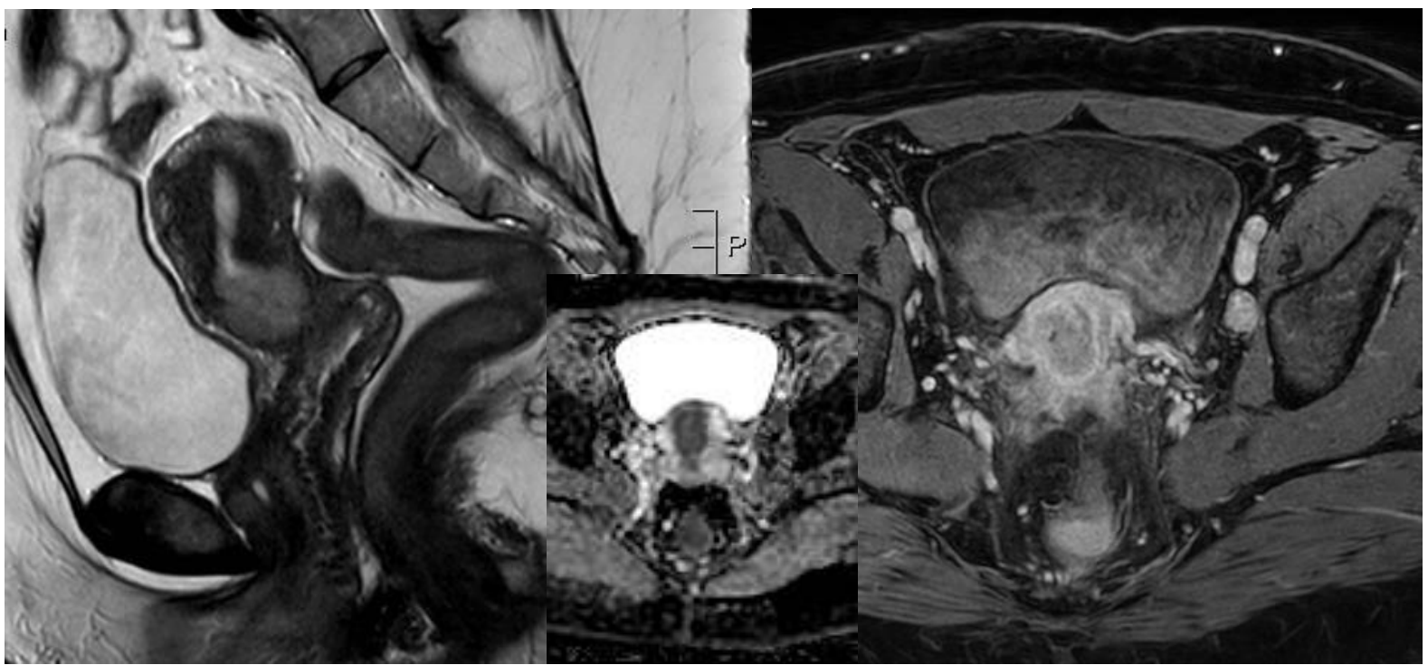
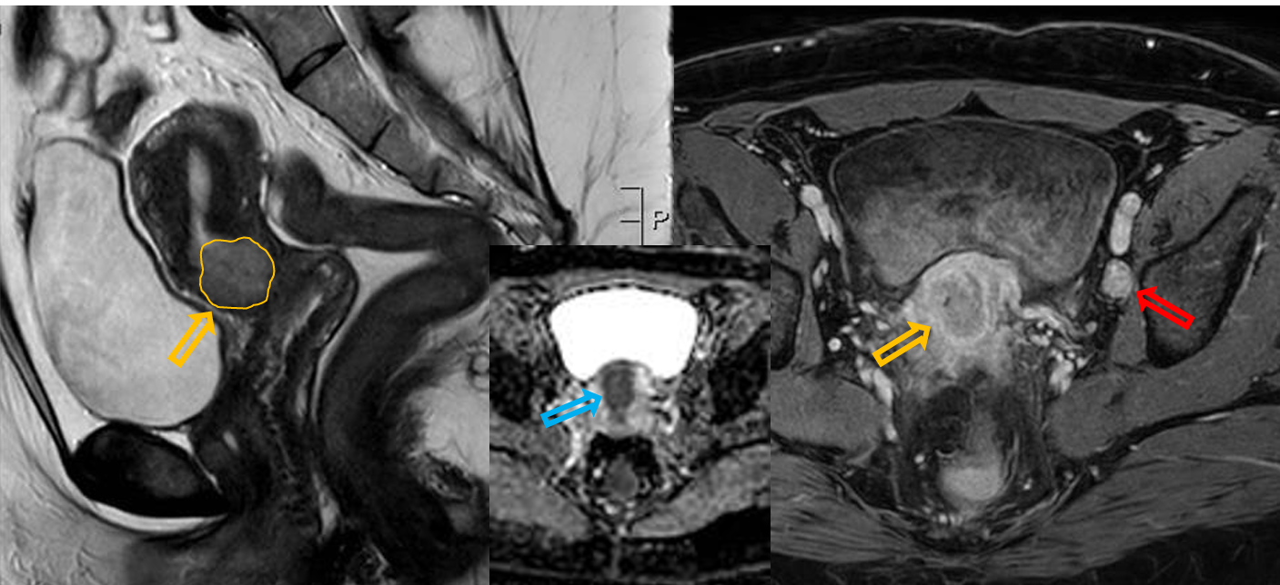
Patient history: vaginal bleeding, pelvic pain, colposcopy-guided cervical biopsy identified cervical cancer
Modality: left image – T2W sagittal MRI; middle image – ADC map axial MRI; right image – postcontrast T1W fatsat, axial MRI (iv. gadolinium)
Region: pelvis
Radiologic sign: Contrast-enhancing, cervical mass infiltrating the myometrium (yellow arrow), showing diffusion restriction (blue arrow) and T2-hyperintensity. A pathologically enlarged right iliac lymph node is visible (red arrow).
Diagnosis: cervical carcinoma with locoregional lymphadenopathy
14.5
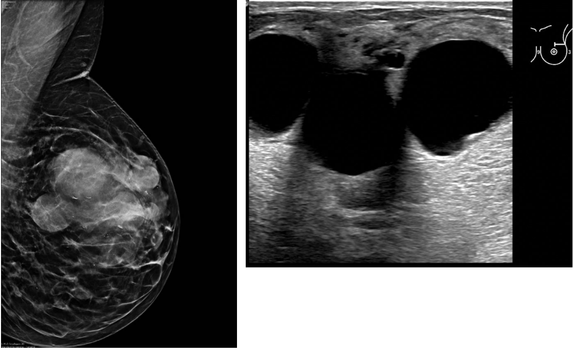
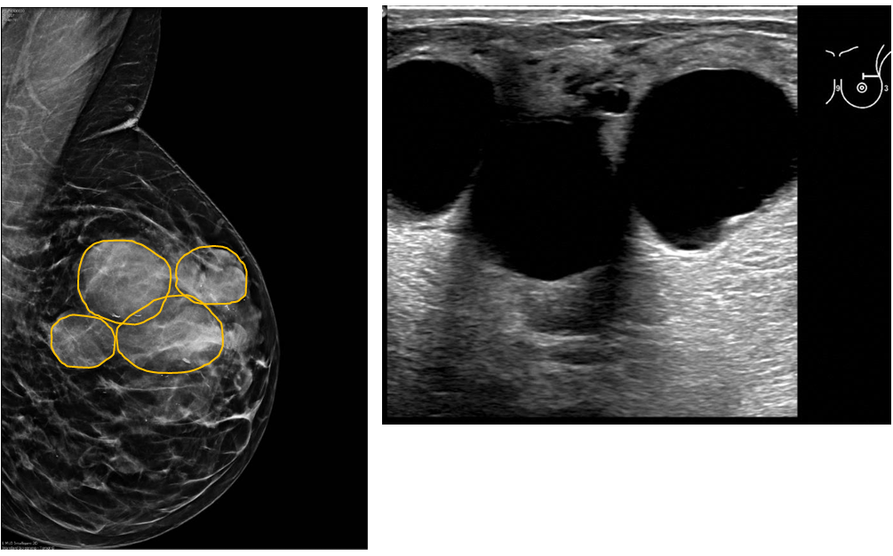
Patient history: palpable left breast masses in a 48-year-old patient
Modality: left image – mammogram of left breast in MLO view; right image – US of left breast (upper lateral quadrant), high frequency (7-12 MHz) linear transducer
Region: left breast
Radiologic sign:: Multiple, well-defined masses in the left breast that show up as anechoic lesions with acoustic enhancement on the ultrasound.
Diagnosis multiple cysts in the left breast
14.6
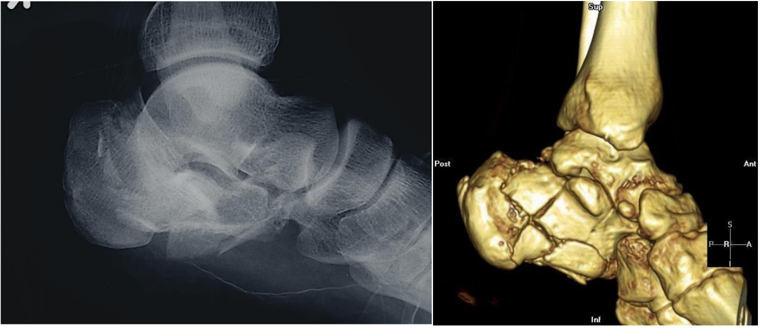
Patient history: a history of a fall from a height
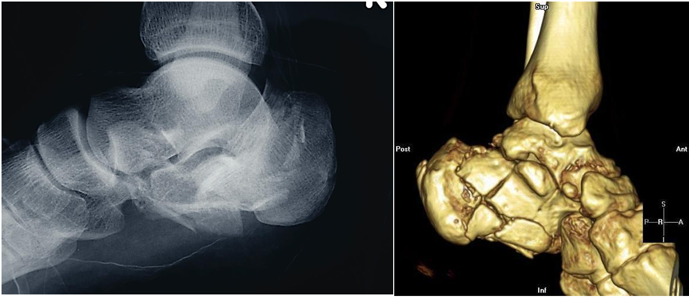
Modality: ankle X-ray – lateral view (left image), CT 3D reconstruction (right image)
Region: left ankle
Radiologic sign: abrupt discontinuity of the cortex of the calcaneus, lucent fracture lines
Diagnosis: comminuted fracture of the calcaneus (Lover’s fracture or Don Juan-fracture)
15.1
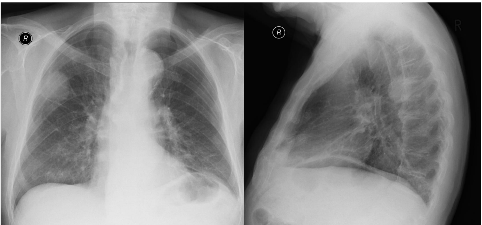
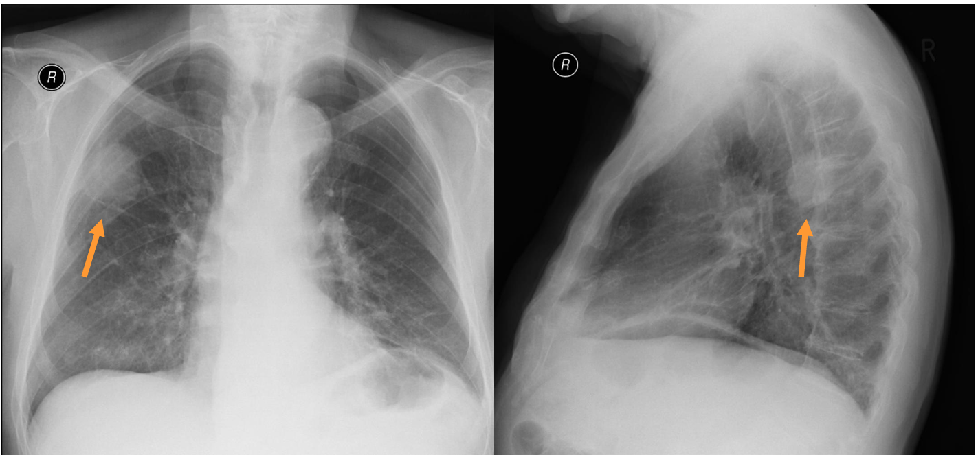
Patient history: chest screening in an asymptomatic middle-aged patient
Modality: left– PA chest radiograph right – left lateral chest radiograph
Region:chest
Radiologic sign: peripheral, well-circumscribed, novel (compared to older films) mass in the right upper lobe
Diagnosis: lung adenocarcinoma
15.2
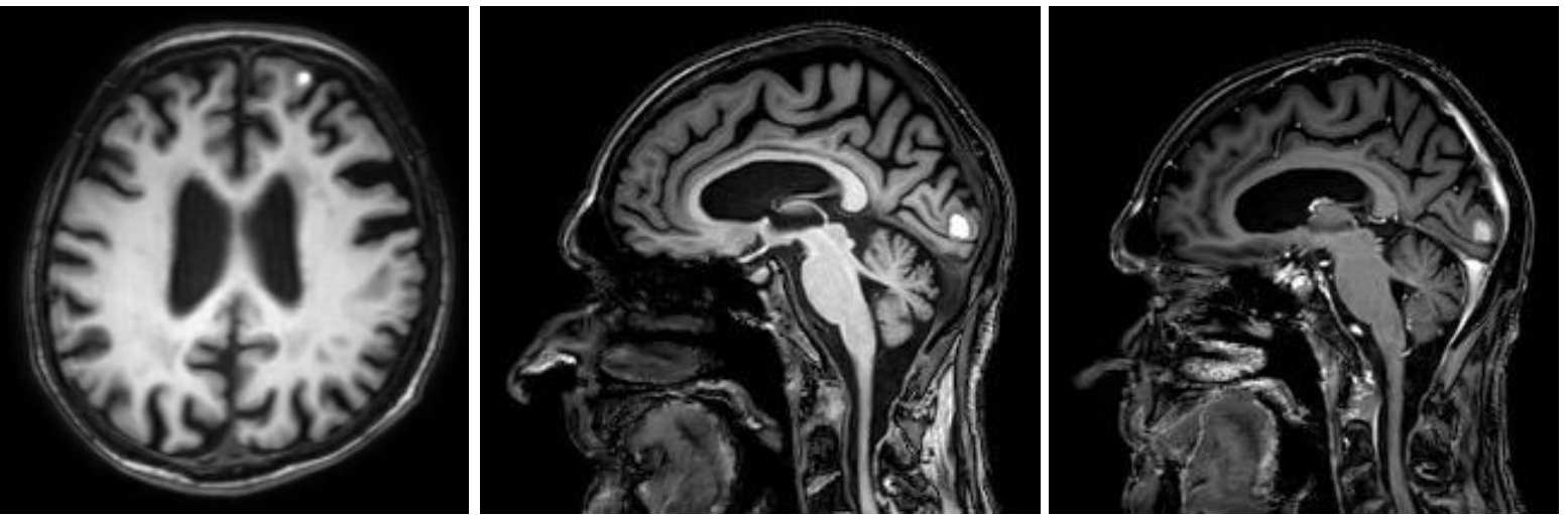
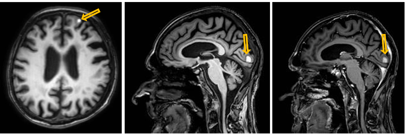
Patient history: melanoma malignum, at present headache and epileptic seizure
Modality: left image – non-enhanced T1W MRI, axial plane; middle image – non-enhanced T1W MRI, sagittal plane; right image – postcontrast T1W MRI, sagittal plane (iv. gadolinium)
Region: skull
Radiologic signs: multiple contrast-enhancing, T1 hyperintense intra-axial space-occupying lesions in the left frontal and occipital lobes (yellow arrows)
Diagnosis: multiple brain metastases from malignant melanoma
15.3
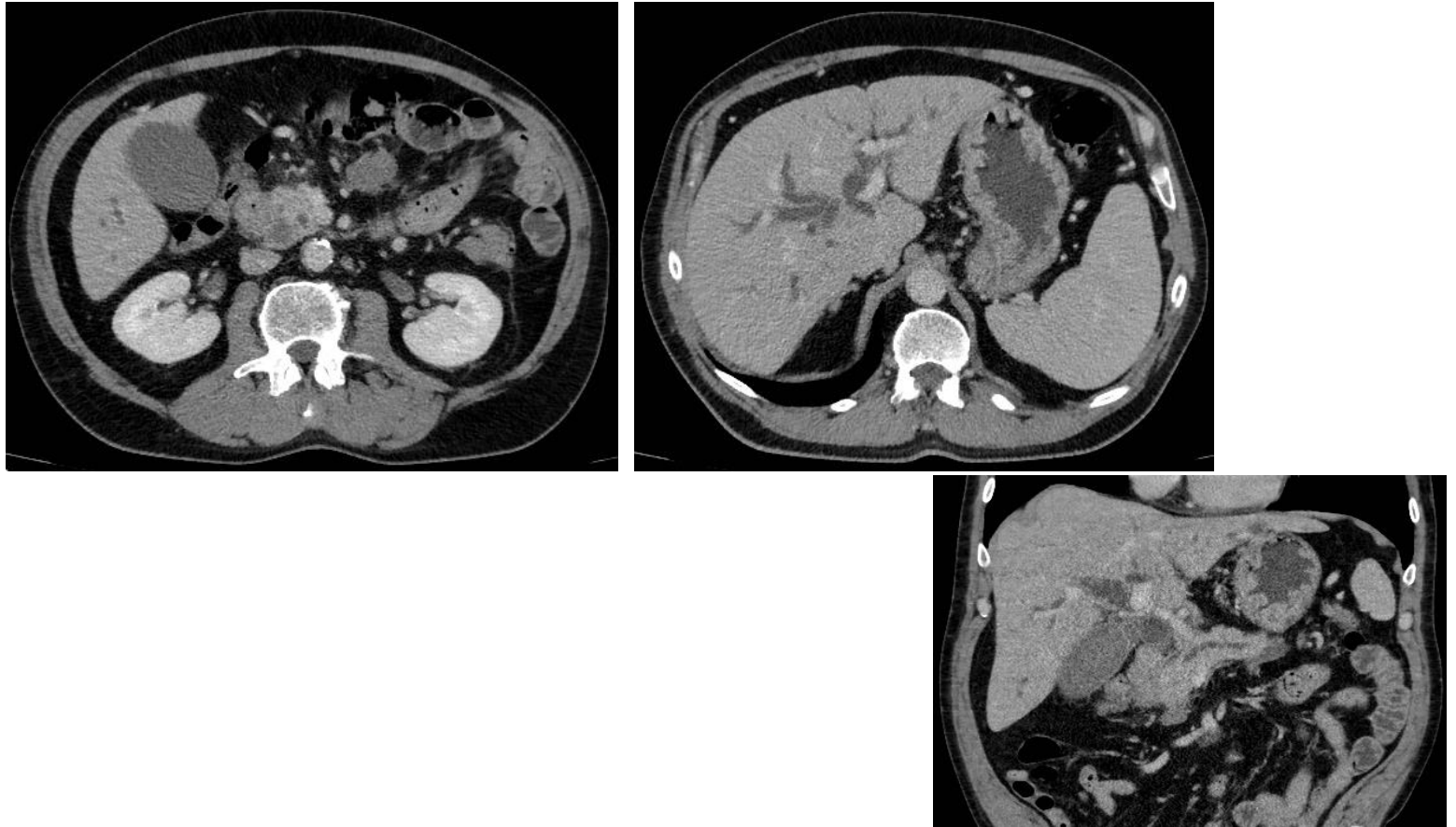
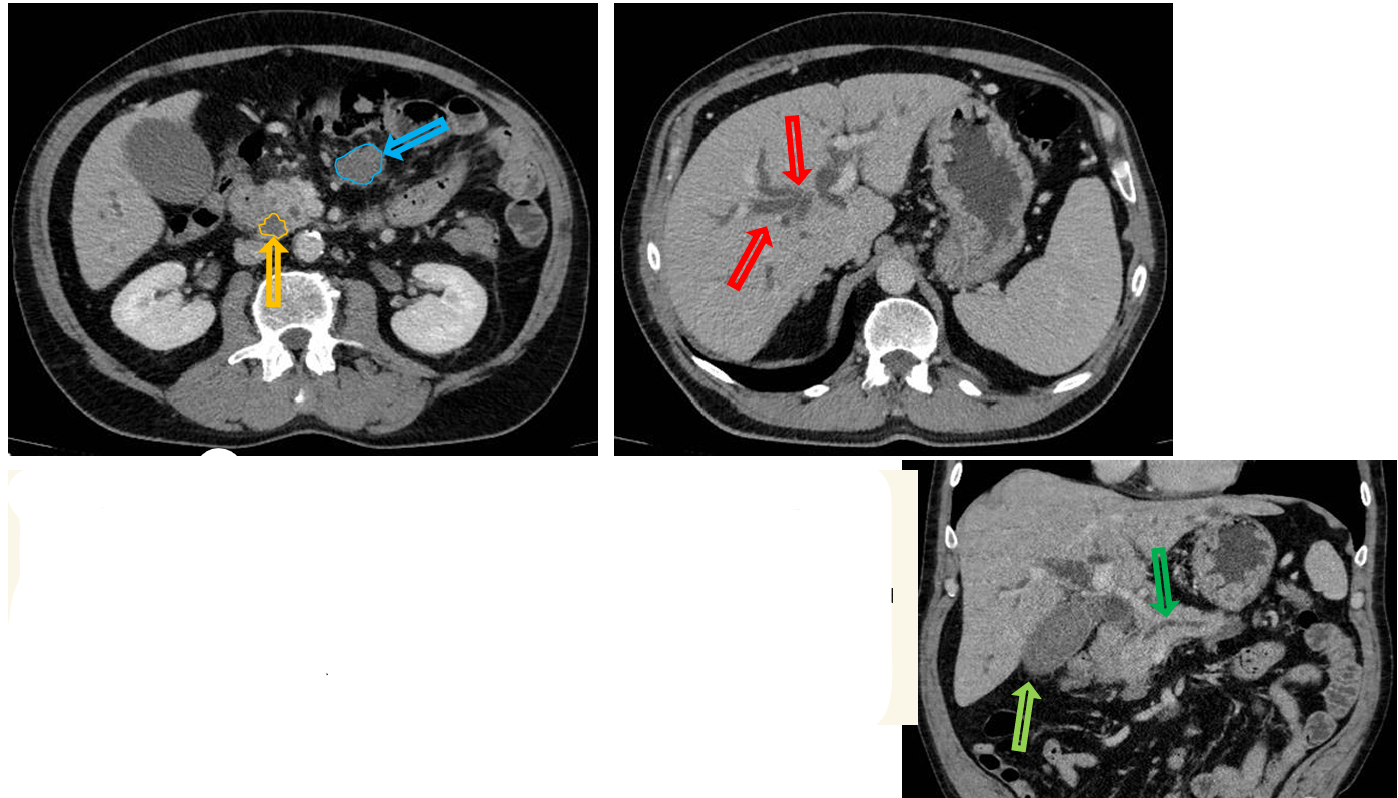
Patient history: fatigue, weight loss and jaundice in a middle-aged male patient
Modality: contrast enhanced CT (venous phase), left and right – axial planes, bottom – coronal plane reconstruction (iv. iodine-based contrast agent)
Region: upper abdomen
Radiologic sign: mass in the pancreas head that is hypodense in the venous phase (yellow area); dilated hypodense intrahepatic bile ducts (red arrows, normally not visible), gallbladder distended with fluid, hydropic (light green arrow), pancreatic duct also dilated due to the obstruction (dark green arrow). Enhancing soft tissue mass with lobulated margin in the mesenterium (blue arrow).
Diagnosis: pancreas adenocarcinoma with regional lymphadenopathy, distended bile ducts and pancreatic duct
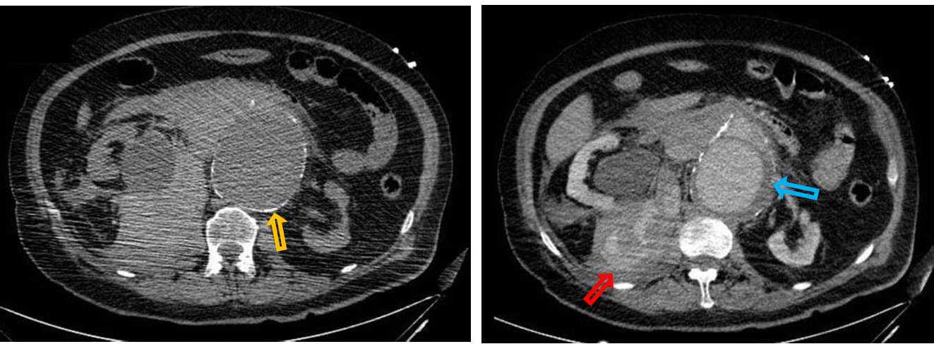
15.4
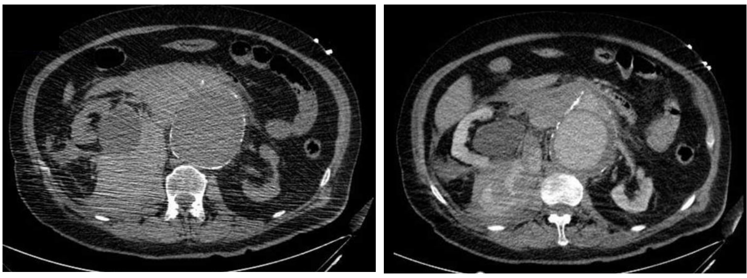
Patient history: acute abdominal pain, hypotension, heart palpations and pulsatile abdominal mass in an elderly patient Modality: left: Non-enhanced CT, right: Postcontrast CT (venous phase), iv. iodine-based contrast agent
Region: abdomen, retroperitoneum
Radiologic sign: Dilated abdominal aortic aneurysm with wall calcifications (yellow arrow) and hypodense wall thrombus (blue arrow), retroperitoneal fluid collection with active extravasation of contrast material on postcontrast scan (fluid becomes denser, its average density is 60-80 HU, which is characteristic for fresh hematoma) (red arrow). The large hematoma pushes the right kidney ventrally.
Diagnosis: abdominal aortic aneurysm rupture
15.5
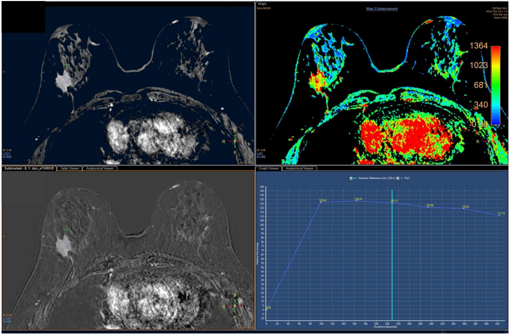
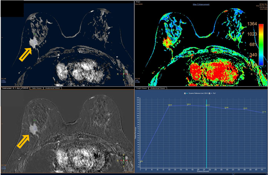
Patient history: known invasive ductal carcinoma, staging MR
Modality: dynamic contrast enhanced MRI (eThrive – upper left image), dynamic subtraction MRI (lower left image); contrast agent: iv. gadolinium; right images show the kinetics of contrast enhancement (highest rate of washout signed with red color) Region: breast, axial plane
Radiologic sign: Irregular, spiculated mass in the right breast (yellow arrow), with intense contrast enhancement. The kinetic enhancement curve is characterized by a washout pattern.
Diagnosis : invasive breast carcinoma in the right breast, no multiplicity or contralateral breast lesion
15.6
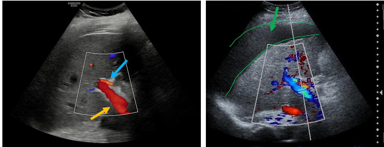
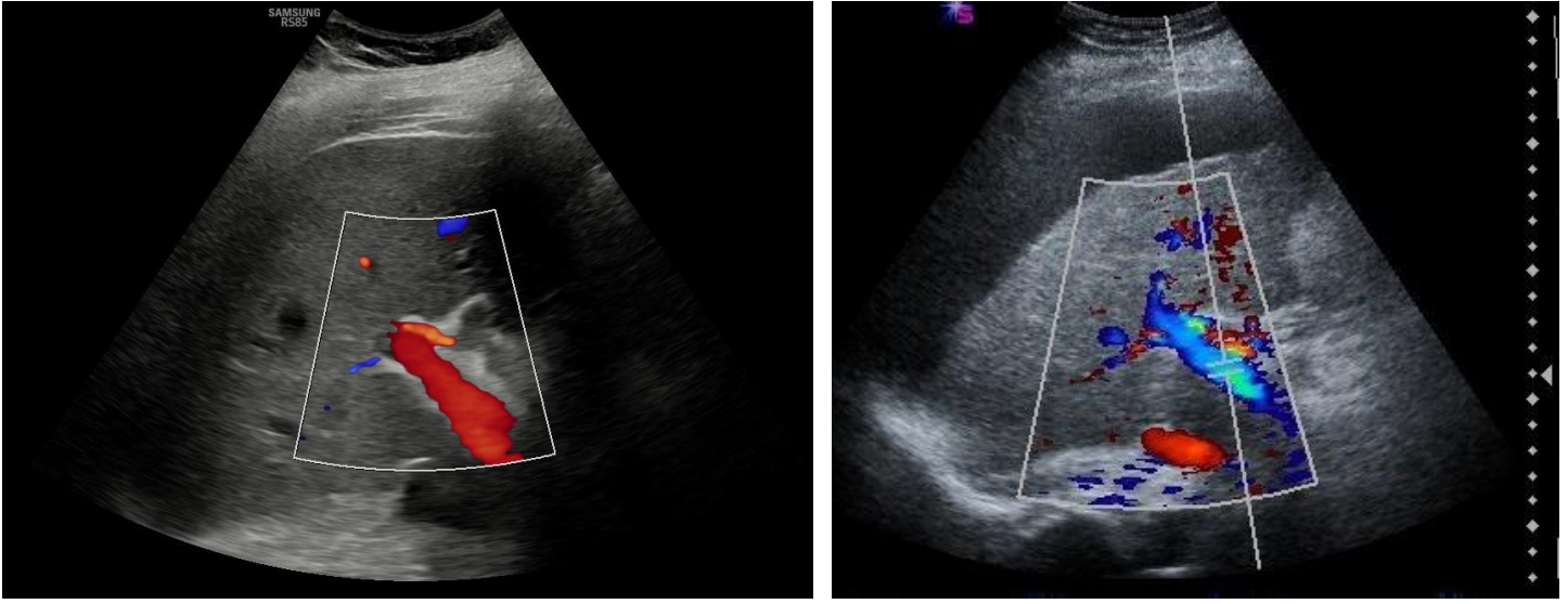
Patient history: routine abdominal ultrasonography (left); years of heavy alcohol consumption, at present fatigue and enlarging abdominal circumference (right)
Modality: abdominal ultrasound, 3-5 MHz convex transducer
Region: abdomen, right upper quadrant
Radiologic sign: left image – normal hepatopetal flow in the portal vein (yellow arrow); blue arrow signs the narrower hepatic artery adjacent to portal vein; right image – reversed, hepatofugal flow in the portal vein in a liver cirrhotic patient due to increased hepatic resistance; perihepatic fluid (ascites) is also observable on the same image (green arrow and outline).
Diagnosis: reversed portal flow and ascites in liver cirrhosis