bio 142 female reproductive lab
 Call Kai
 Learn
Learn Practice Test
 Spaced Repetition
 Match
Match Flashcards
 Knowt Play
1/58

There's no tags or description
Looks like no tags are added yet.
Name | Mastery | Learn | Test | Matching | Spaced | Call with Kai | Chat |
|---|
No analytics yet
Send a link to your students to track their progress
59 Terms
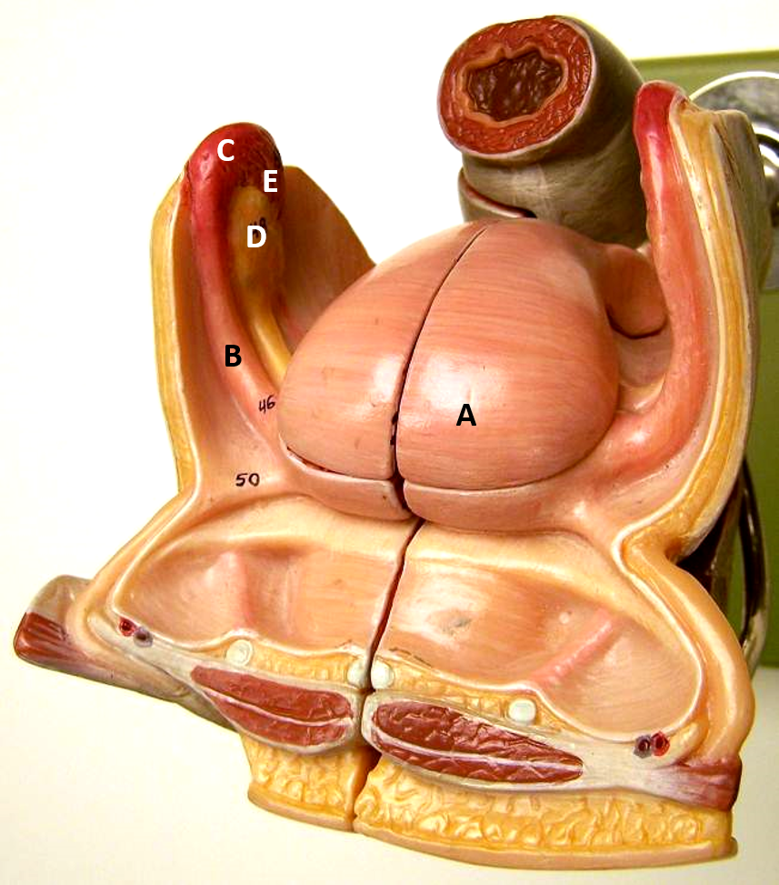
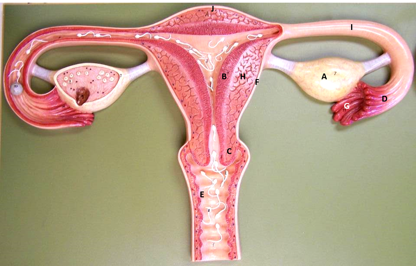
A
uterus
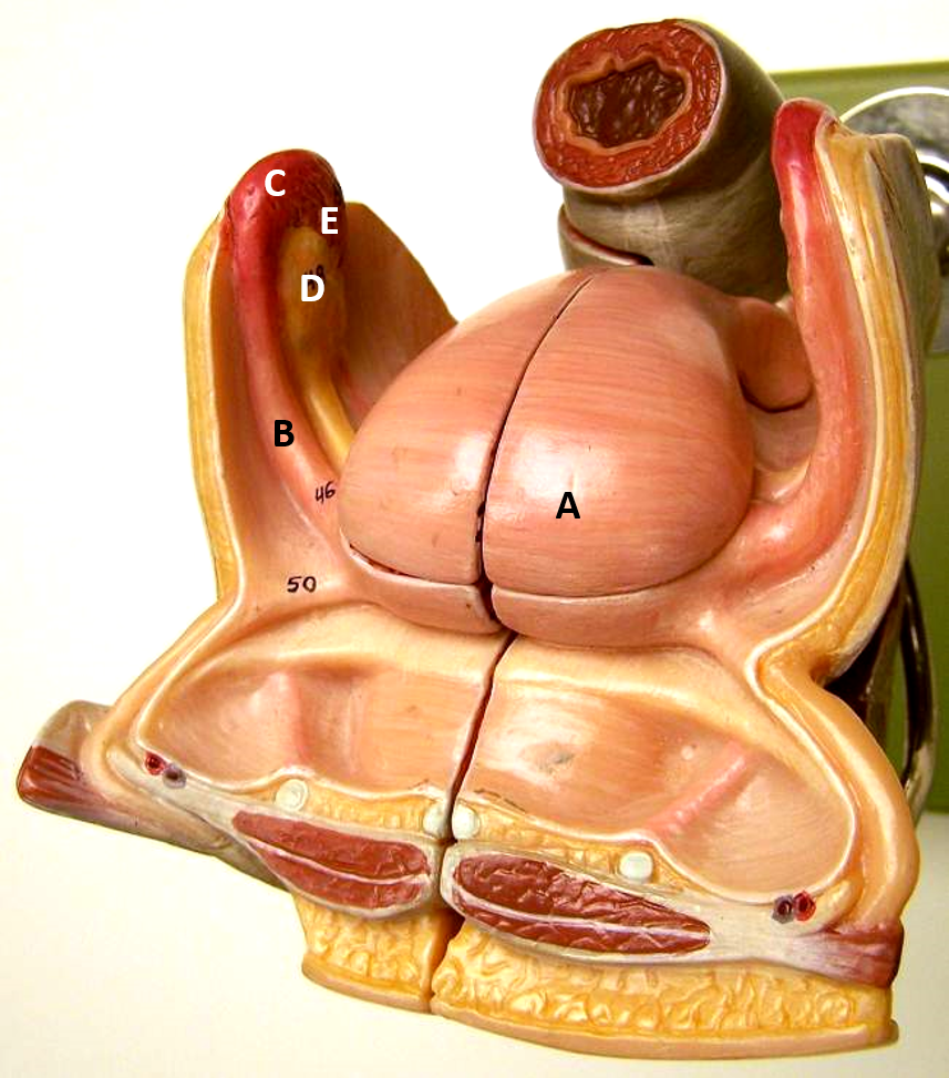
b
fallopian tube
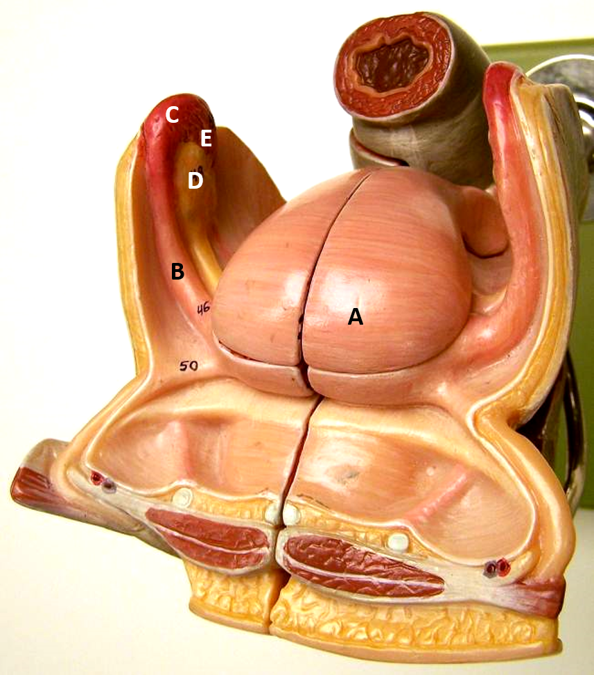
c
infundibulum
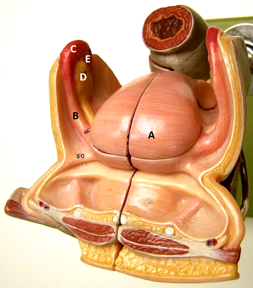
d
ovary
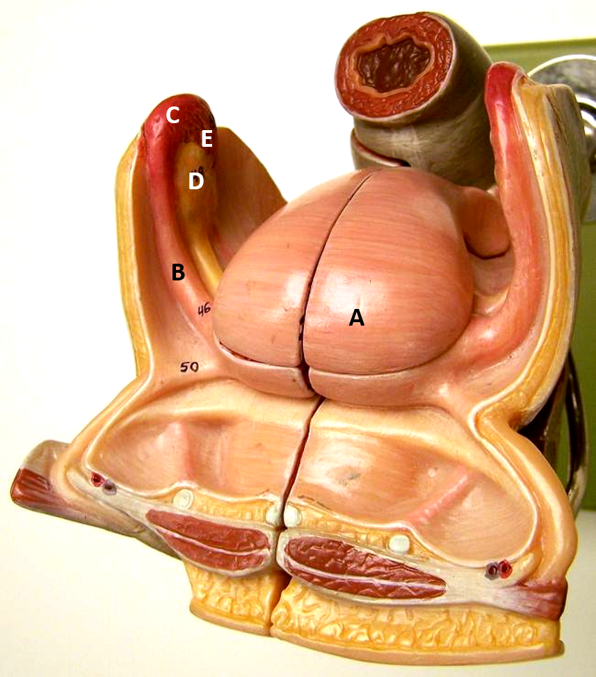
e
fimbriae
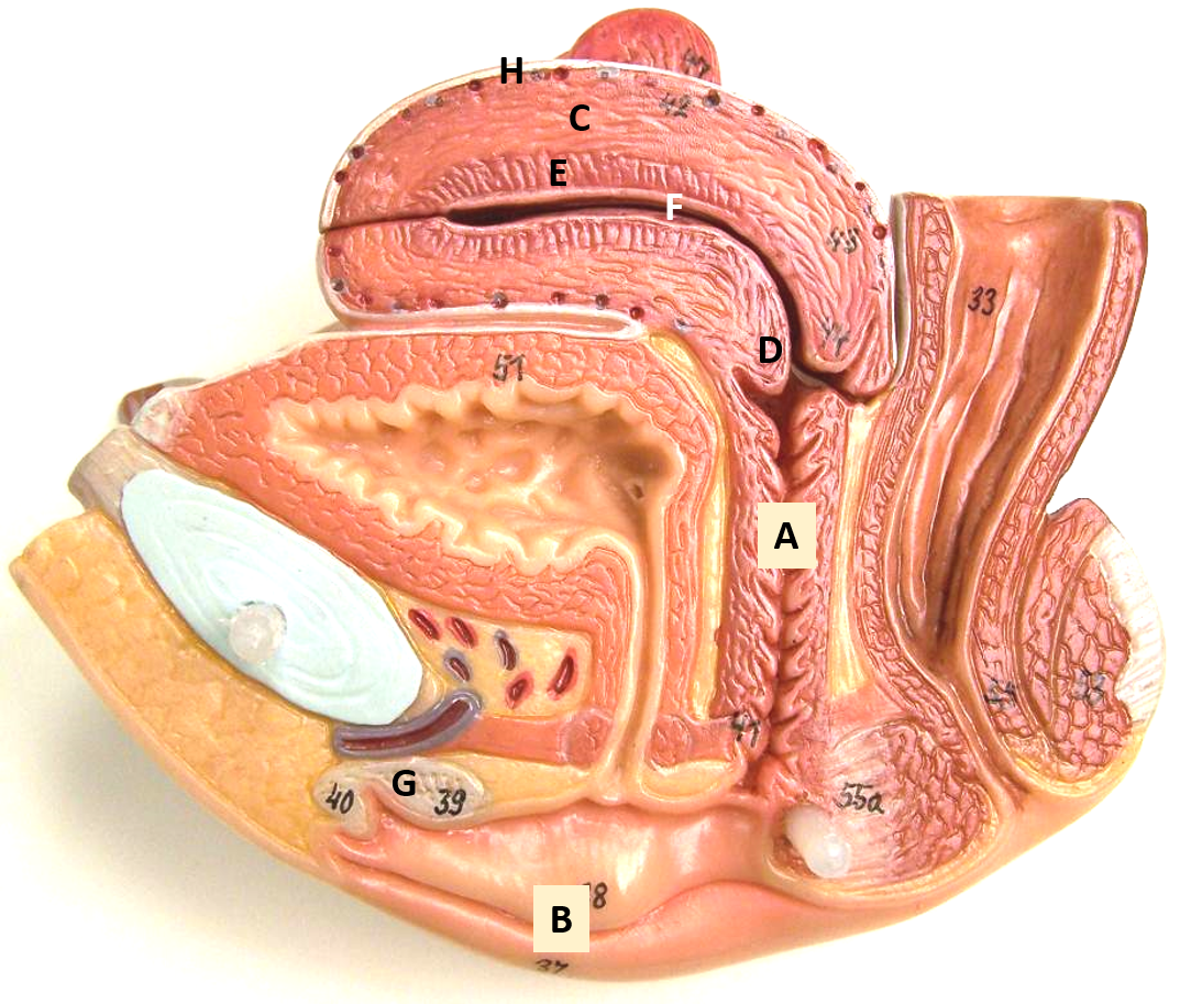
a
vagina
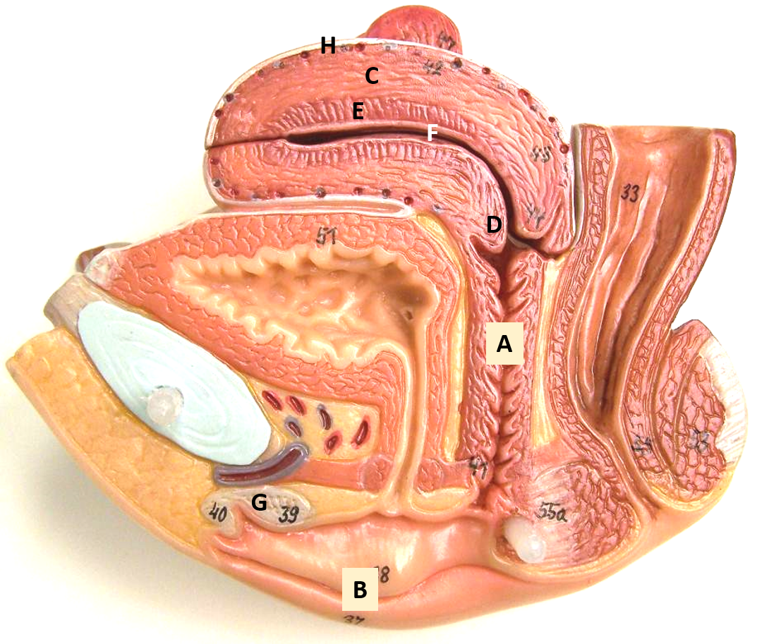
b
vulva
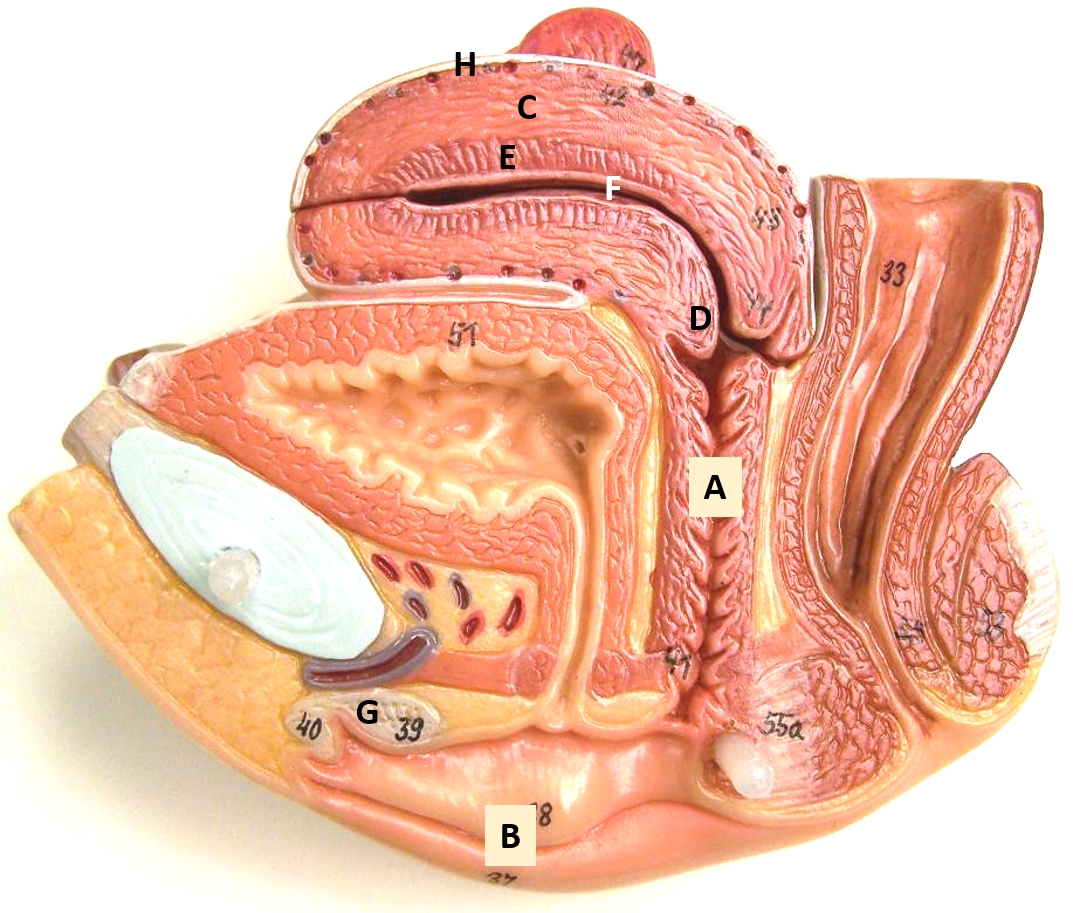
c
myometrium
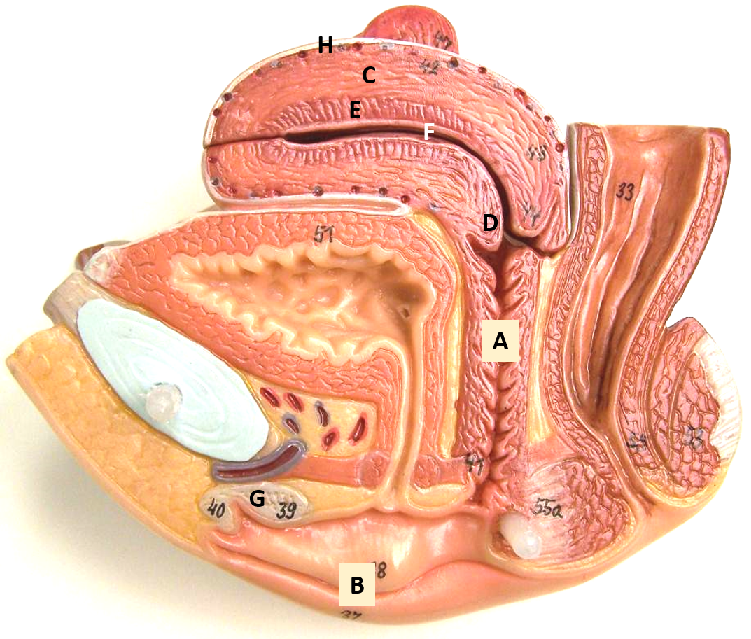
d
cervix
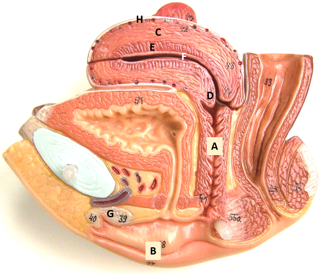
e
endometrium
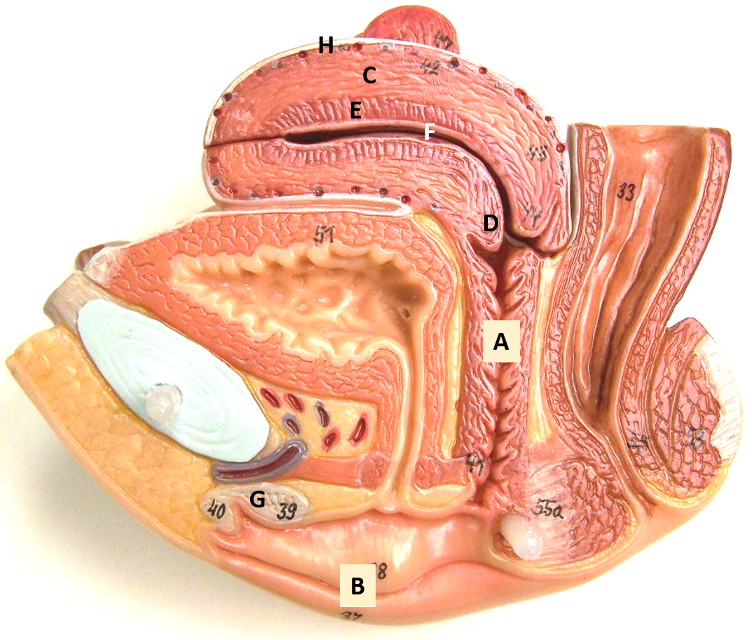
f
uterus
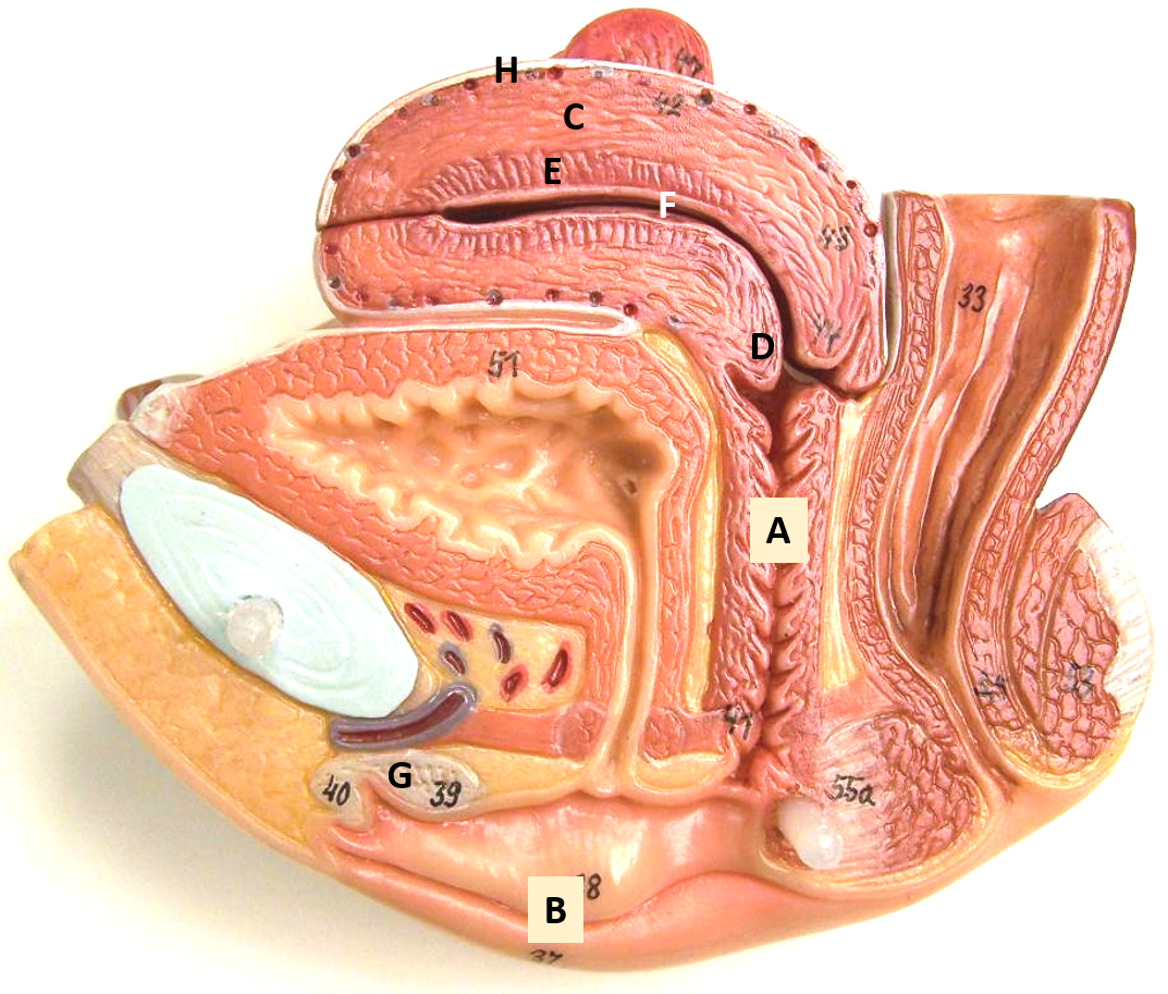
g
clitoris
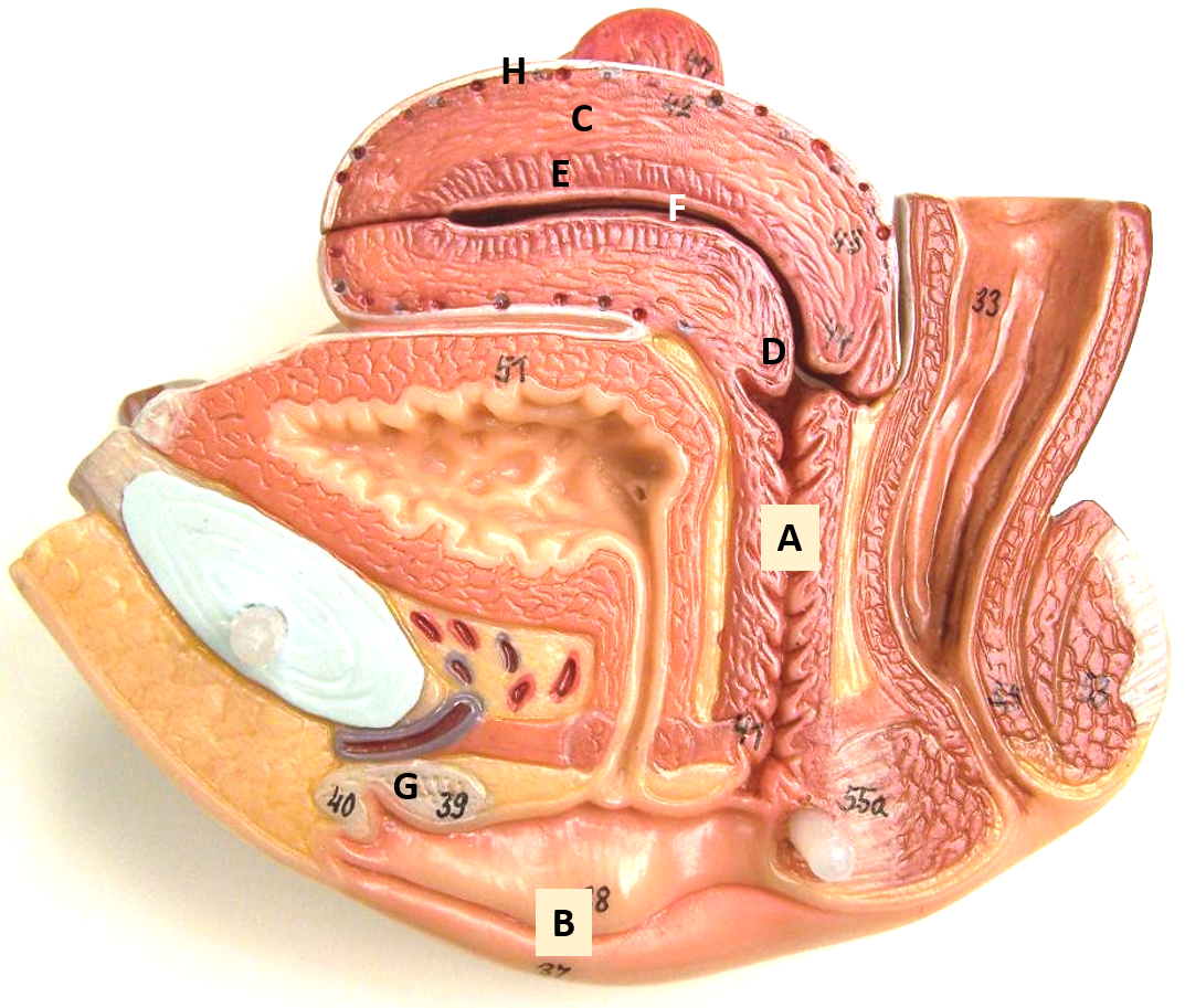
h
perimetrium
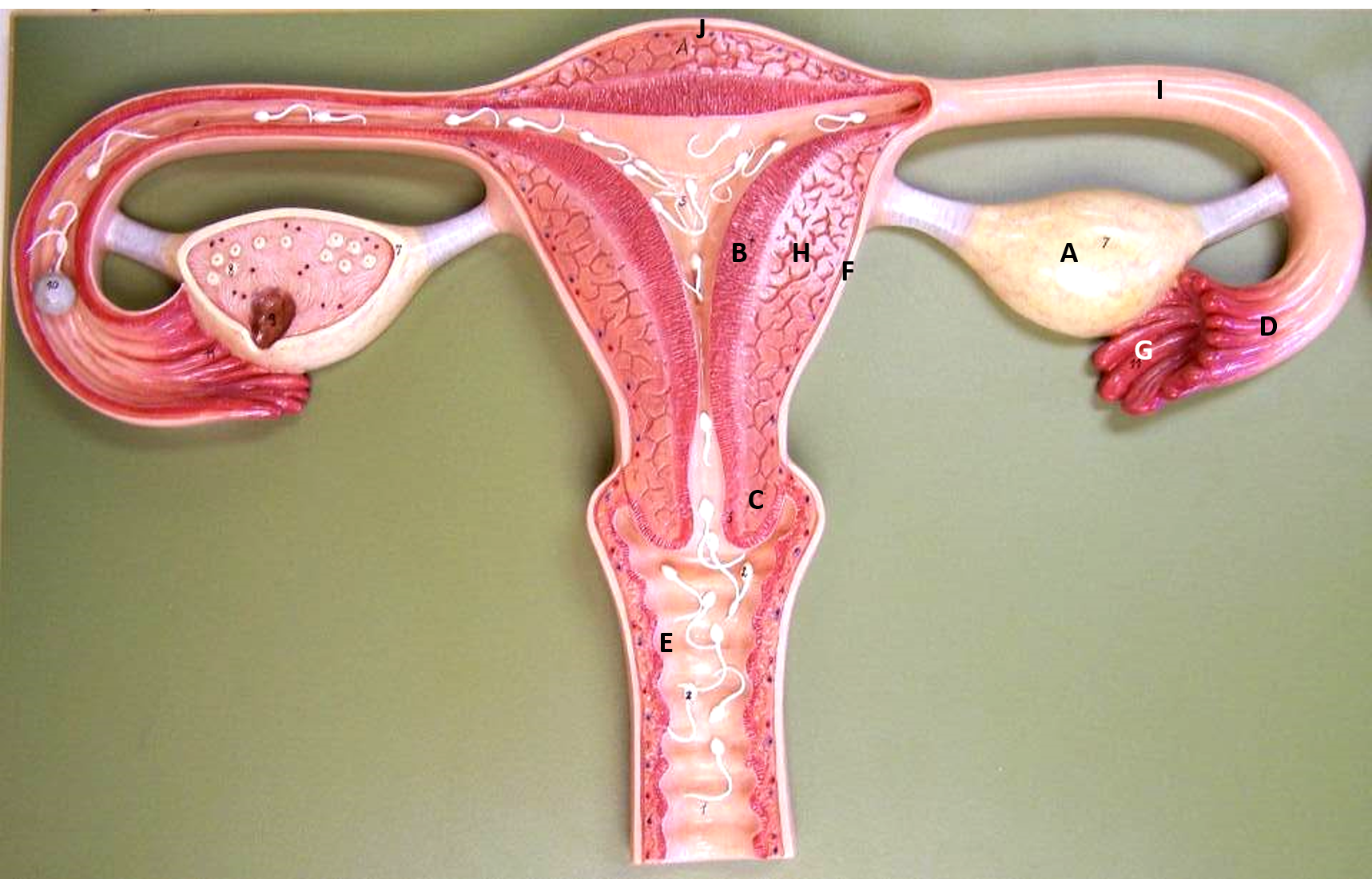
a
ovary
b
endometrium
c
cervix
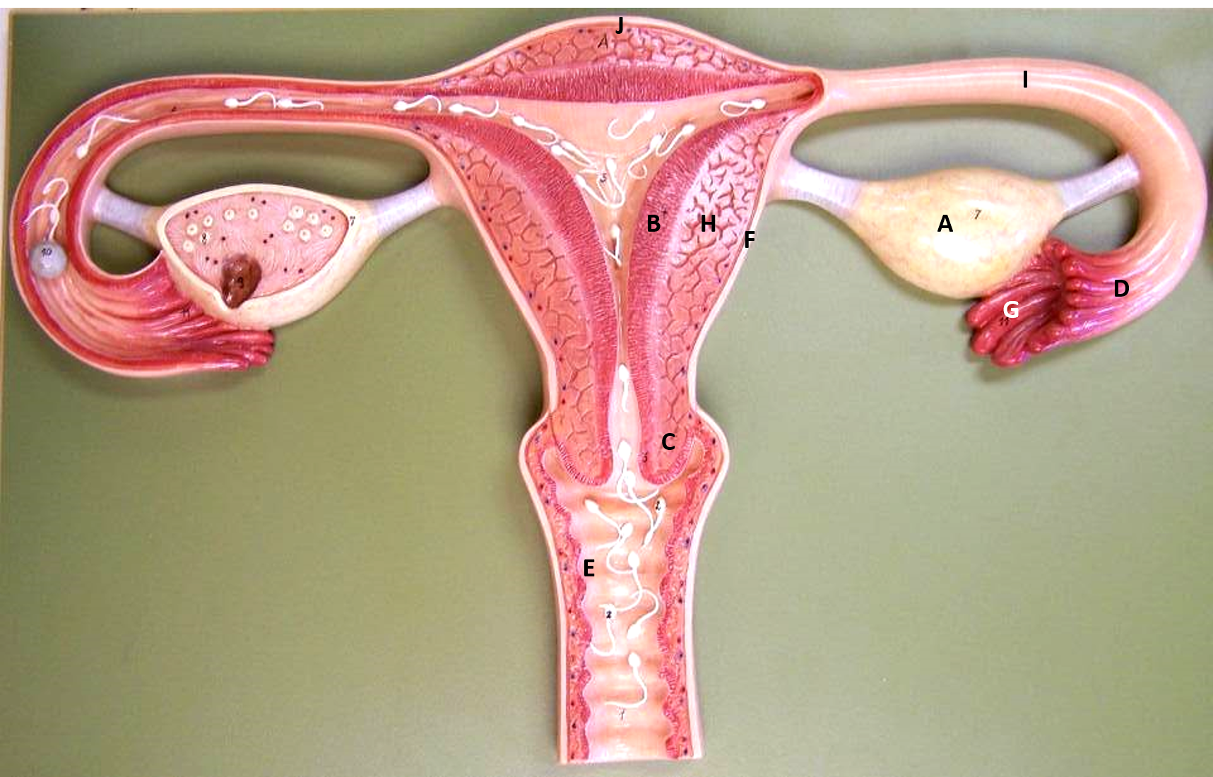
d
infundibulum
e
vagina
f
perimetrium
g
fimbriae
h
myometrium
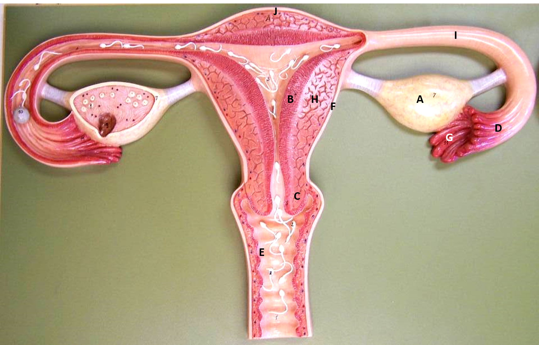
i
uterine tube
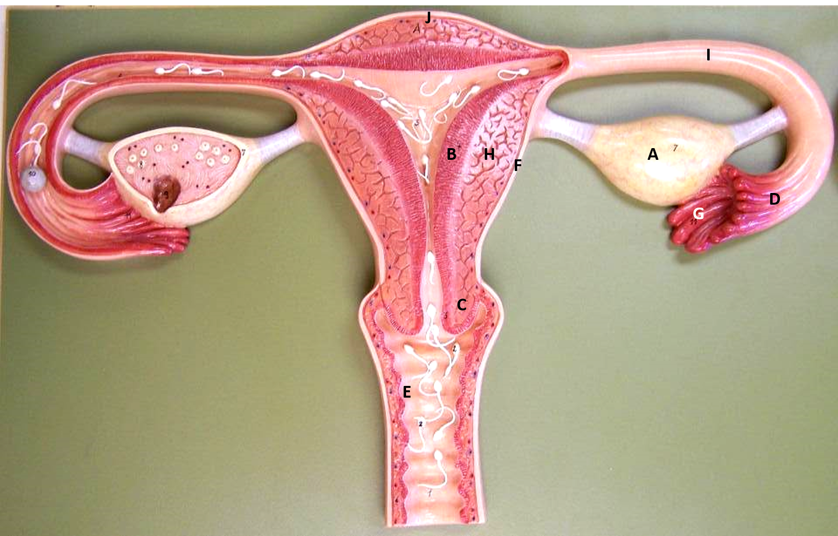
J (jelly)
fundus
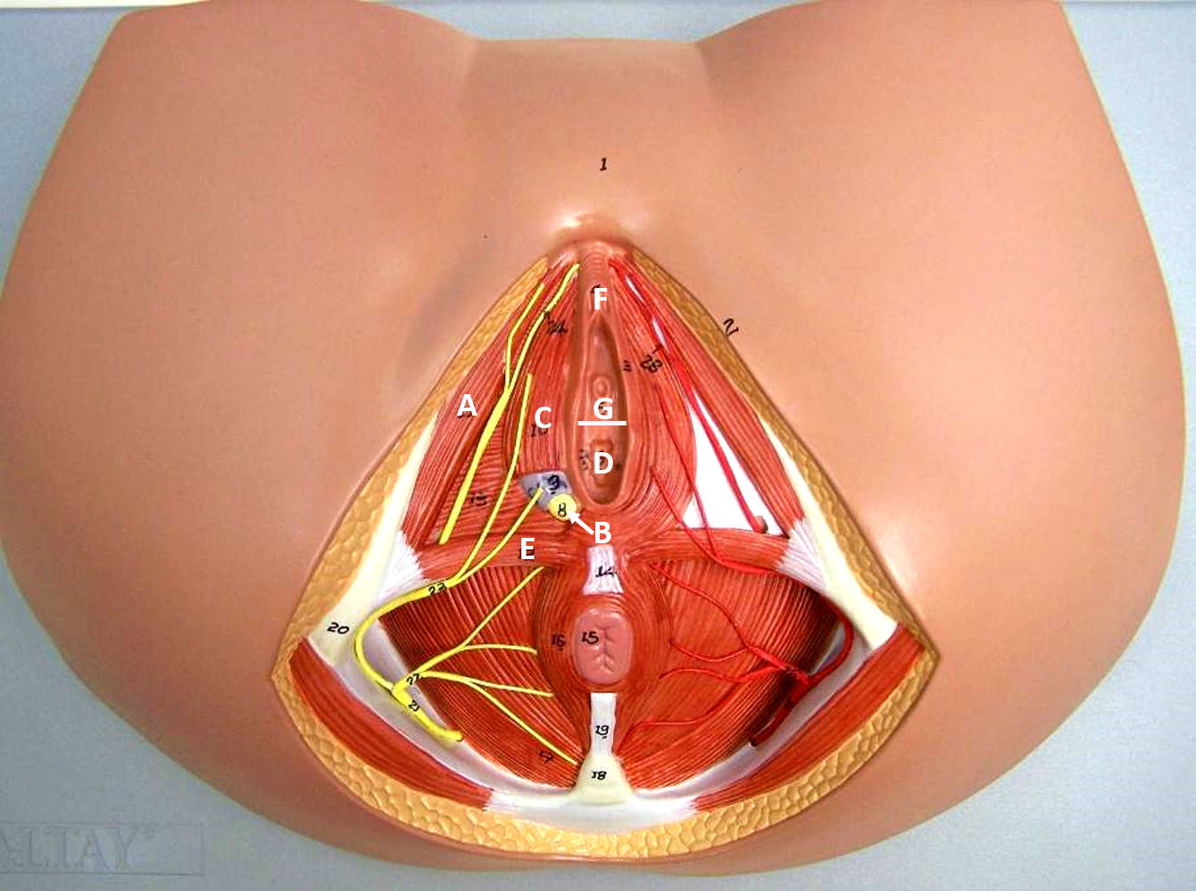
a
ischiocavernosus
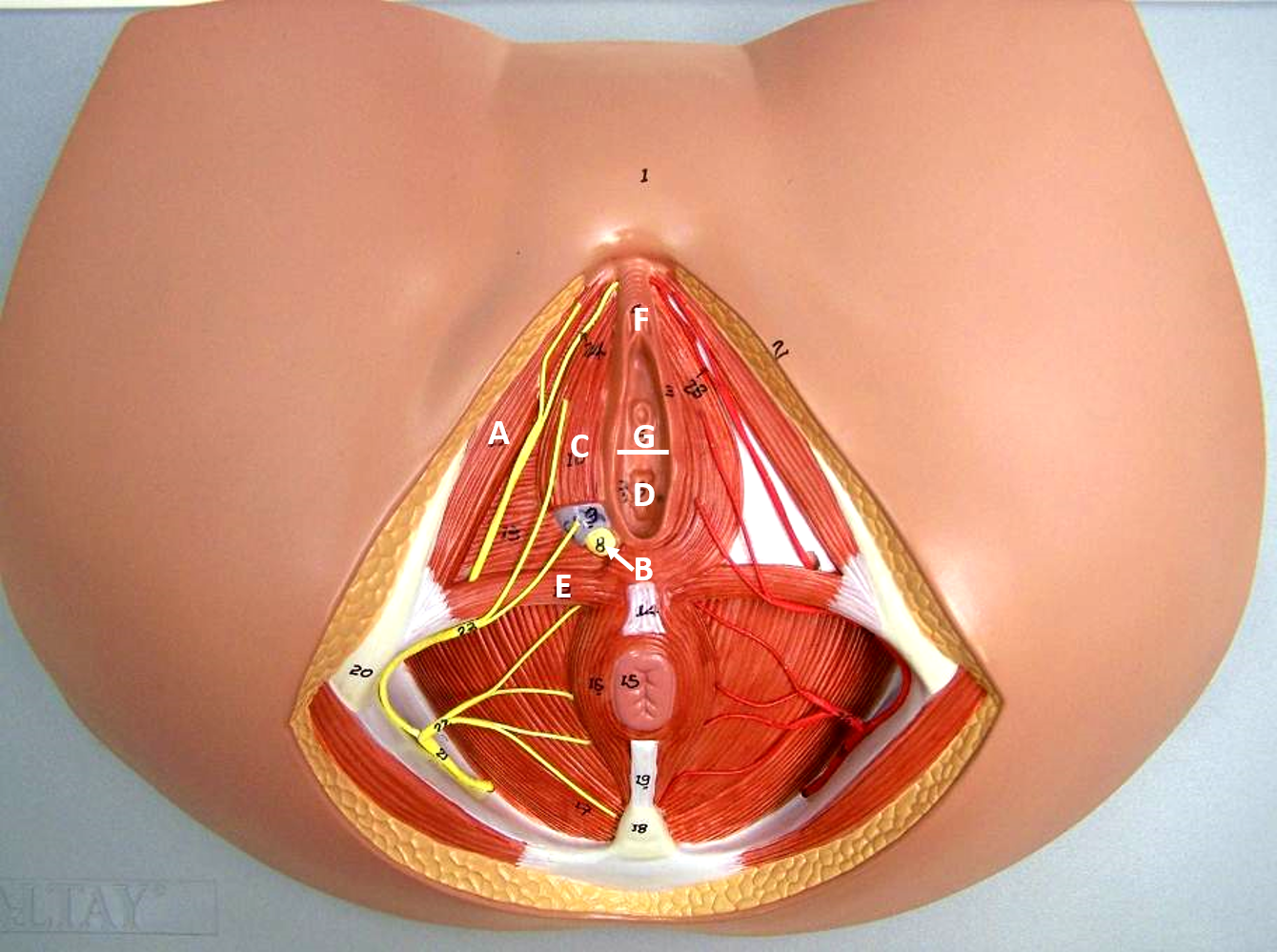
b
greater vestibular gland
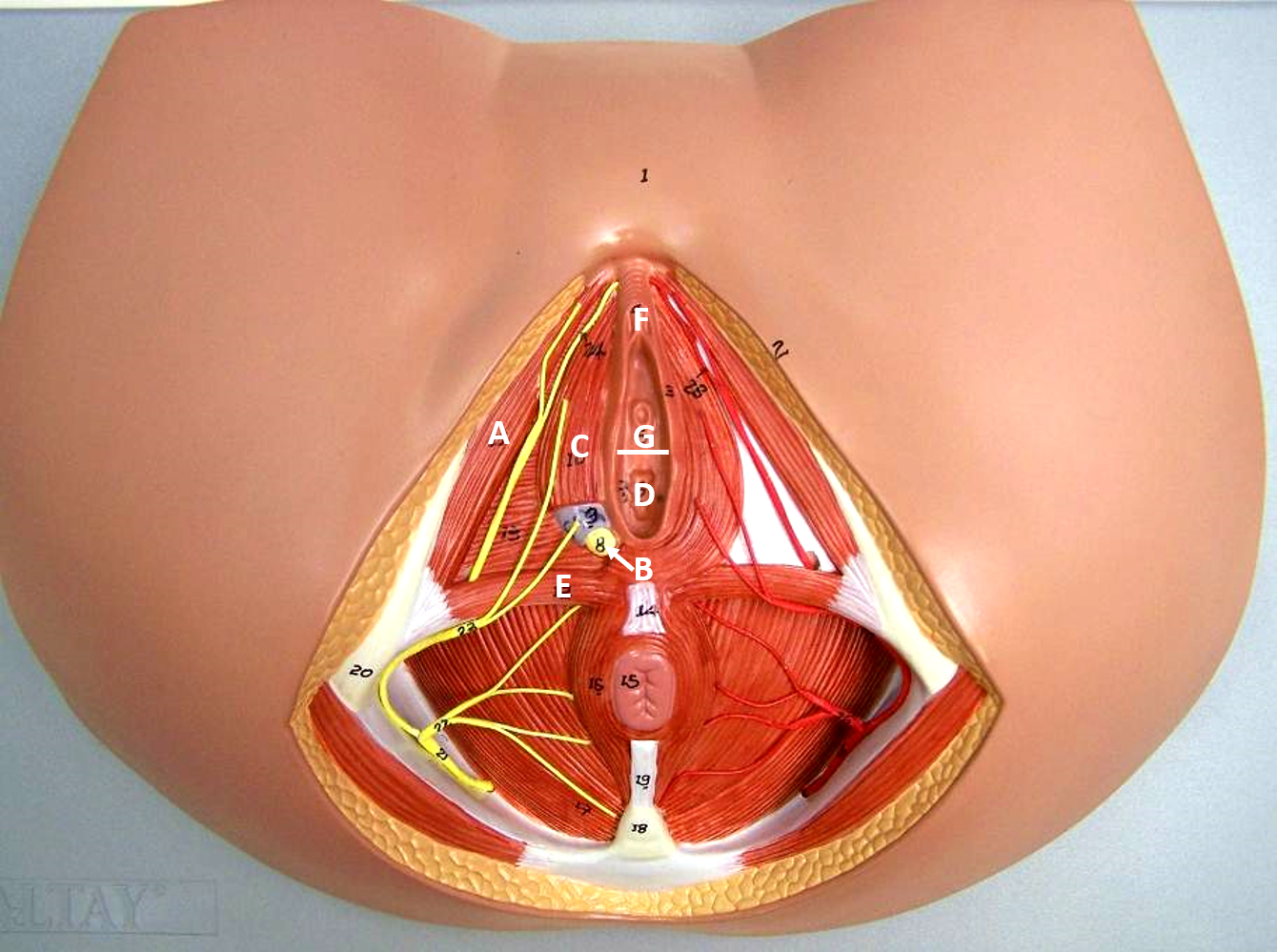
c
bulbospongiosus
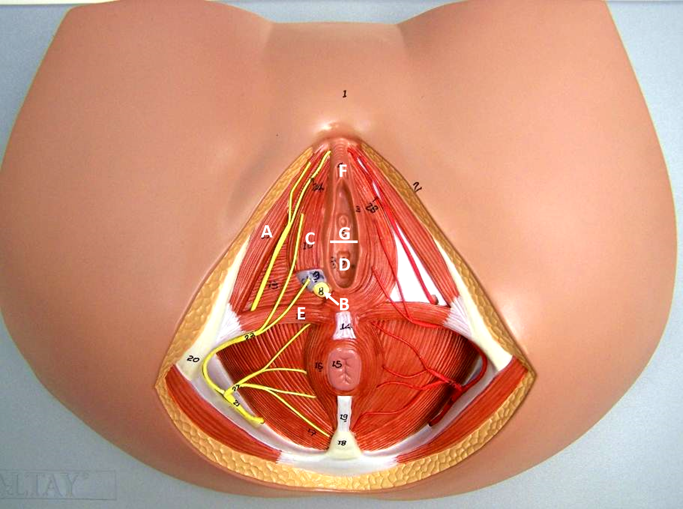
d
vaginal orifice
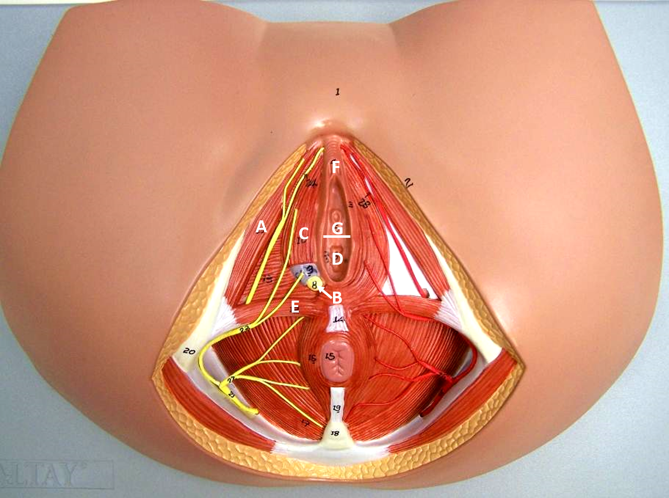
e
transverse perinei
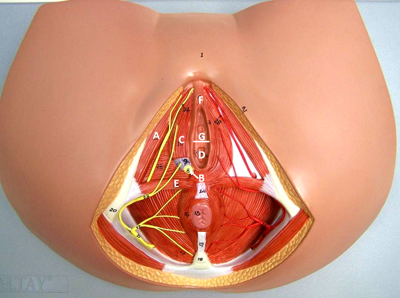
f
clitoris
g
vestibule
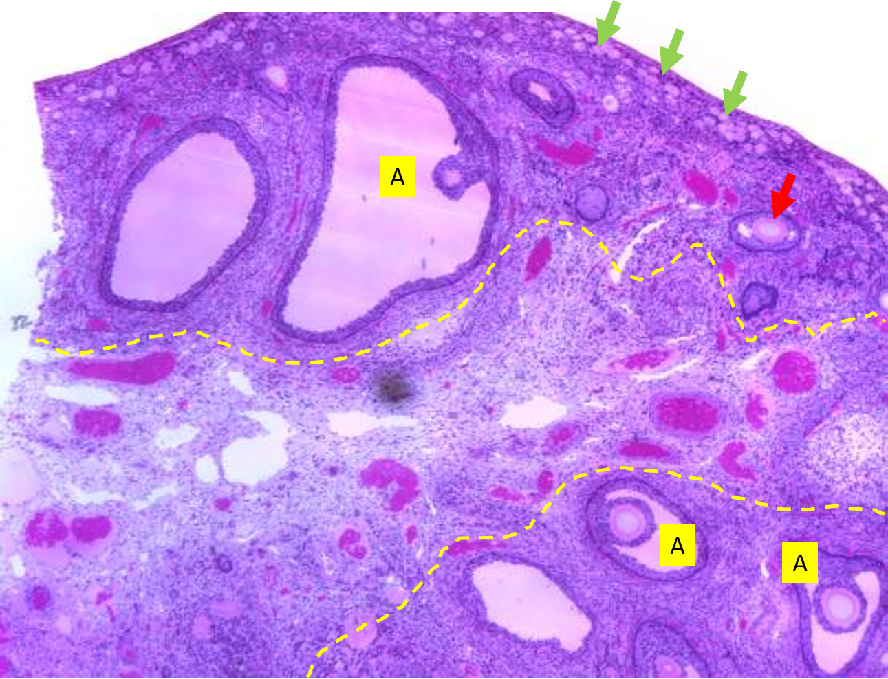
The region between the two dotted yellow lines is the ___
of the ovary.
It is full of blood vessels and connective tissue
medulla
The area at top and bottom of the image is the outer region of the ovary called the ___
It contains many oocytes in follicles in various stages of development.
cortex
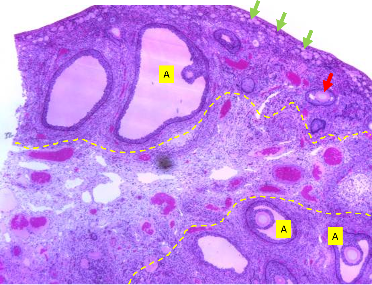
The structures labeled A are ___ follicles.
These follicles are close to releasing their oocyte.
mature
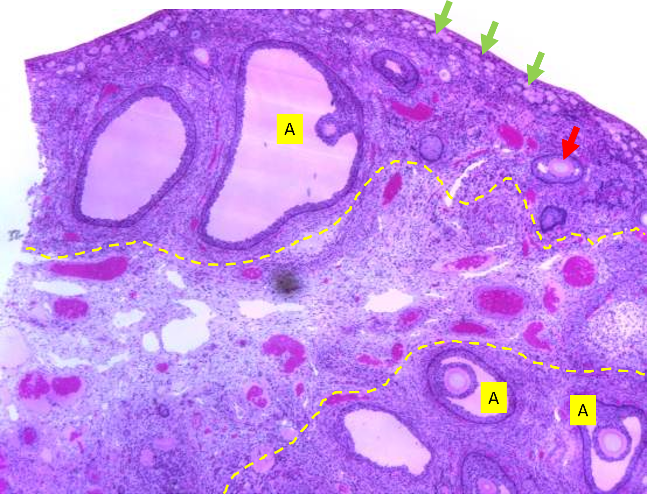
The structures at the tip of the green arrows are ___
follicles
These are dormant.
primordial
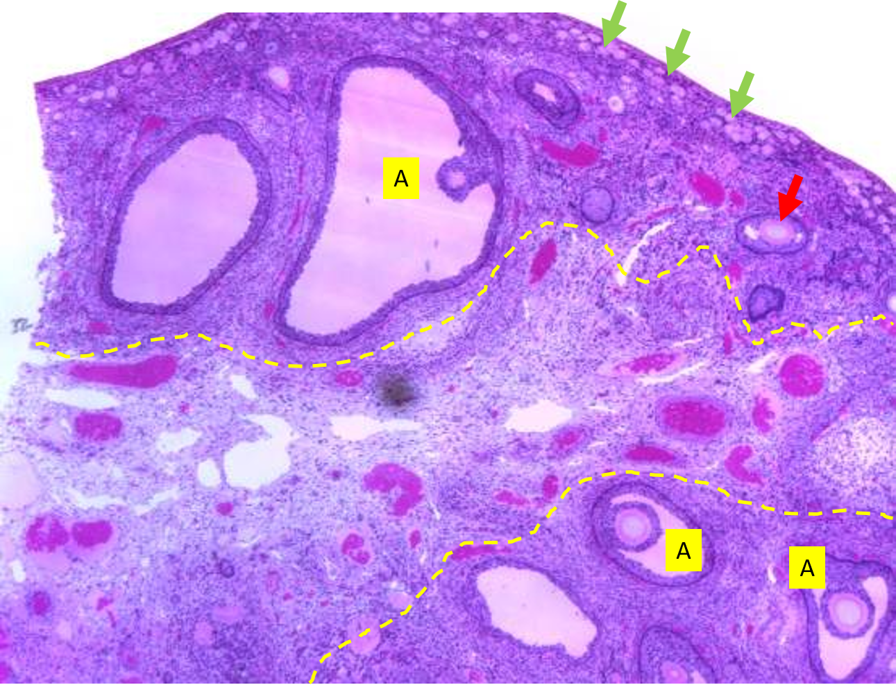
The structure at the tip of the red arrow is a
secondary
follicle.
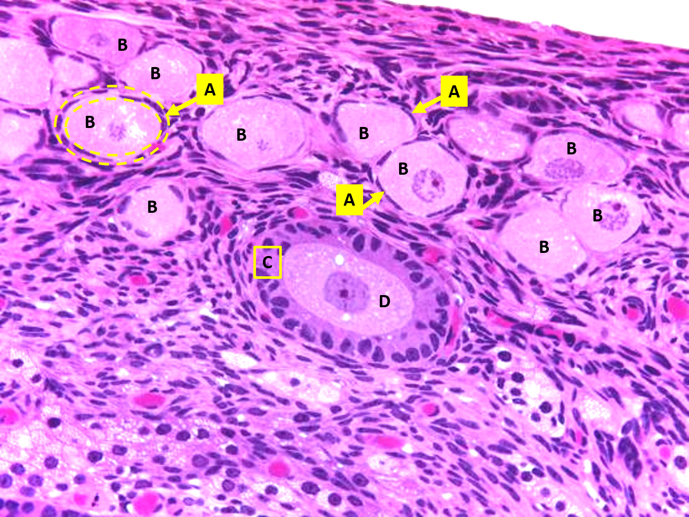
This image is from the
cortex of the ovaries
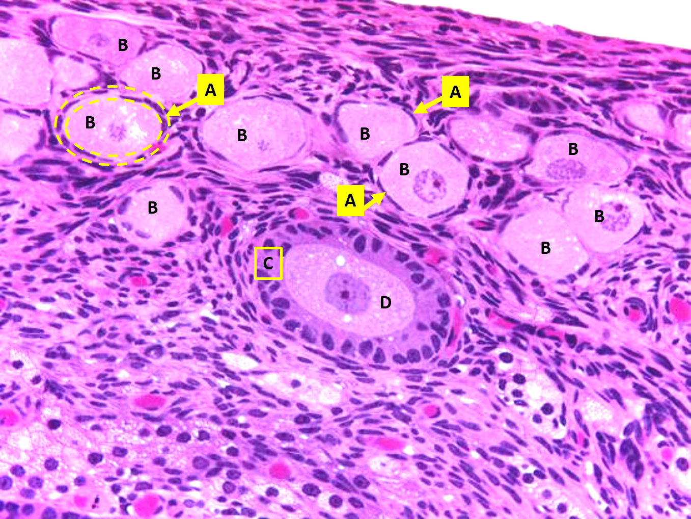
The cells marked B are
primary oocytes
what follicles are primary oocytes found in
primordial
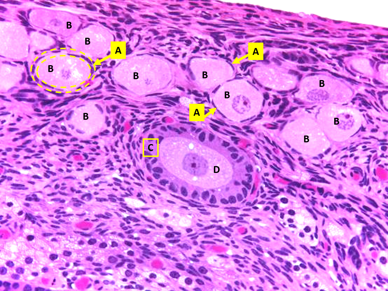
The cell marked D is a
primary oocyte
what follicle are primary
oocyte found in
primary follicles
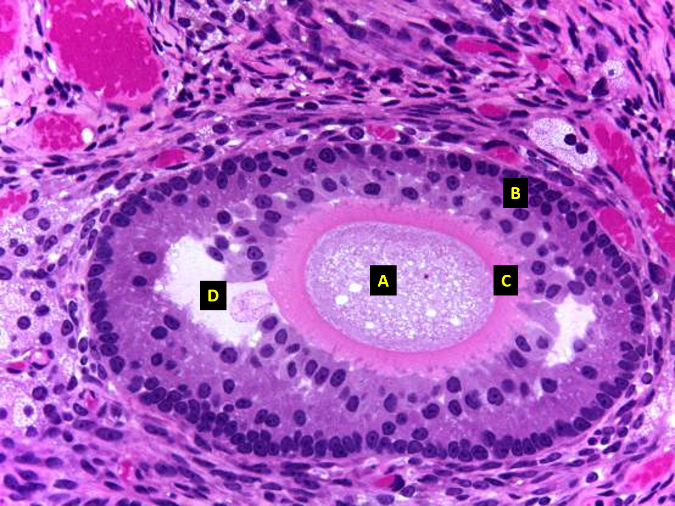
Structure A is a
primary oocyte inside a secondary follicle
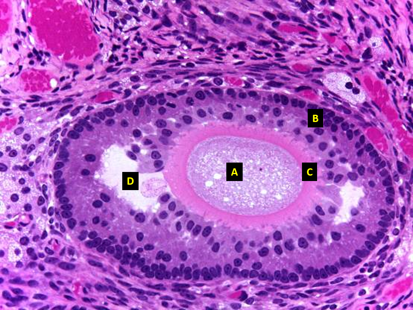
B
granulosa cells
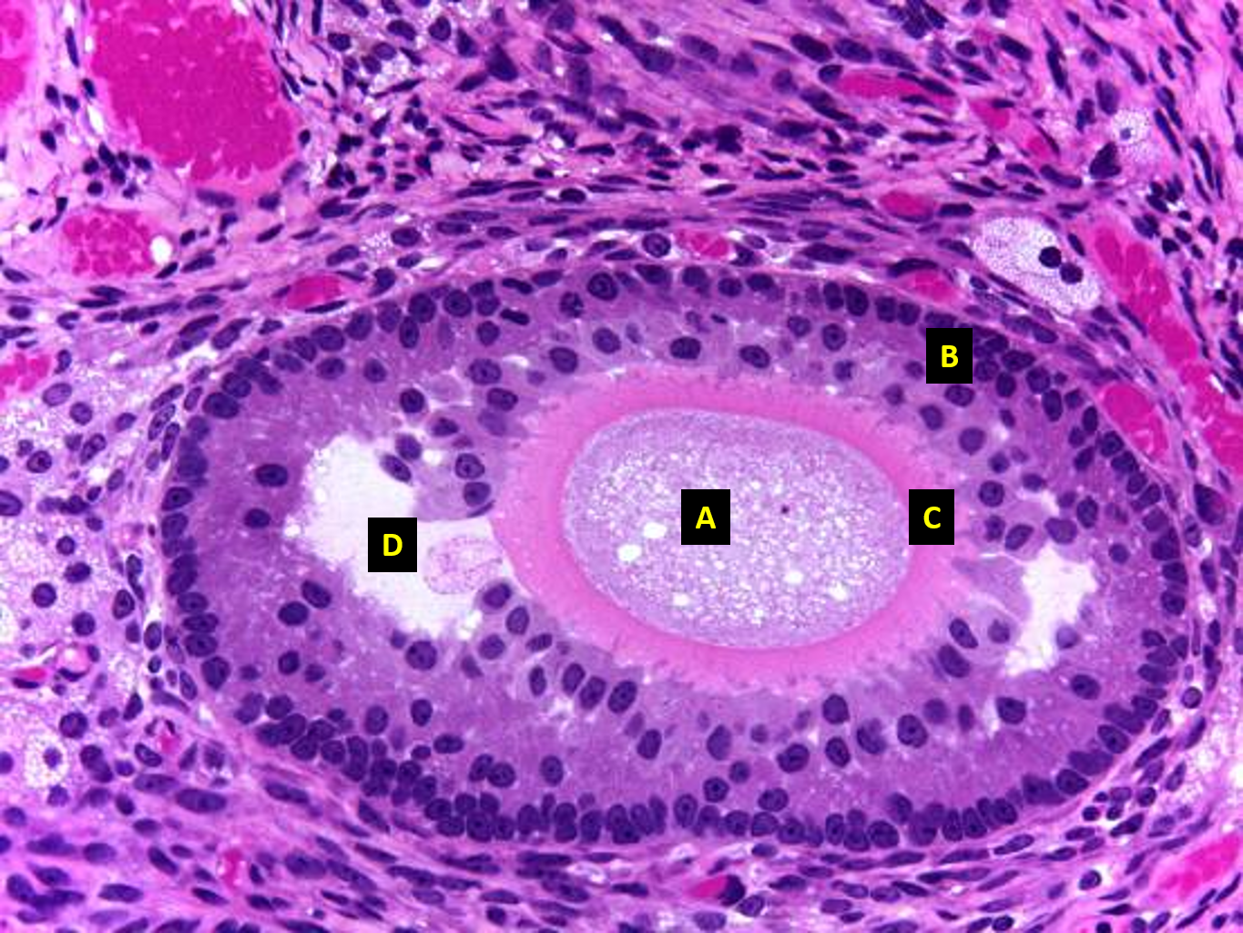
C
zona pelucida
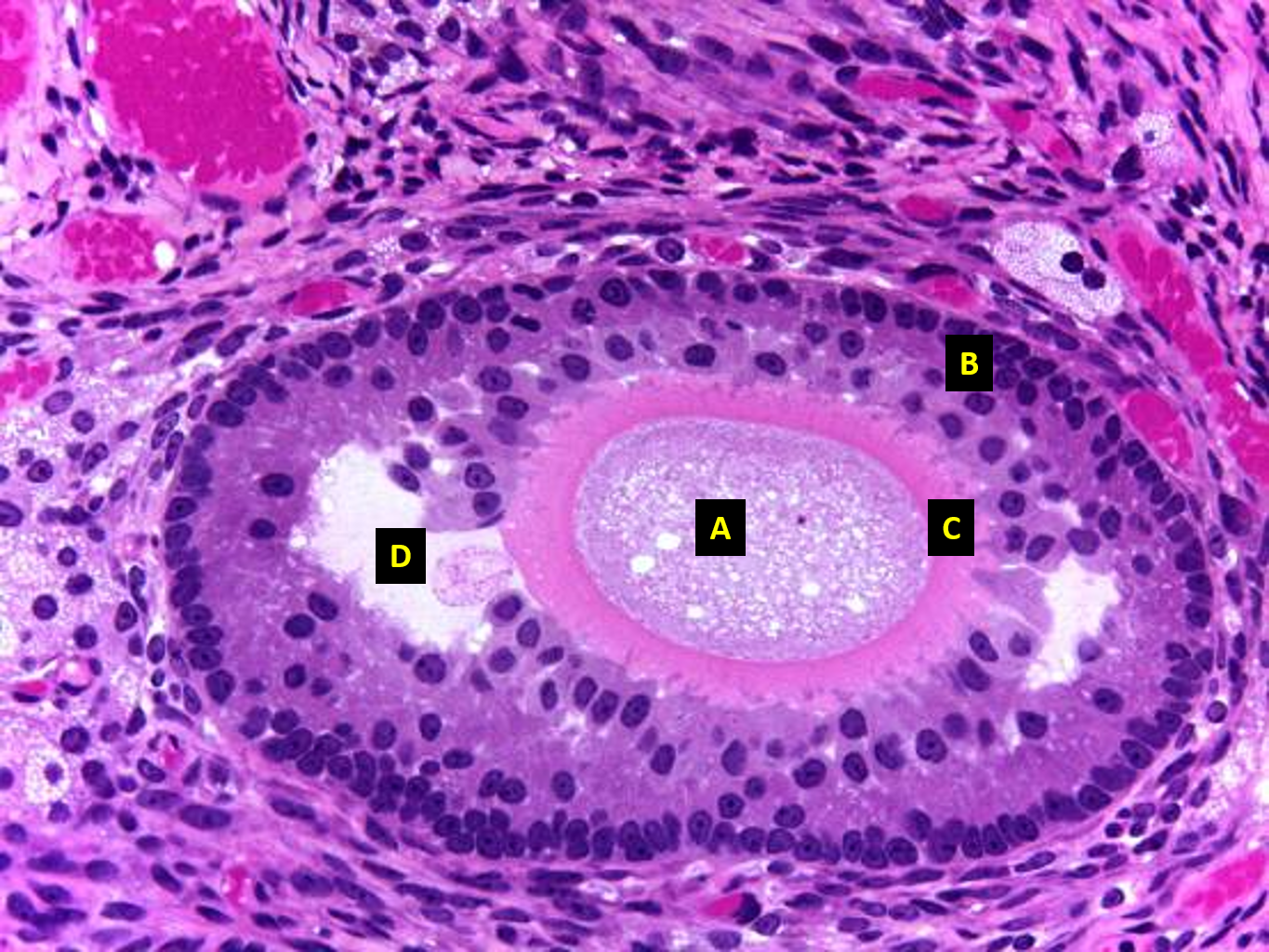
D
antrum
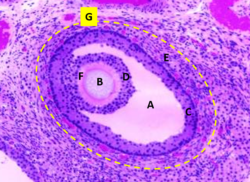
cell b
secondary oocyte
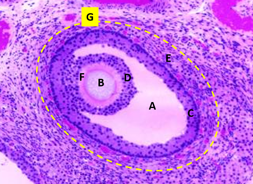
G
tertiary follicle
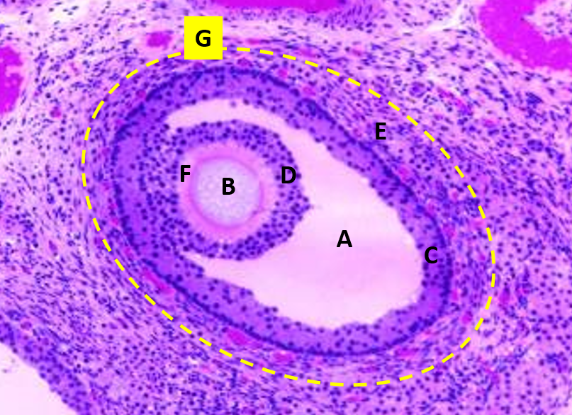
f
zona pellucida
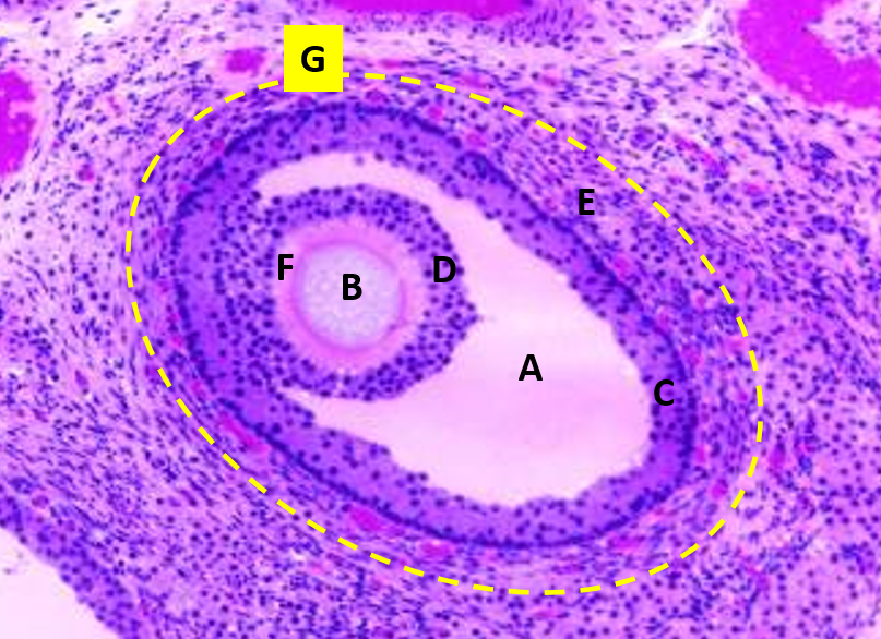
d
corona radiata
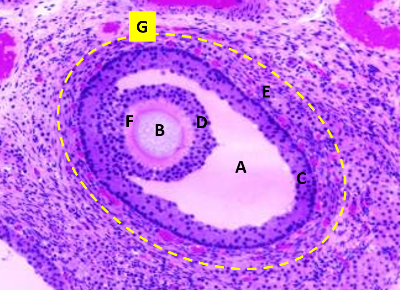
a
antrum
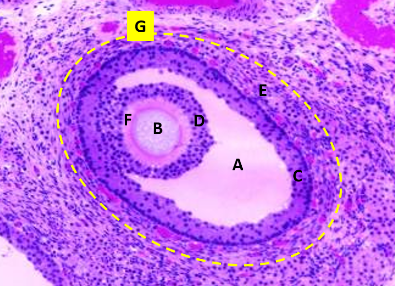
c
follicular cells
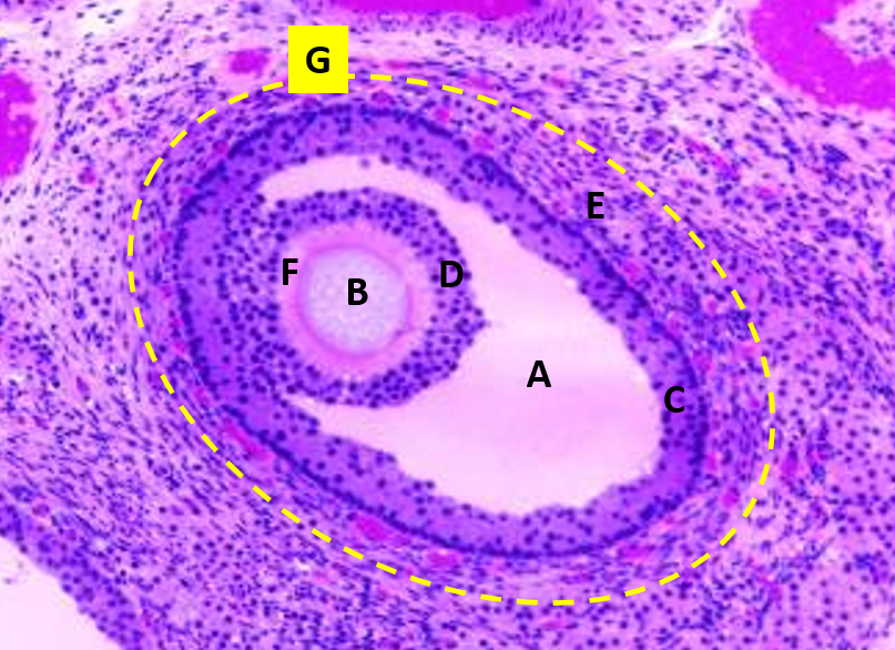
E
thecal cells
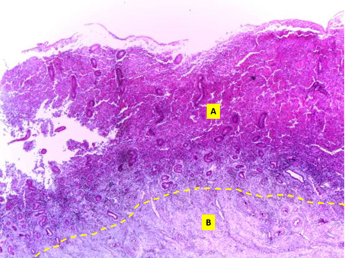
a
endometrium layer of the uterus.
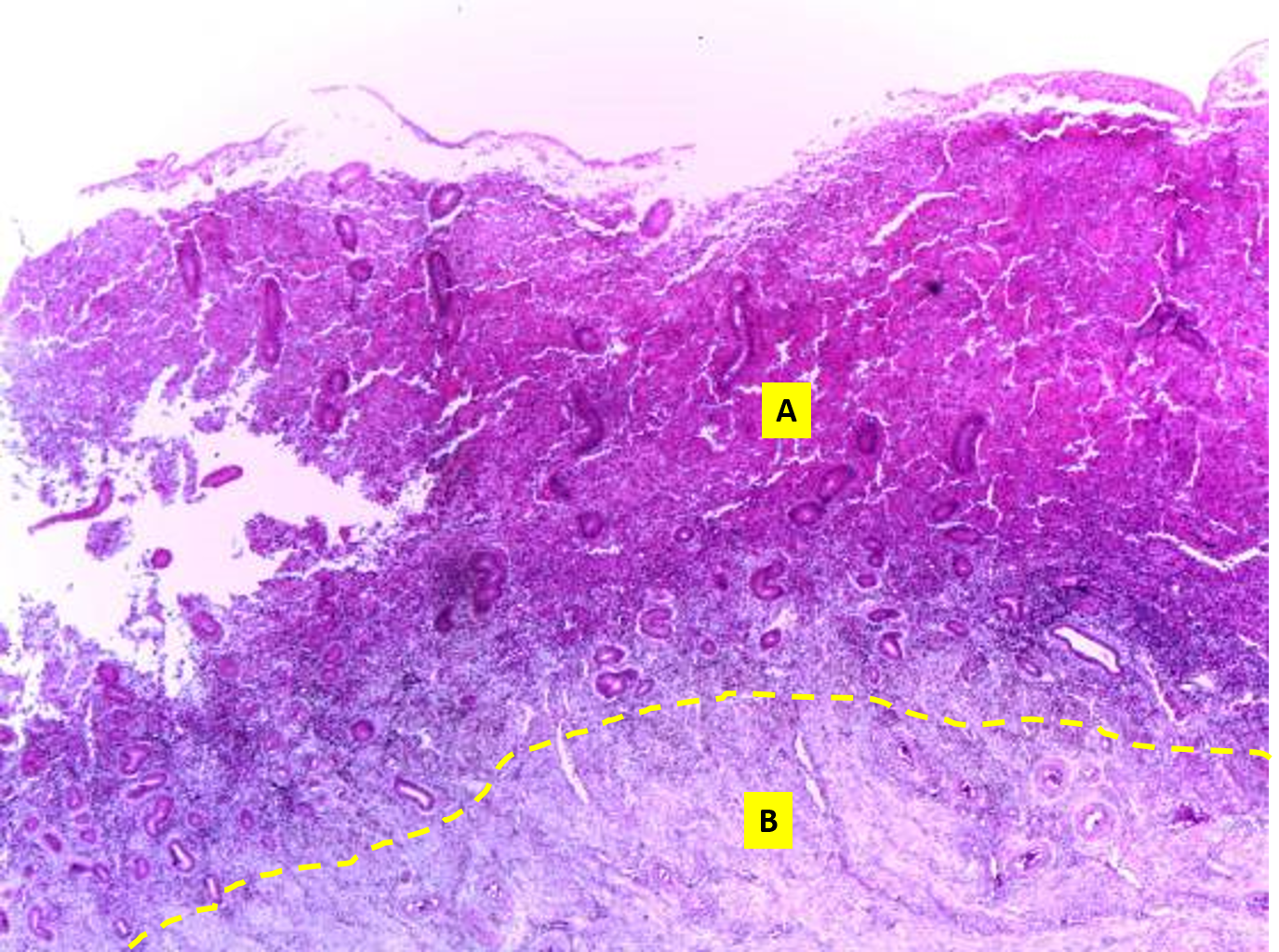
b
myometrium layer of the uterus.
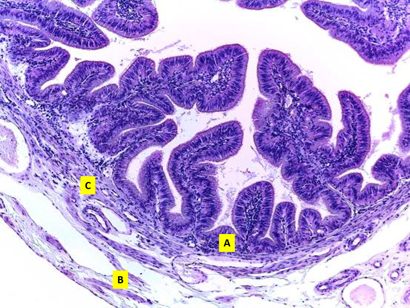
This organ is the
uterine tube
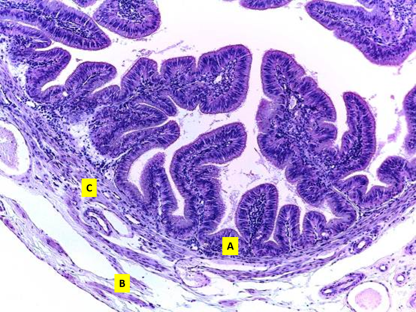
a
mucosa
what is the mucosa composed of
simple columnar
epithelium with cilia.
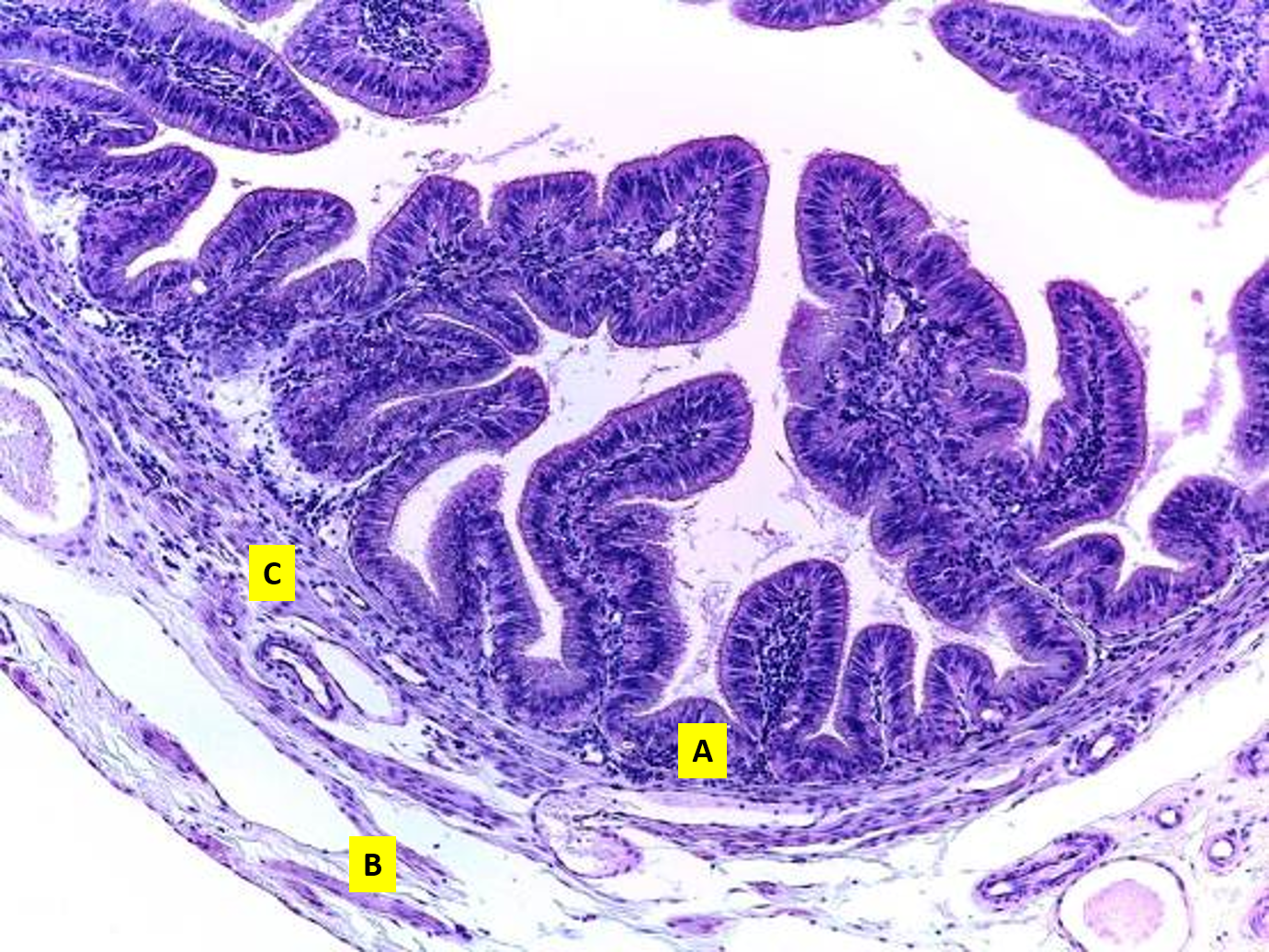
c
muscular coat
what is the muscular coat composed of
smooth muscle
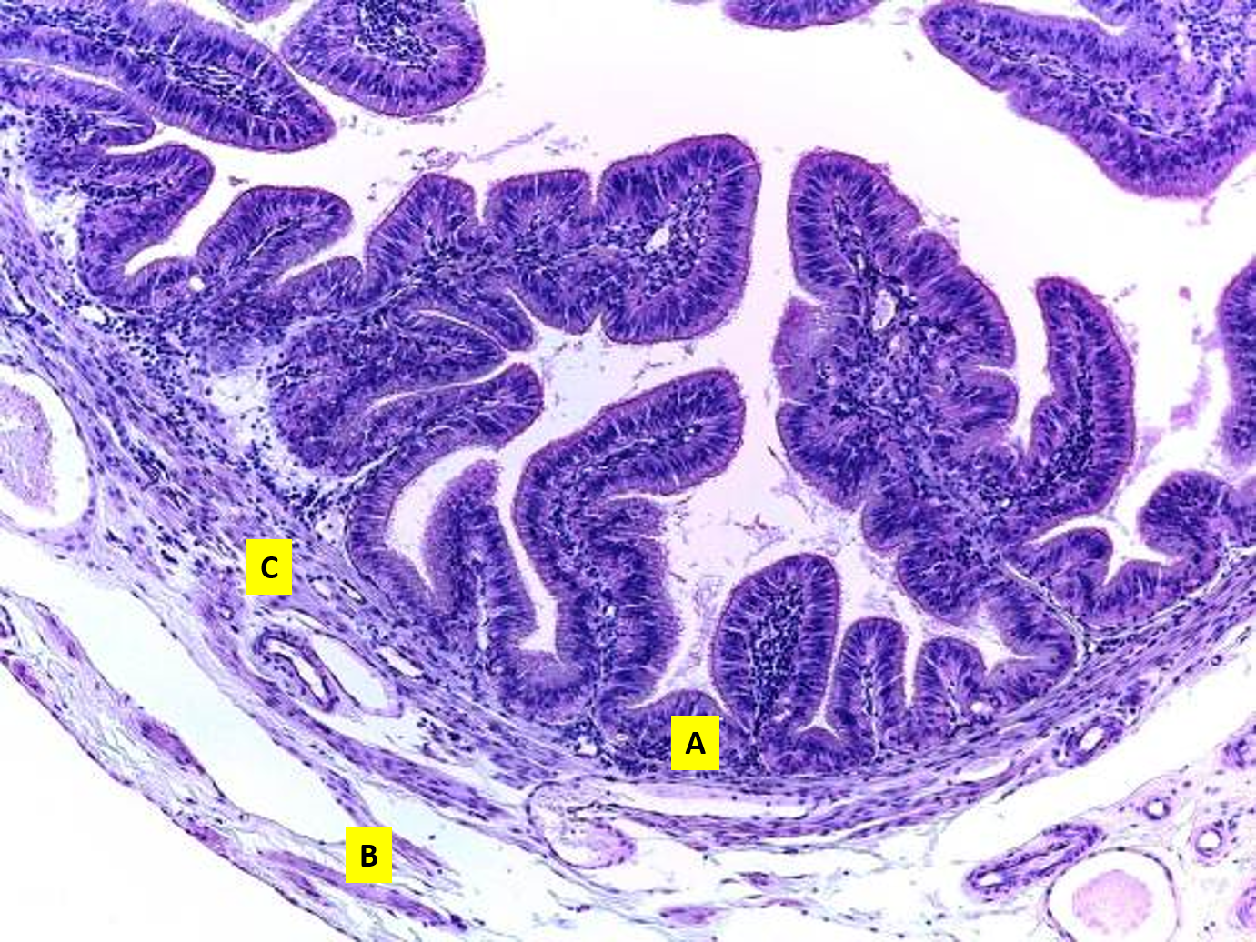
b
serosa coat