Parasitology visual exam
1/31
There's no tags or description
Looks like no tags are added yet.
Name | Mastery | Learn | Test | Matching | Spaced |
|---|
No study sessions yet.
32 Terms
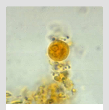
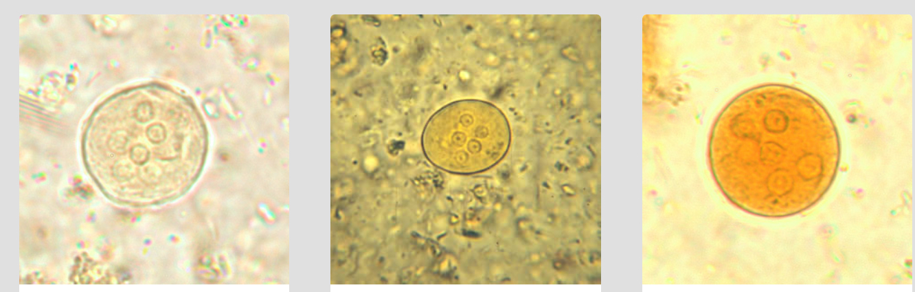
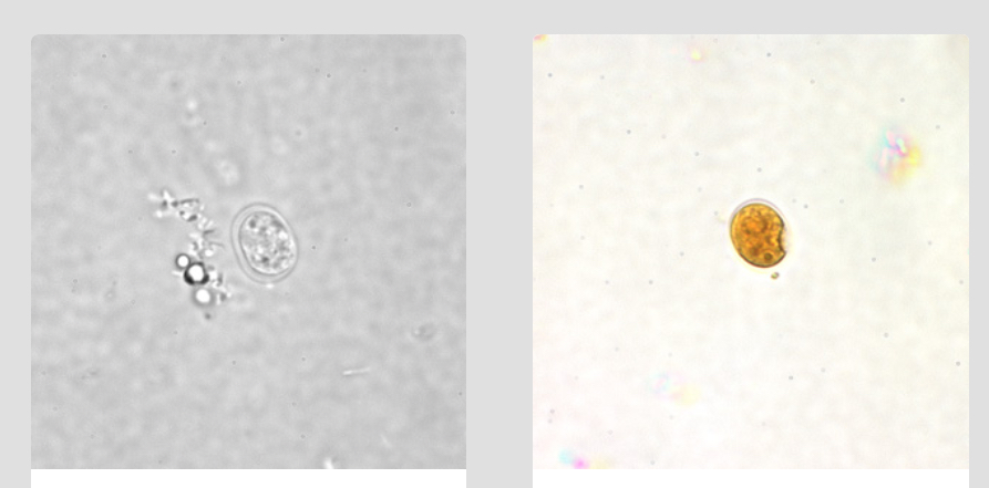
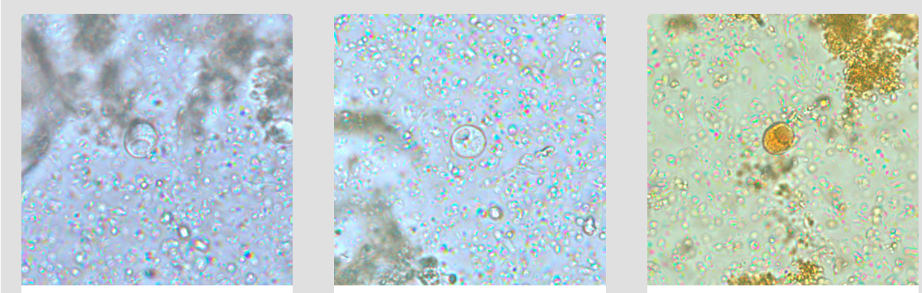
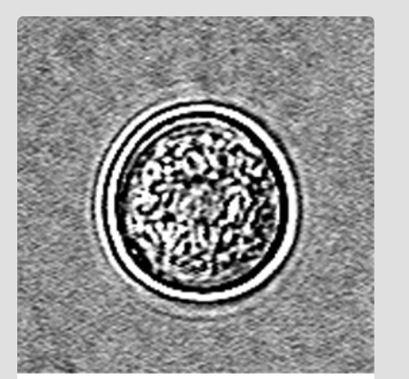
E. Histolytica/E. Dispar cyst unstained wet mount
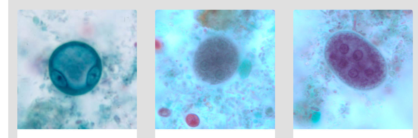
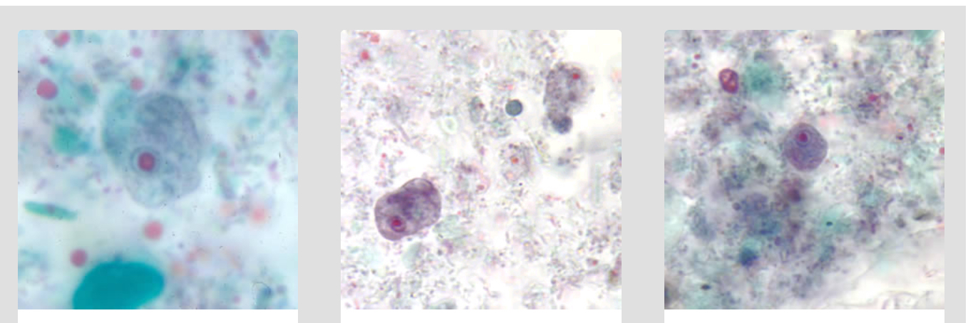
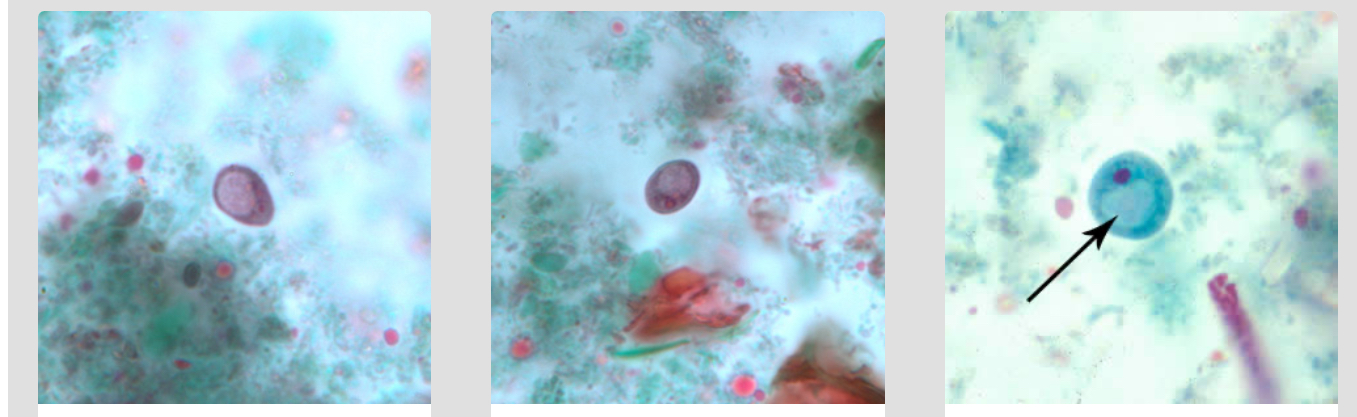
E. Histolytica/E. Dispar Cysts stained w/ trichromatic
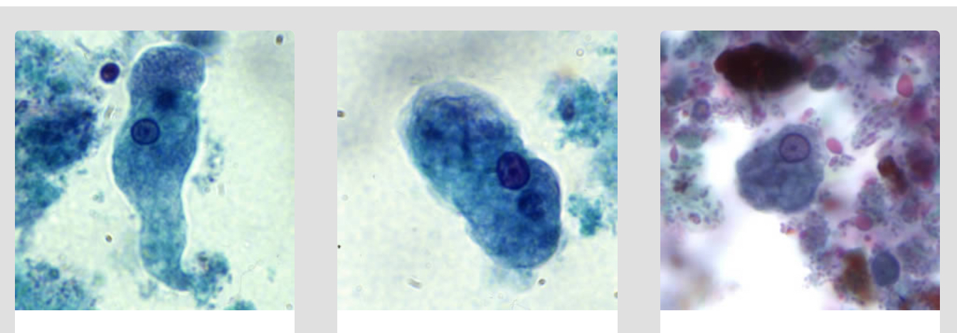
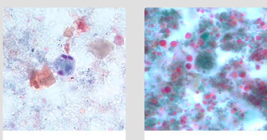
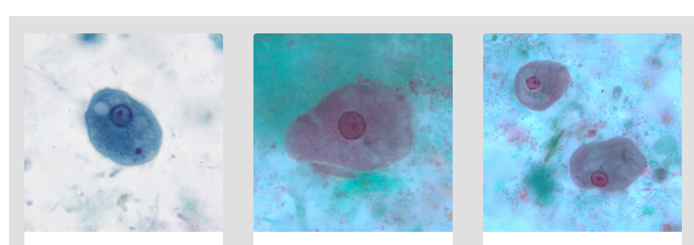
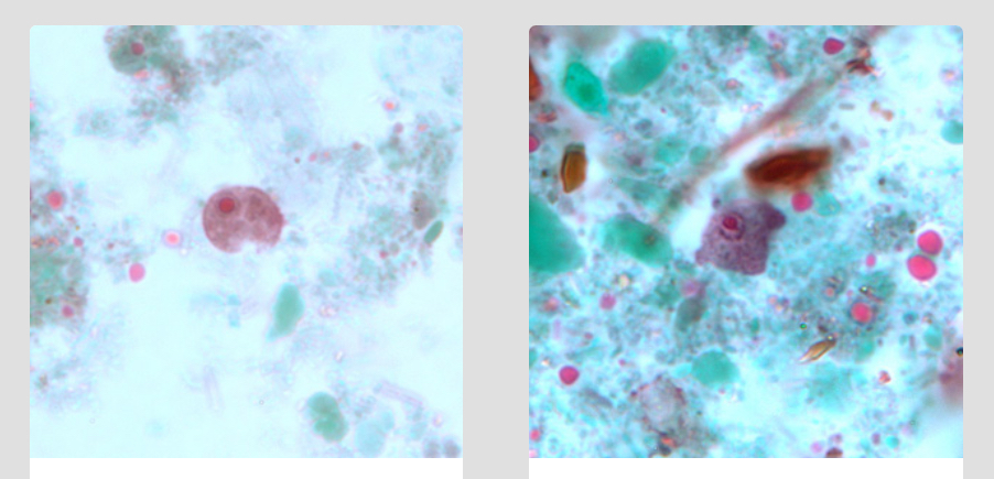
E. Histolytica/E. Dispar trophozoites in direct wet mount
E. Histolytica/E. Dispar trophozoites stained w/ trichromatic
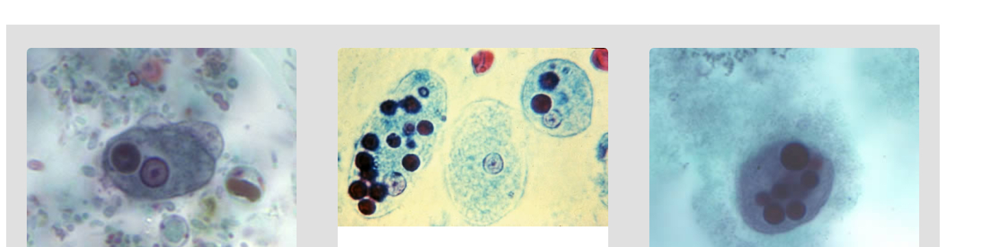
E. Histolytica trophozoites w/ ingested RBCs
NOTE: not generally E. Dispar (but can occur occasionally, not a distinguishing factor)
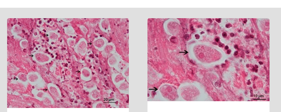
E. histolytica trophozoites in tissue stained with hematoxylin and eosin (H&E).
E. Hartmanni cyst wet mount stained w/ iodine
E. Hartmanni cyst stained w/ trichrome
Note: blunt ends of chromatoid bodies
E. Hartmanni trophozoites stained w/ trichrome
E. Coli cyst in concentrated wet mount
E. Coli cysts stained w/ trichrome
E. Coli trophozoites stained with trichrome
E. Nana cysts in concentrated wet mounts
E. Nana cysts stained w/ trichrome
E. Nana trophozoites stained w/ trichrome
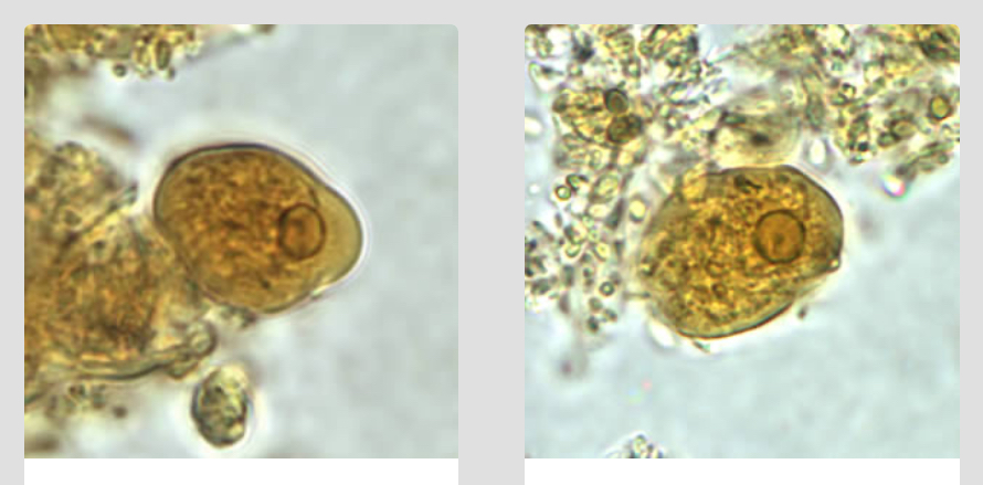
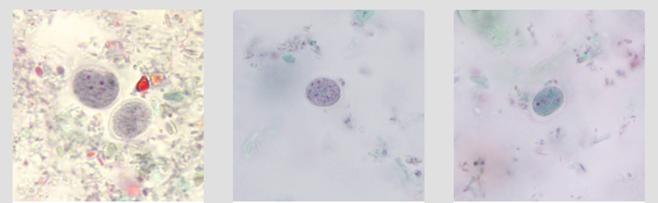
Iodamoeba buetschlii cysts in concentrated wet mounts.
Iodamoeba buetschlii cysts stained w/ trichrome
Iodamoeba buetschlii trophozoites stained w/ trichrome
Naegleria fowleri cysts. Naegleria fowleri does not form cysts in human tissue.
Naegleria fowleri trophozoites.
There are two forms of trophozoites in Naegleria fowleri: ameboid and ameboflagellate, the latter of which is only rarely found in humans (within CSF).
Acanthamoeba spp. cyst
Acanthamoeba spp. Trophozoites

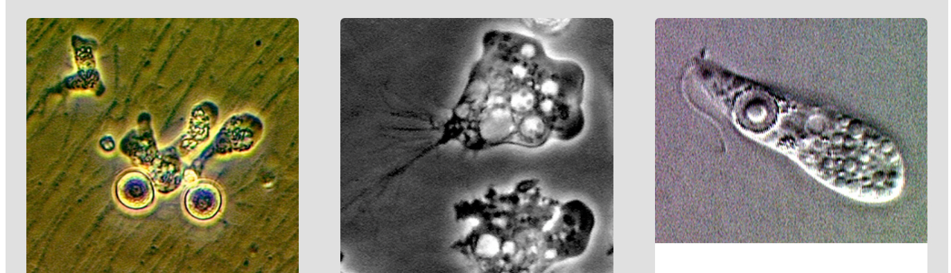
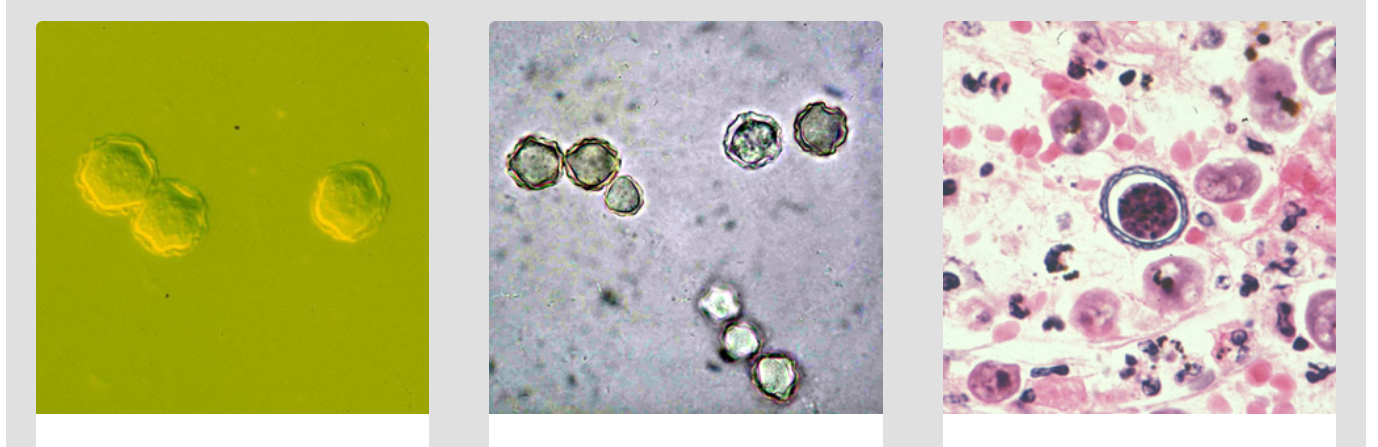
Balamuthia mandrillaris cysts
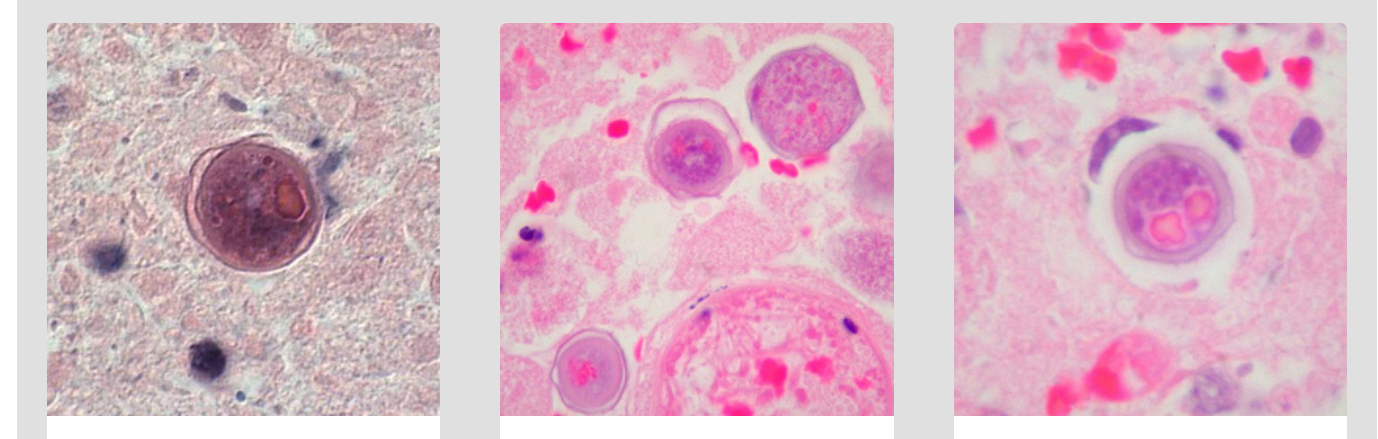
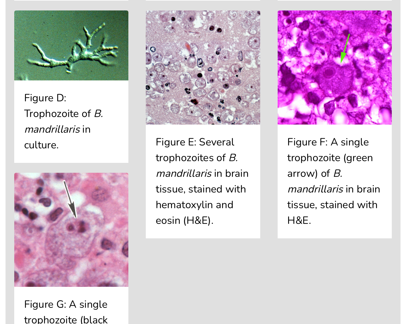
Balamuthia mandrillaris trophozoites

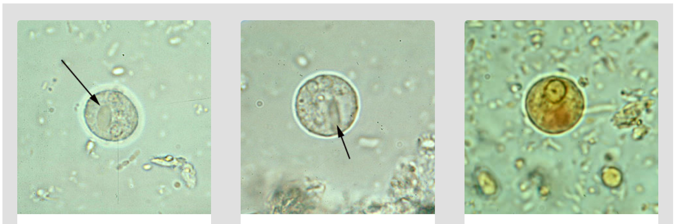
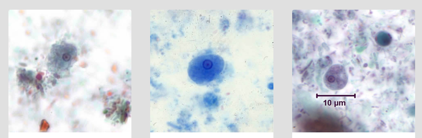
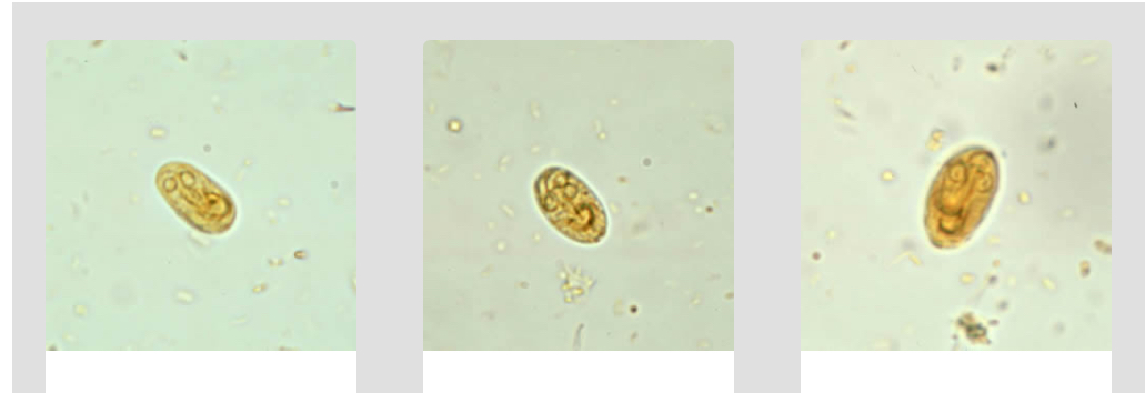
Giardia duodenalis cysts in wet mounts stained with iodine.
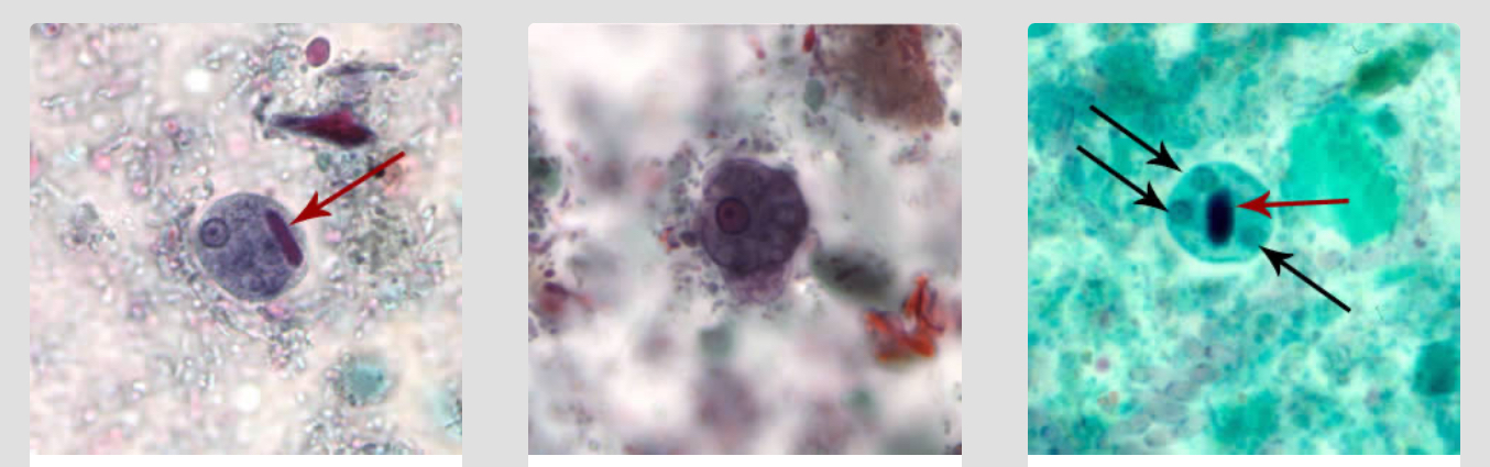
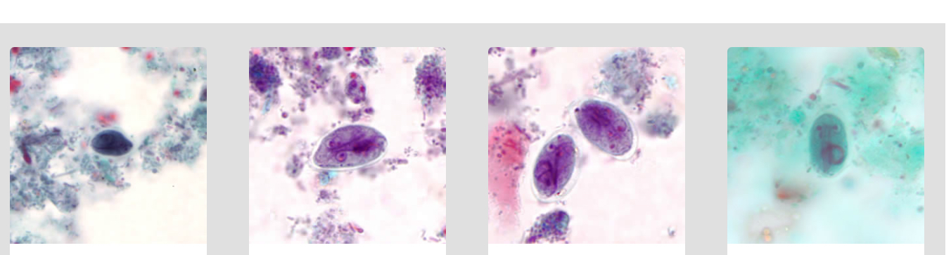
Giardia duodenalis cysts in trichrome stain
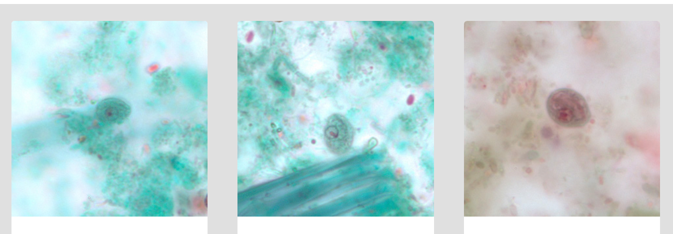
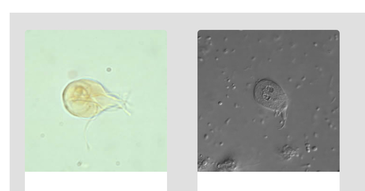
G. duodenalis trophozoites in wet mounts.
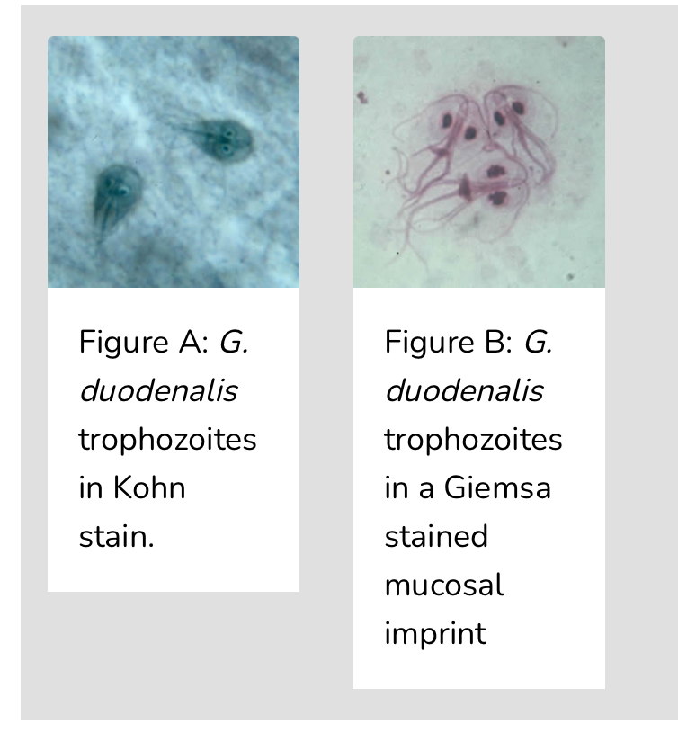
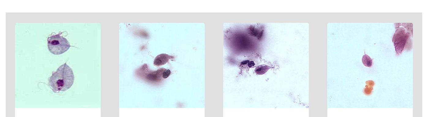
G. duodenalis trophozoites stained with trichrome.
This Image is other unique stains
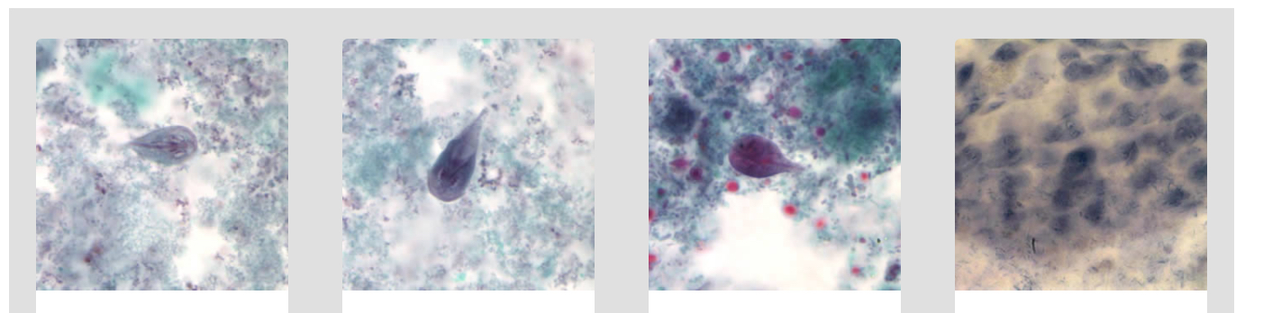
Trichomonas vaginalis trophozoites.
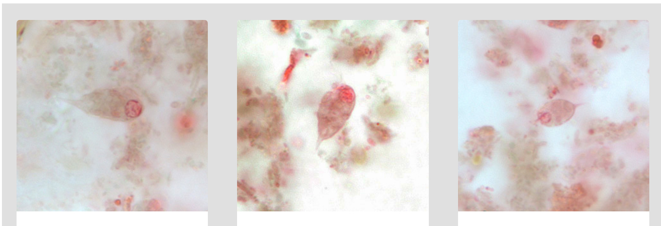
Chilomastix mesnili trophozoites, trichrome stain.
Chilomastix mesnili cysts, trichrome stain